- Etymology
The term “bullous” is derived from the Latin word bulla, meaning bubble or blister, describing the large, air-filled spaces characteristic of this condition. - AKA
Bullae; Giant bullous disease; Vanishing lung syndrome (in extensive cases). - What is it?
Bullous emphysema is a subtype of emphysema characterized by the presence of bullae, which are large air-filled spaces (>1 cm in diameter) that result from alveolar wall destruction. These bullae can displace normal lung parenchyma and impair lung function. - Caused by
- Most common cause
- Cigarette smoking (chronic inhalation of toxic substances).
- Other causes include
- Alpha-1 antitrypsin deficiency: A genetic predisposition linked to panlobular emphysema.
- Chronic lung infections: Persistent inflammation can accelerate bulla formation.
- Idiopathic: Cases with no identifiable cause.
- Mechanical stress: Repeated high pressures, as in ventilator-associated trauma.
- Predisposition in young, thin men: Young, thin males with narrow anteroposterior chest diameters are at increased risk of bullous formation and spontaneous pneumothorax due to exaggerated mechanical forces in the apices (see below).
- Most common cause
- Resulting in
- Loss of lung elastic recoil and reduced ventilation in the affected areas.
- Compression of adjacent functional lung parenchyma, leading to hypoxemia.
- Increased risk of complications such as spontaneous pneumothorax.
- Structural changes
- Large airspaces due to destruction of alveolar walls.
- Loss of functional capillary-alveolar surface area.
- Potential distortion of lung anatomy due to the size and location of bullae.
- Pathophysiology
- Bullae result from coalescence of multiple adjacent destroyed alveoli.
- Progressive destruction is driven by protease-antiprotease imbalance, chronic inflammation, and oxidative stress.
- Pathology
- Bullae are thin-walled structures composed of fibrous tissue and lined by remnants of alveolar epithelium.
- Found in association with surrounding emphysematous changes, particularly in paraseptal emphysema.
- Special Considerations in Young, Thin Men
- Narrow AP Chest Dimensions:
- The acute angle of the lung apex in narrow-chested individuals focuses mechanical forces over a smaller area of lung tissue.
- This creates higher transpulmonary pressure and stress in the apices during respiratory cycles, predisposing this region to alveolar wall destruction and bullous formation.
- Mechanical Stress:
- With reduced structural support from the chest wall and increased local forces, apical alveoli experience exaggerated strain, leading to progressive bullous changes.
- Risk of Spontaneous Pneumothorax:
- Thin-walled bullae near the pleura are more prone to rupture under stress, causing air leakage into the pleural space and pneumothorax.
- Narrow AP Chest Dimensions:
- Diagnosis
- Clinical
- Symptoms: Dyspnea, reduced exercise tolerance, and symptoms of pneumothorax if rupture occurs.
- Signs: Hyperinflation of the chest, diminished breath sounds, or hyperresonance on percussion.
- Radiology
- CXR: Large, air-filled lucencies with thin walls, commonly in the upper lobes.
- CT: Best modality for evaluating bullae size, distribution, and associated complications.
- Clinical
- Radiology Detail
- CXR
- Findings
- Hyperlucent areas with well-defined thin walls, often seen in the apices.
- Signs of hyperinflation and diaphragm flattening in severe cases.
- Associated Findings
- Signs of pneumothorax in case of rupture.
- Findings
- CT
- Parts
- Bullae commonly occur in association with paraseptal or centrilobular emphysema.
- Size
- Bullae are >1 cm in diameter and can grow to occupy a significant portion of a lobe.
- Shape
- Rounded or irregular.
- Position
- Predominantly in the upper lobes but can occur throughout the lungs.
- Exaggerated transpulmonary pressure in the lung apices predisposes this region to bullous formation.
- Character
- Thin-walled, air-filled spaces that retain some remnants of alveolar wall structure (e.g., fibrous tissue). While they lack the vascular markings seen in emphysema, they are not entirely devoid of internal components.
- Time
- Progressive enlargement over time, potentially leading to rupture.
- Associated Findings
- Adjacent lung compression, pneumothorax, or infection.
- Parts
- Distinction Between Bullae and Cysts
- Bullae:
- Develop from alveolar wall destruction in emphysema.
- Have thin fibrous walls with remnants of alveolar structure.
- Typically lack a true epithelial lining.
- Arise from preexisting lung tissue.
- Cysts:
- Thin-walled, air- or fluid-filled structures with no internal architecture.
- Lined by epithelium or fibrous tissue.
- Can arise from congenital or acquired processes, distinct from emphysematous destruction.
- Bullae:
- CXR
- Pulmonary Function Tests (PFTs)
- Typical findings of airflow obstruction:
- Reduced FEV1/FVC ratio.
- Increased residual volume and total lung capacity.
- Decreased DLCO in severe cases.
- Typical findings of airflow obstruction:
- Recommendations
- Smoking cessation to prevent progression.
- Monitor for complications such as pneumothorax.
- Consider surgical interventions (e.g., bullectomy) in patients with symptomatic or large bullae compressing healthy lung tissue.
- Key Points and Pearls
- Bullous emphysema is commonly associated with paraseptal emphysema but can occur with other subtypes.
- Exaggerated physical forces in the apices, including increased transpulmonary pressure and mechanical stress, predispose these regions to bullous formation.
- Young, thin men with narrow chest dimensions are at particular risk for bullae and spontaneous pneumothorax.
- CT imaging is the gold standard for evaluating bullae size, extent, and impact on adjacent lung.
- Bullectomy or lung volume reduction surgery can be considered for large, symptomatic bullae.

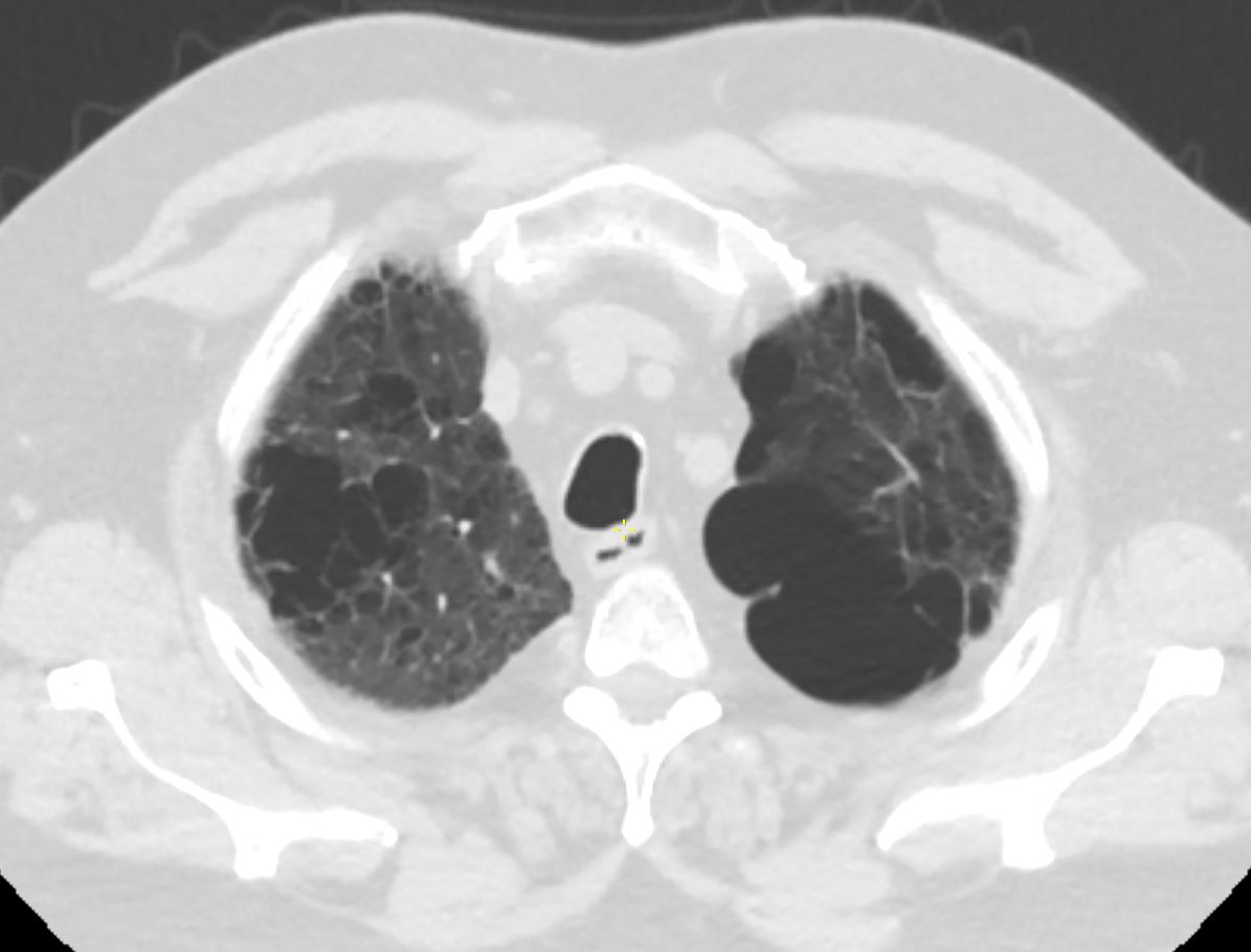
CT scan in the axial plane of a 64- year-old man with emphysema shows bilateral apical bullous lung disease,
Ashley Davidoff MD TheCommonVein.Net 136440

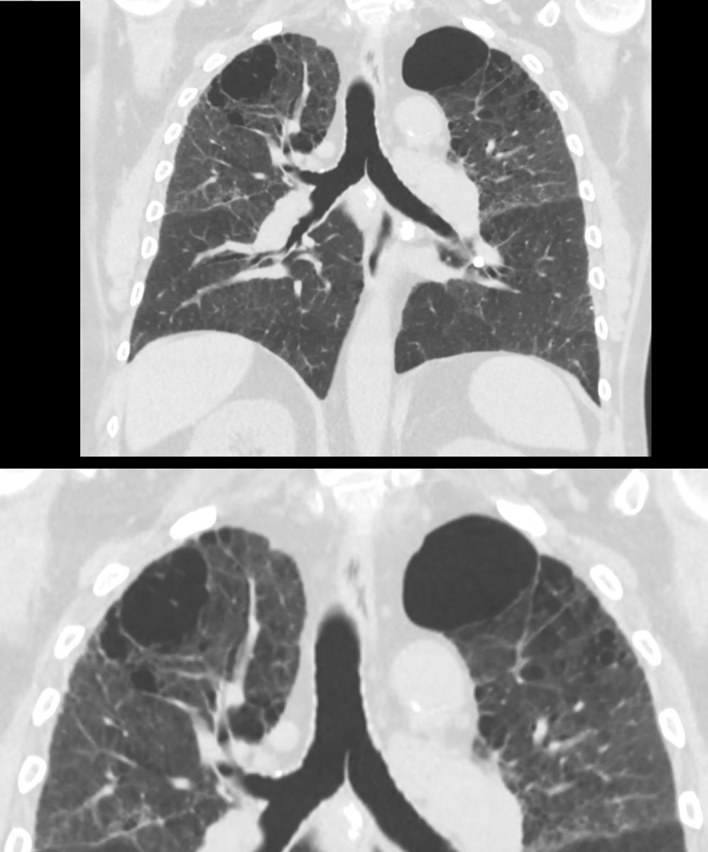
CT scan in the coronal plane of a 64- year-old man with emphysema shows bilateral apical bullous lung disease, magnified in the lower image
Ashley Davidoff MD TheCommonVein.Net 136439c

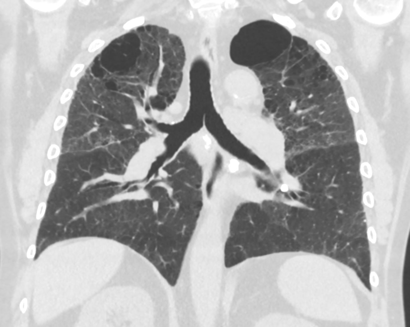
CT scan in the coronal plane of a 64- year-old man with emphysema shows bilateral apical bullous lung disease
Ashley Davidoff MD TheCommonVein.net 136439
