
- 60 yo male smoker with
- severe psoriasis,
- h/o hypercalcemia,
- s/p STEMI 1
- drug eluting stent to LAD, 8 years prior
- obesity, HTN, hyperlipidemia,
- aflutter
- s/p ablation of cavotricuspid isthmus,
- echo
- EF 40%,
- RV mod dil
- Lungs
- Persistent diffuse ground glass opacities
- Lung bx revealed desquamative interstitial pneumonitis (DIP).
- PFTs 1
- mild restriction and mild diffusion limitation, slight worsening
- DIP overall fairly stable and
- likely active at a low grade level in the presence of ongoing heavy smoking.
Normal CXR DIP
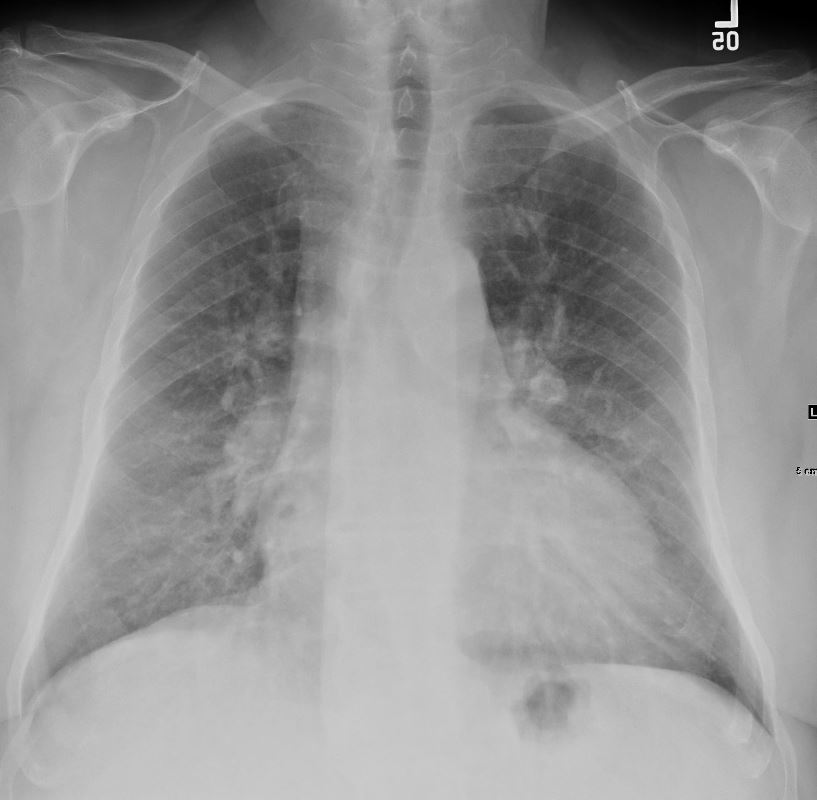
60-year-old male smoker with a history of progressive dyspnea. Frontal chest Xray shows mild increase in lower lobe density likely reflecting overlying soft tissues of the breasts (known obesity) rather than the diffuse ground glass changes noted on subsequent CT which were more prominent in the upper lobes
Pathology confirmed a diagnosis of DIP
Ashley Davidoff MD TheCommonVein.net 253Lu 136003
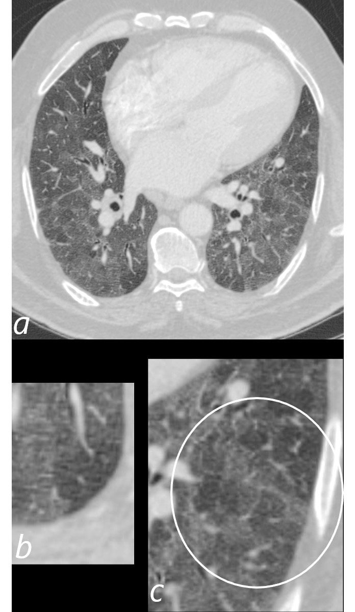
DIP Diffuse Ground Glass Changes in the Upper Lobes
Small Secondary Lobules
Some Centrilobular Nodules
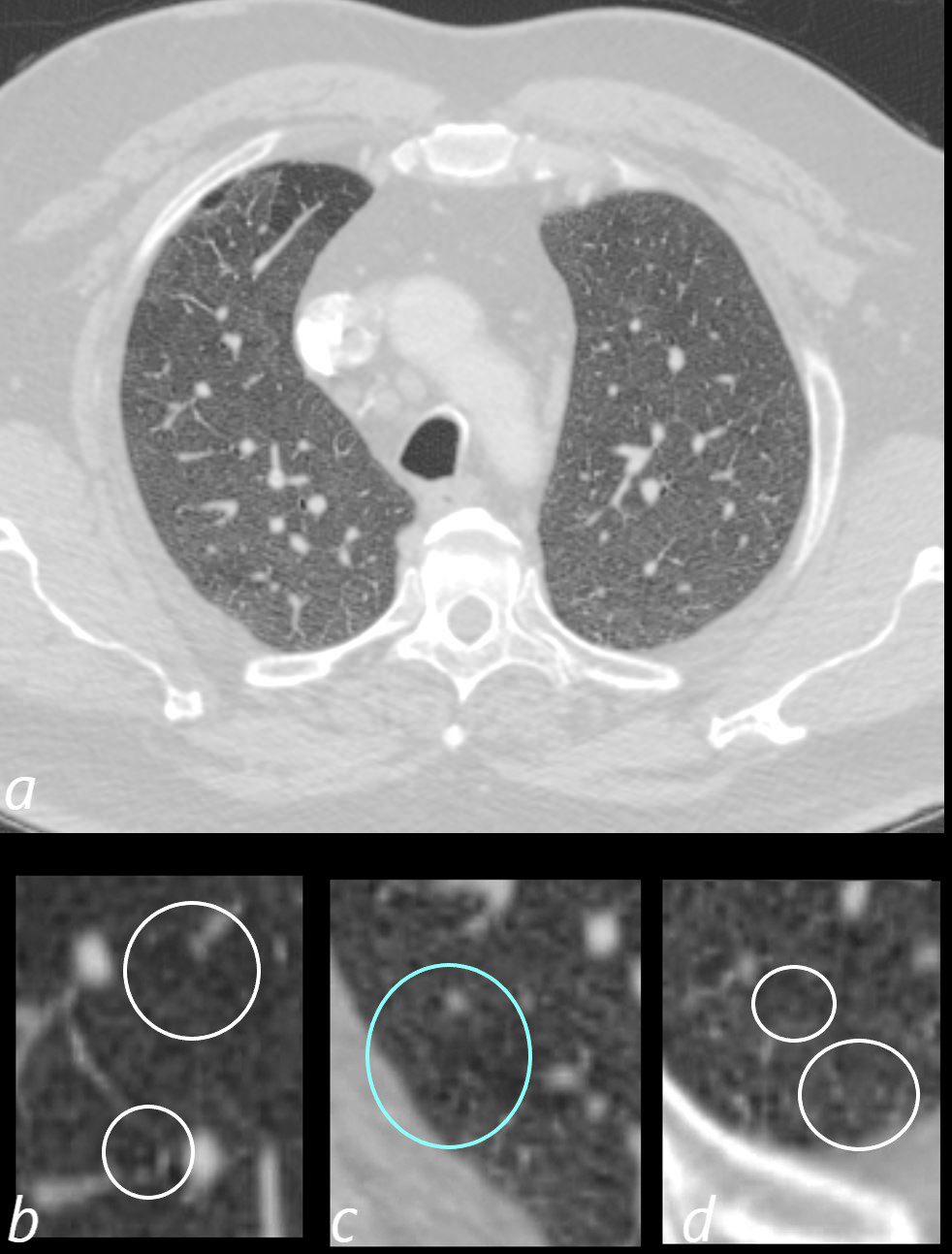
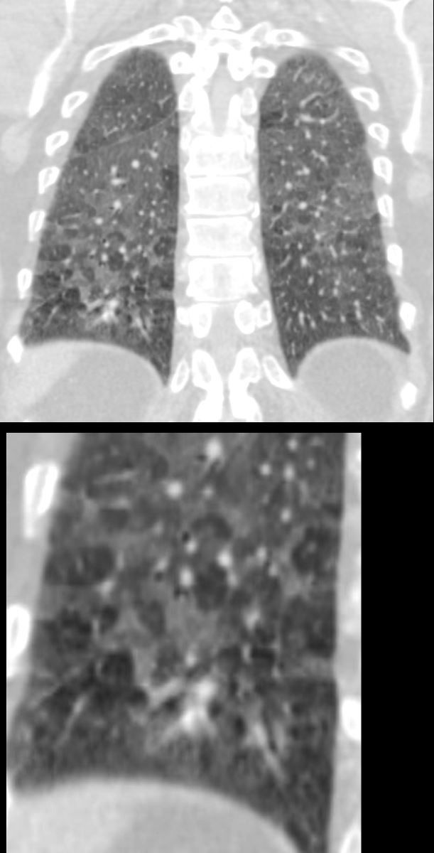
60-year-old male smoker with a history of progressive dyspnea. Axial CT through the upper lung fields shows diffuse ground glass changes with minimal heterogeneity. The secondary lobules appear relatively small with slightly thickened septa and some have prominent centrilobular nodules (b and c within the white rings) suggesting small airway disease Part of the anterior segment of the right upper lobe is spared.
Pathology confirmed a diagnosis of DIP
Ashley Davidoff MD TheCommonVein.net 253Lu 136004cL
DIP
Diffuse Patchy Ground Glass Changes and
Sparing of the Anterior Segment of the Right Upper Lobe
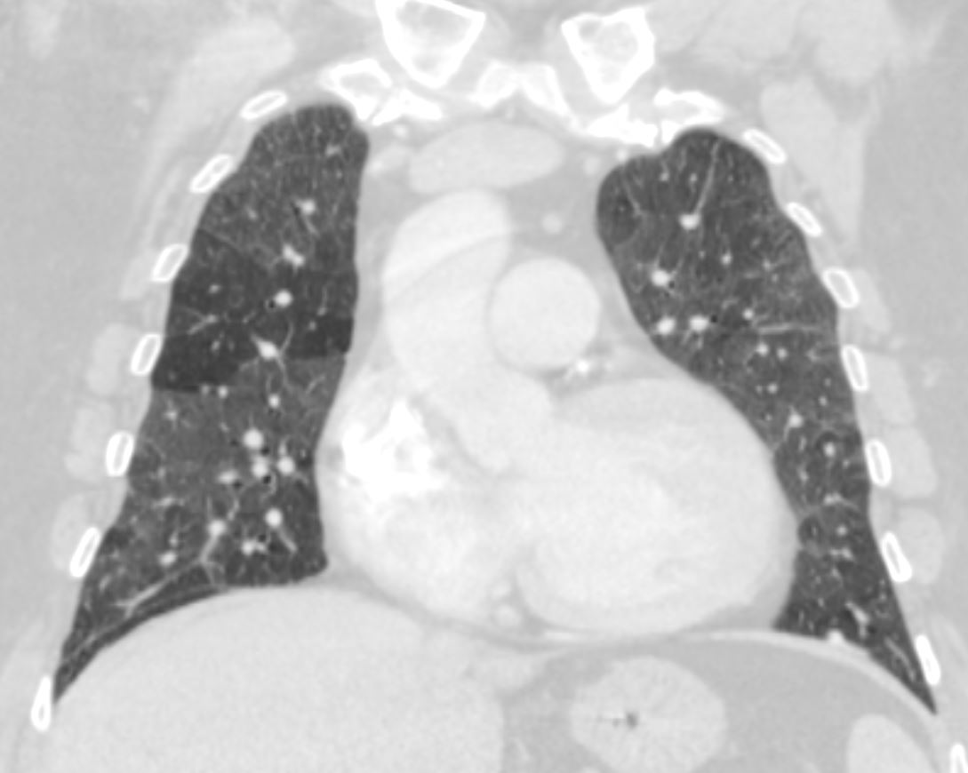
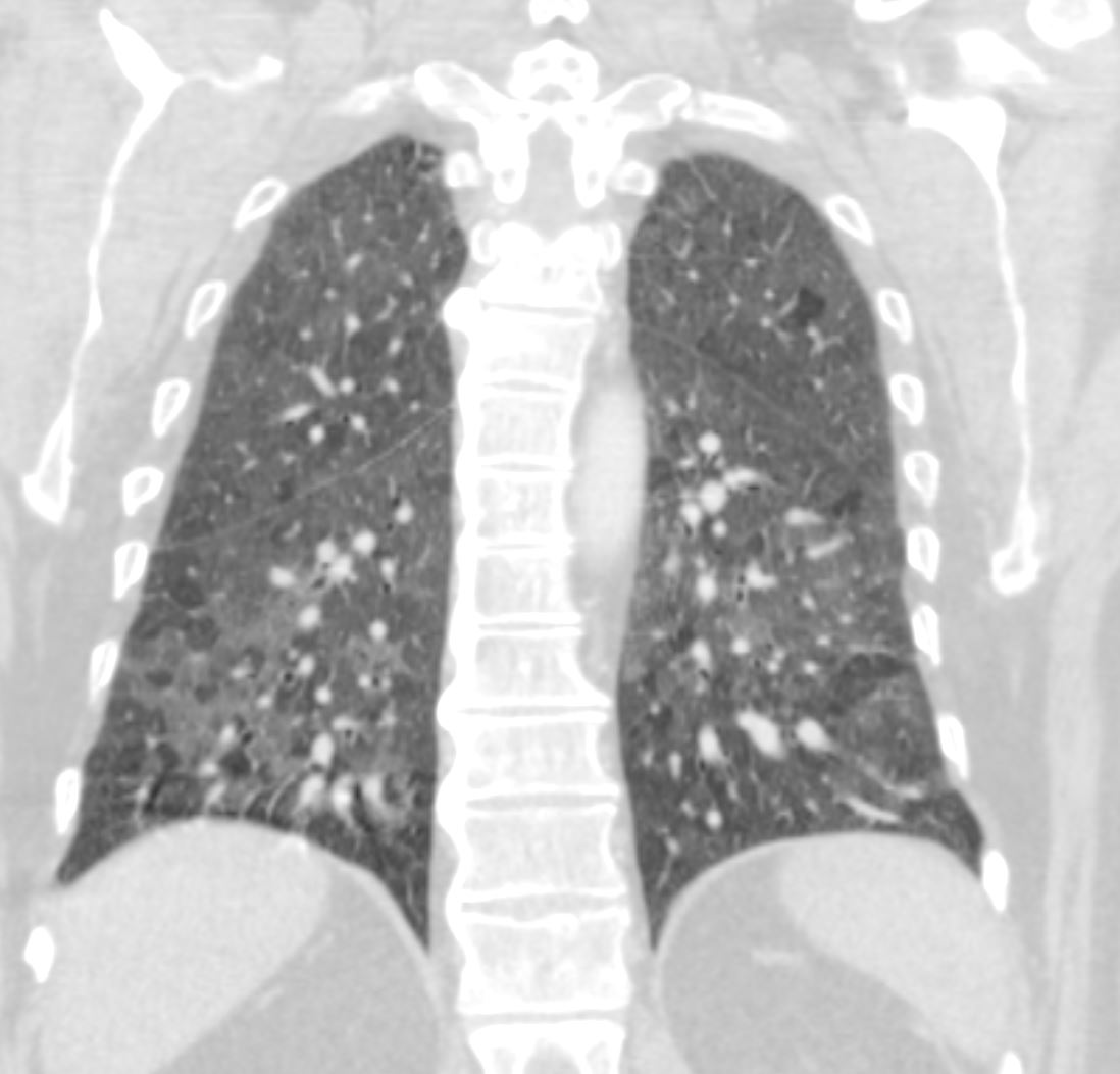
60-year-old male smoker with a history of progressive dyspnea. Coronal CT through the mid lung fields at the level of the left ventricle shows diffuse patchy ground glass changes and mosaic attenuation. There is sparing of the anterior segment of the right upper lobe.
Pathology confirmed a diagnosis of DIP
Ashley Davidoff MD TheCommonVein.net 253Lu 136011
Diffuse Patchy Ground Glass Changes and
Sparing of the Anterior Segment of the Right Upper Lobe
Mosaic Attenuation
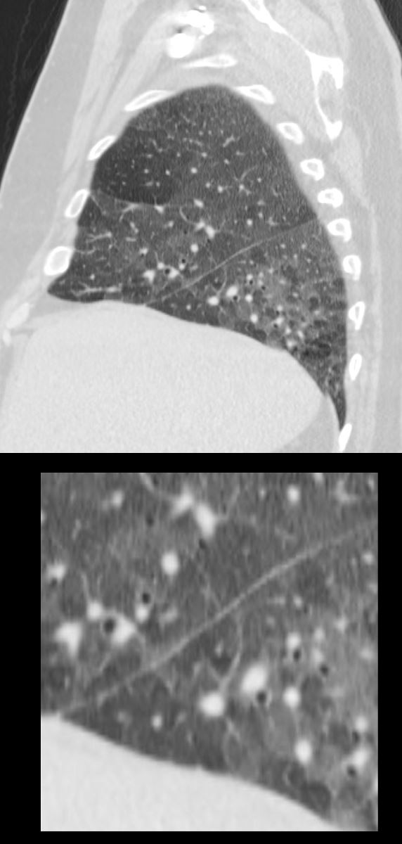
60-year-old male smoker with a history of progressive dyspnea. Sagittal CT through the lateral right mid lung shows diffuse patchy ground glass changes and mosaic attenuation with greater heterogeneity of the lower lobe. There is sparing of the anterior segment of the right upper lobe.
A few of the secondary lobules show prominent centrilobular nodules reflecting a small airways component but the predominant pattern is an alveolar pattern
Pathology confirmed a diagnosis of DIP
Ashley Davidoff MD TheCommonVein.net 253Lu 136015c
60-year-old male smoker with a history of progressive dyspnea. Axial CT through the mid lung fields at the level of the carina shows diffuse ground glass changes with mild heterogeneity. The secondary lobules appear relatively small with slightly thickened septa suggesting small airway disease The anterior segment of the right upper lobe is relatively spared.
Pathology confirmed a diagnosis of DIP
Ashley Davidoff MD TheCommonVein.net 253Lu 136006
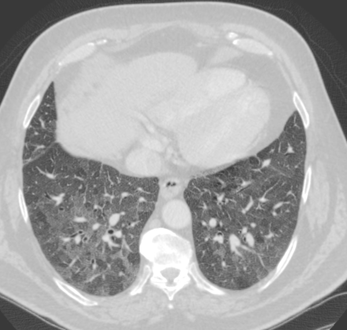
60-year-old male smoker with a history of progressive dyspnea. Axial CT through the lower lung fields at the level of the left atrium shows diffuse ground glass changes with more prominent heterogeneity and mosaic attenuation. The secondary lobules appear relatively small with slightly thickened septa.
Pathology confirmed a diagnosis of DIP
Ashley Davidoff MD TheCommonVein.net 253Lu 136007
Heterogeneous Ground Glass Changes in the Lower Lung Fields with Mosaic Attenuation
60-year-old male smoker with a history of progressive dyspnea. Axial CT through the lower lung fields at the level of the left atrium shows diffuse ground glass changes with more prominent heterogeneity and mosaic attenuation (b and c). Some of secondary lobules are expanded, with some with slightly thickened septa and prominent centrilobular nodules likely indicating small airway involvement (c, white ring).
Pathology confirmed a diagnosis of DIP
Ashley Davidoff MD TheCommonVein.net 253Lu 136008cL
DIP
Heterogeneous Ground Glass Changes in the Lower Lung Fields with Mosaic Attenuation
60-year-old male smoker with a history of progressive dyspnea. Axial CT through the lower lung fields show diffuse ground glass changes with more prominent heterogeneity and mosaic attenuation. The secondary lobules are normal in size.
Pathology confirmed a diagnosis of DIP
Ashley Davidoff MD TheCommonVein.net 253Lu 136010
Diffuse Ground Glass Changes with
Patchy Changes More Prominent at the Lung Bases
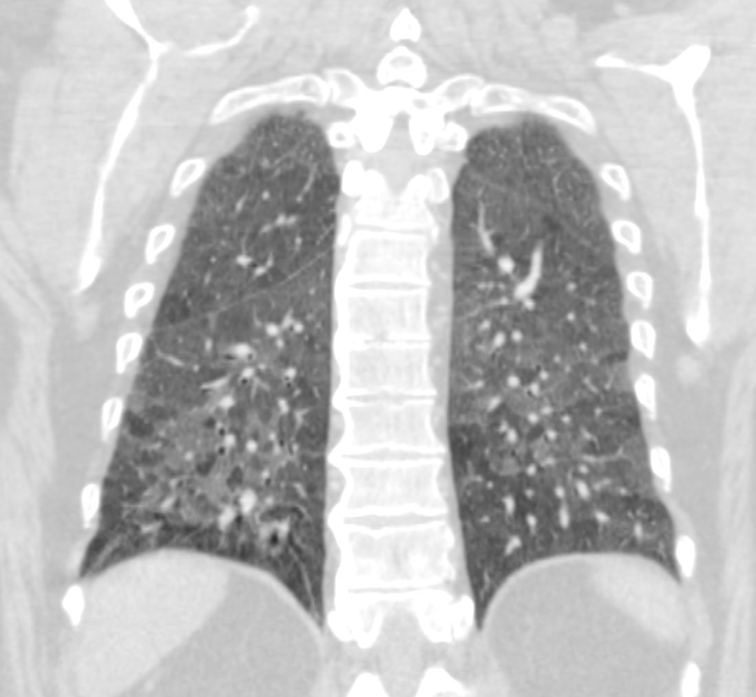
60-year-old male smoker with a history of progressive dyspnea. Coronal CT through the posterior lung fields at the level of the vertebral column shows extensive patchy ground glass changes and mosaic attenuation. The lower lobes show more prominent parenchymal heterogeneity.
Pathology confirmed a diagnosis of DIP
Ashley Davidoff MD TheCommonVein.net 253Lu 136012
60-year-old male smoker with a history of progressive dyspnea. Coronal CT through the posterior lung fields at the level of the vertebral column shows extensive patchy ground glass changes and mosaic attenuation. The lower lobes show more prominent parenchymal heterogeneity.
Pathology confirmed a diagnosis of DIP
Ashley Davidoff MD TheCommonVein.net 253Lu 136013
Desquamative Interstitial Pneumonia Diffuse Ground Glass Changes with Patchy Changes more Prominent at the Lung Bases
60-year-old male smoker with a history of progressive dyspnea. Coronal CT through the posterior lung fields at the level of the vertebral column shows extensive patchy ground glass changes and mosaic attenuation. A few of the secondary lobules show prominent centrilobular nodules reflecting a small airways component but the predominant pattern is an alveolar pattern
Pathology confirmed a diagnosis of DIP
Ashley Davidoff MD TheCommonVein.net 253Lu 136014c
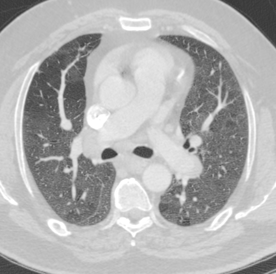
Desquamative Interstitial Pneumonia –
Mediastinal Adenopathy
60-year-old male smoker with a history of progressive dyspnea. Axial CT through the level of the left pulmonary artery shows borderline enlarged mediastinal lymph nodes.
Pathology of the lung confirmed a diagnosis of DIP
Ashley Davidoff MD TheCommonVein.net 253Lu 136017
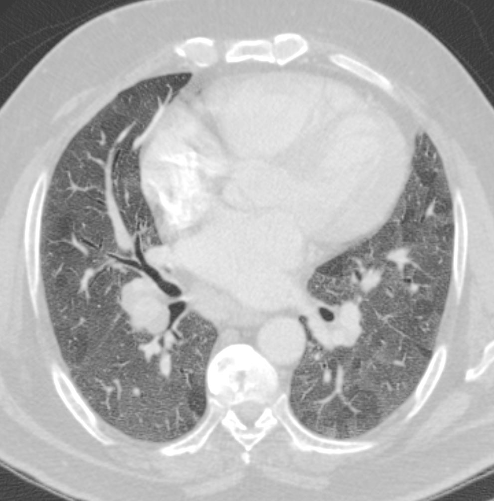
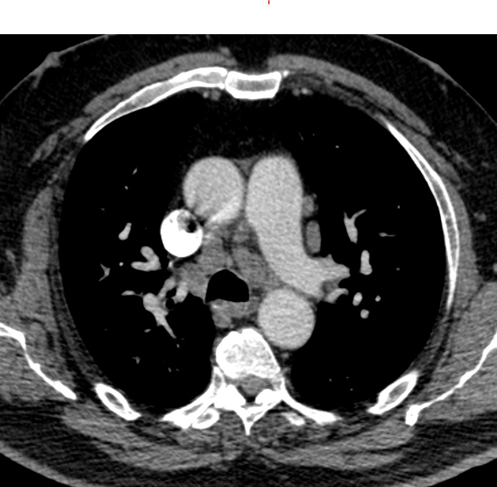
Borderline Enlarged MPA
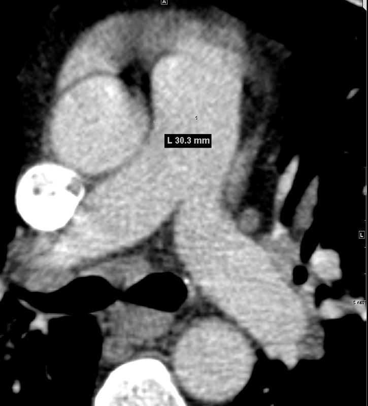
60-year-old male smoker with a history of progressive dyspnea. Axial CT through the level of the main pulmonary artery shows borderline enlarged MPA at 30.3mms
Pathology of the lung confirmed a diagnosis of DIP
Ashley Davidoff MD TheCommonVein.net 253Lu 136018