
Active
- Cavitation:
- result from
- necrosis and liquefaction of
- lung parenchyma.
- result from
- Consolidation:
- dense, homogeneous opacities
- reflecting the accumulation of inflammatory cells, bacteria, and cellular debris,
- Miliary TB:
- disseminates through the bloodstream,
- Tree-in-Bud Appearance:
- associated with bronchogenic spread of infection and may be seen in active TB.
- Pleural Effusion:
Consolidation
Necrotizing Pneumonia Left Upper Lobe
38-year-old male with HIV
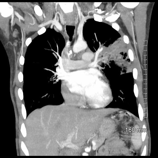
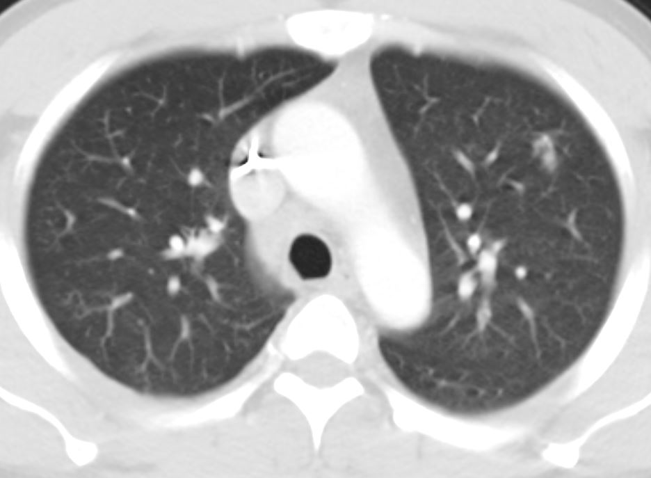
38-year-old male with HIV presents with cough CT scan in the coronal plane shows a focal consolidation in the left upper lobe.
Lab tests confirmed a diagnosis of TB
Ashley Davidoff MD TheCommonVein.net 256Lu 136093c
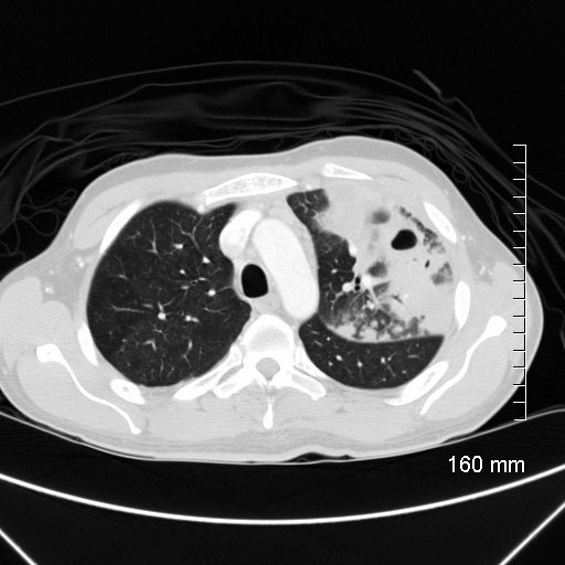
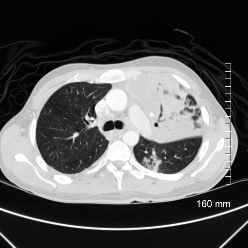
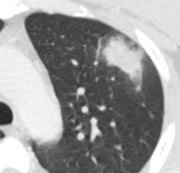
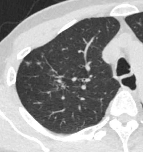
38-year-old male with HIV presents with cough CT scan in the axial plane shows a focal necrotizing consolidation in the left upper lobe.
Lab tests confirmed a diagnosis of TB
Ashley Davidoff MD TheCommonVein.net 256Lu 136098
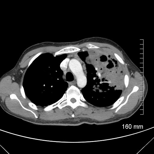
38-year-old male with HIV presents with cough CT scan in the axial plane shows a focal necrotizing consolidation in the left upper lobe. Regional lymphadenopathy in the mediastinum is noted
Lab tests confirmed a diagnosis of TB
Ashley Davidoff MD TheCommonVein.net 256Lu 136099
38-year-old male with HIV presents with cough CT scan in the axial plane shows a focal necrotizing consolidation in the left upper lobe. Reginal lymphadenopathy in the mediastinum is noted
Lab tests confirmed a diagnosis of TB
Ashley Davidoff MD TheCommonVein.net 256Lu 136104
28-year-old immigrant with cough
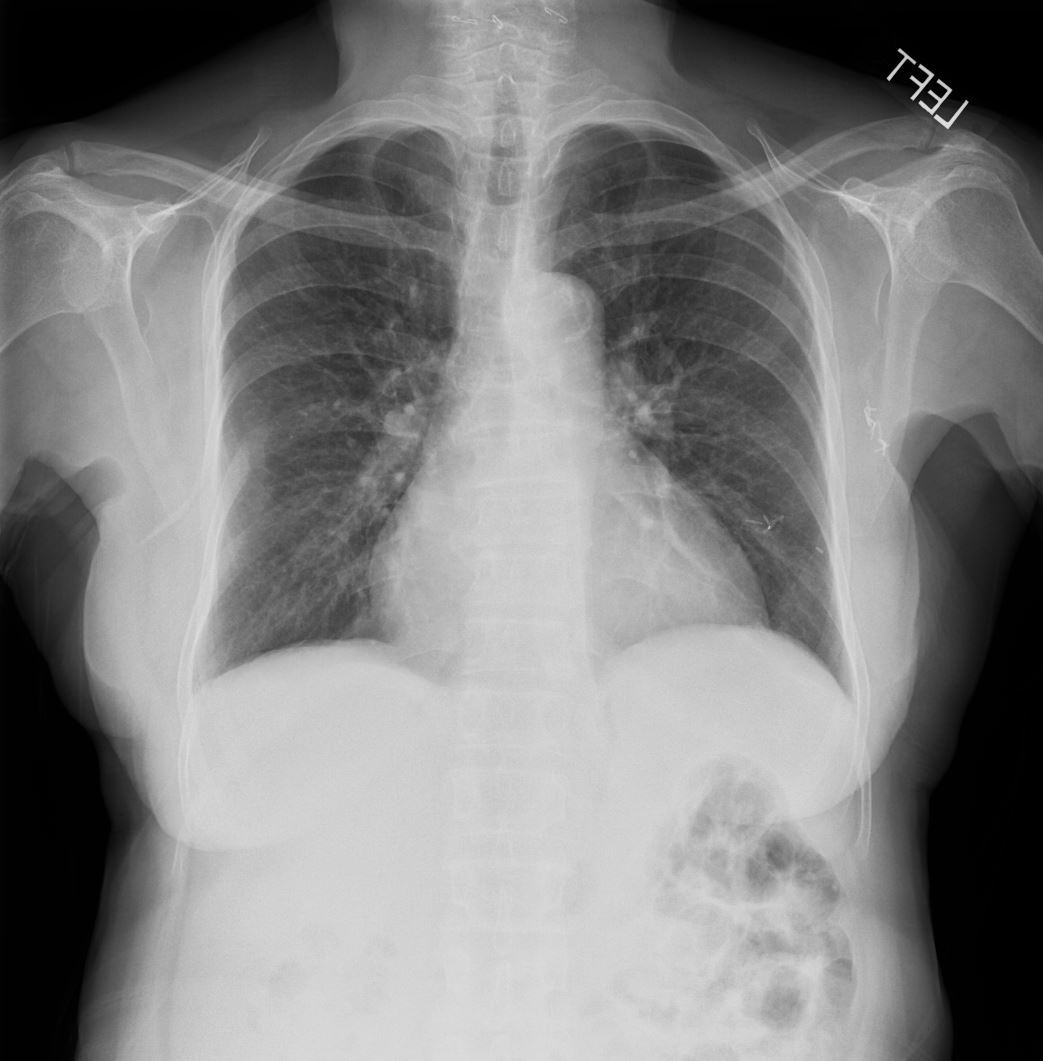
CXR – Reactivation TB Cavitating Pneumonia – Left Upper Lobe
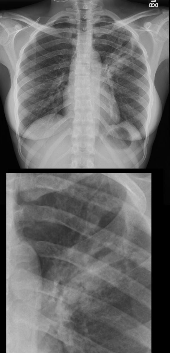
Frontal CXR of a 28-year-old immigrant with cough shows a cavitating pneumonia in the left upper lobe (magnified in the lower image)
Lab tests confirmed the diagnosis of TB and the patient was treated with RISE a 4-month treatment regimen of rifapentine-moxifloxacin for mycobacterium tuberculosis.
Ashley Davidoff MD TheCommonVein.net 255Lu 136071c
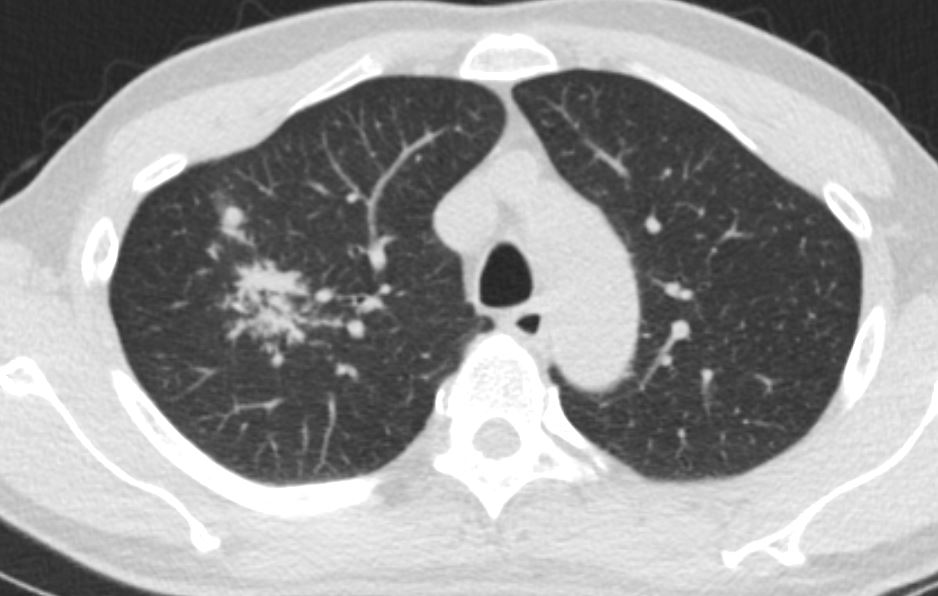
CXR –TB Pneumonia – Left Upper Lobe
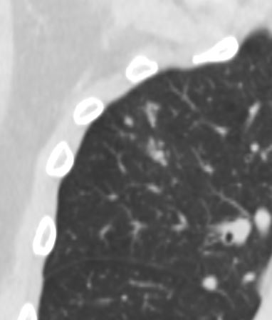
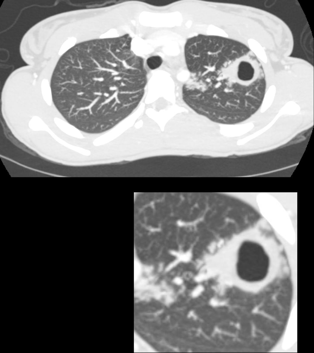
Consolidation Cavitation and Endobronchial Spread
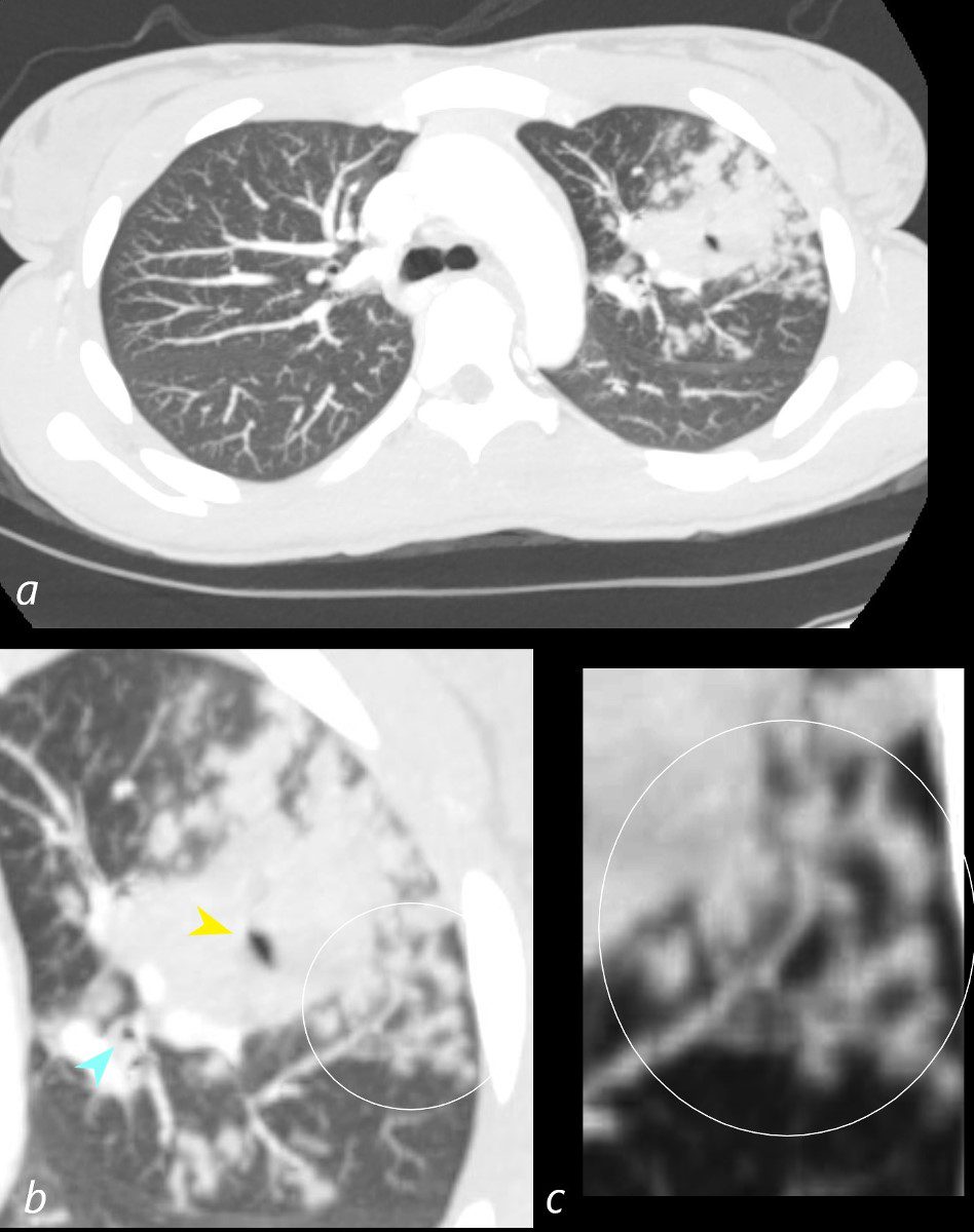
CT scan in the axial plane of the left upper lobe of a 28-year-old immigrant with cough shows a focal subsegmental consolidation with focal cavitation (yellow arrowhead) subtended by a thick-walled subsegmental airway. There are extensive tree in bud changes ringed in white (b and c) indicating transbronchial spread. Lab tests confirmed a diagnosis of TB and the patient was treated with RISE, a 4-month treatment regimen of rifapentine-moxifloxacin for mycobacterium tuberculosis.
Ashley Davidoff MD TheCommonVein.net 255Lu 136075cL
Focal Infiltrate
LUL Infiltrate14 months prior
LUL Nodule
Ashley Davidoff MD TheCommonVein.net
LUL Infiltrate 2 Months Later
Ashley Davidoff MD TheCommonVein.net
LUL Infiltrate 1 Month Later Following Initiation of Treatment
Ashley Davidoff MD TheCommonVein.net
TB presenting as a Right Upper Lobe Infiltrate
Pre Treatment
Ashley Davidoff TheCommonVein.net
Ashley Davidoff TheCommonVein.net
Ashley Davidoff TheCommonVein.net
Ashley Davidoff TheCommonVein.net
Ashley Davidoff TheCommonVein.net
Post Treatment
Ashley Davidoff TheCommonVein.net
Ashley Davidoff TheCommonVein.net
Ashley Davidoff TheCommonVein.net
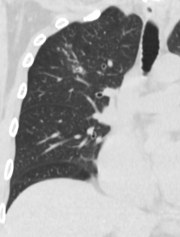
Endobronchial Spread
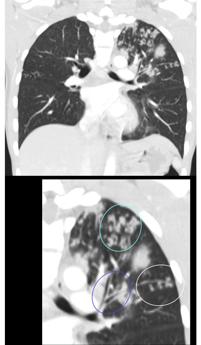
CT scan in the coronal plane of the left upper lobe of a 28-year-old immigrant with cough shows a thickening of the walls of the segmental, (blue circle) and subsegmental airway disease (teal circle ) as well as small airways disease characterised by tree in bud changes (ringed in whit)e These findings indicate transbronchial spread.
Lab tests confirmed a diagnosis of TB and the patient was treated with RISE, a 4-month treatment regimen of rifapentine-moxifloxacin for mycobacterium tuberculosis.
Ashley Davidoff MD TheCommonVein.net 255Lu 136079cL
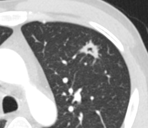
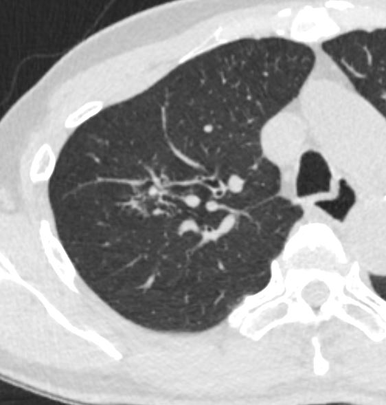
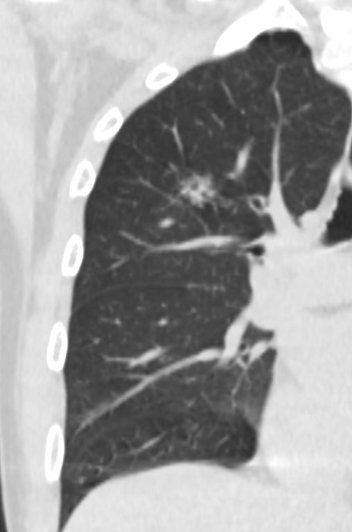
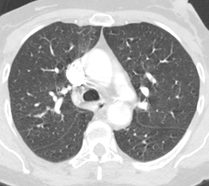
Cavitation
CT scan in the axial plane of the left upper lobe of a 28-year-old immigrant with cough shows a thick walled cavitating mass subtended by a subsegmental thick-walled airway. Lab tests confirmed the diagnosis of TB and the patient was treated with RISE, a 4-month treatment regimen of rifapentine-moxifloxacin for mycobacterium tuberculosis.
Ashley Davidoff MD TheCommonVein.net 255Lu 136074c
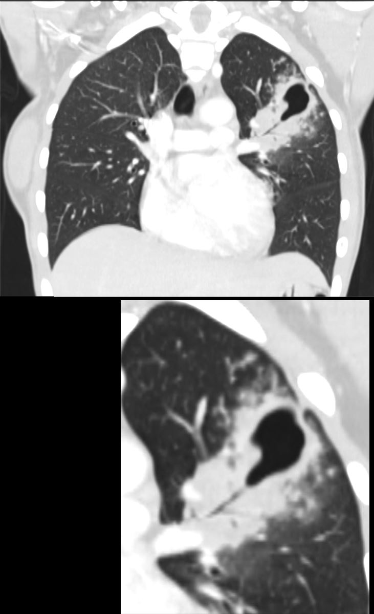
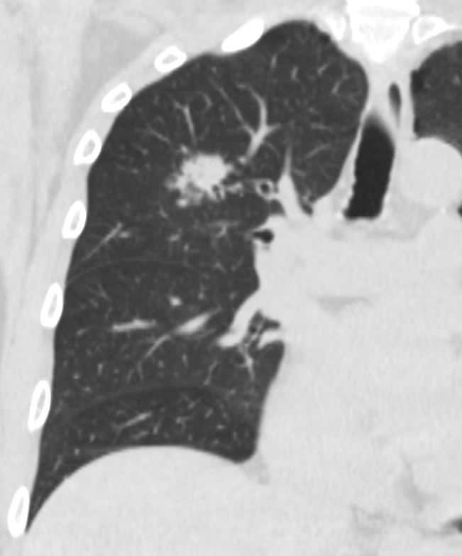
CT scan in the coronal plane of the left upper lobe of a 28-year-old immigrant with cough shows a thick walled cavitating mass subtended by a subsegmental thick-walled airway. Lab tests confirmed the diagnosis of TB and the patient was treated with RISE, a 4-month treatment regimen of rifapentine-moxifloxacin for mycobacterium tuberculosis.
Ashley Davidoff MD TheCommonVein.net 255Lu 136081c
Transbronchial Spread
eft Upper Lobe Airway Disease Segmental Subsegmental and Small Airway Involvement
CT scan in the coronal plane of the left upper lobe of a 28-year-old immigrant with cough shows a thickening of the walls of the segmental, (blue circle) and subsegmental airway disease (teal circle ) as well as small airways disease characterised by tree in bud changes (ringed in whit)e These findings indicate transbronchial spread.
Lab tests confirmed a diagnosis of TB and the patient was treated with RISE, a 4-month treatment regimen of rifapentine-moxifloxacin for mycobacterium tuberculosis.
Ashley Davidoff MD TheCommonVein.net 255Lu 136079cL
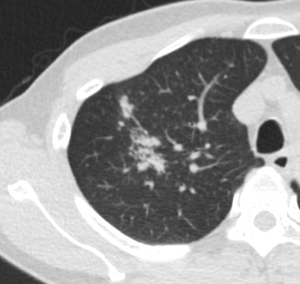
CT scan in the axial plane of the left upper lobe of a 28-year-old immigrant with cough shows a focal subsegmental consolidation with focal cavitation (yellow arrowhead) subtended by a thick-walled subsegmental airway. There are extensive tree in bud changes ringed in white (b and c) indicating transbronchial spread. Lab tests confirmed a diagnosis of TB and the patient was treated with RISE, a 4-month treatment regimen of rifapentine-moxifloxacin for mycobacterium tuberculosis.
Ashley Davidoff MD TheCommonVein.net 255Lu 136075cL
Miliary
Normal CXR and CT 1year Prior
60 year old immunocompromise female
Frontal CXR of a 60 year old immunocompromise female 1 year prior to an episode of miliary TB shows a normal CXR
Ashley Davidoff MD TheCommonVein.net 265Lu 136195
Axial CT of a 60-year-old immunocompromised female 1 year prior to an episode of miliary TB shows a normal examination
Ashley Davidoff MD TheCommonVein.net 265Lu 136196
60-year-old immunocompromise female presents with a
cough and weight loss
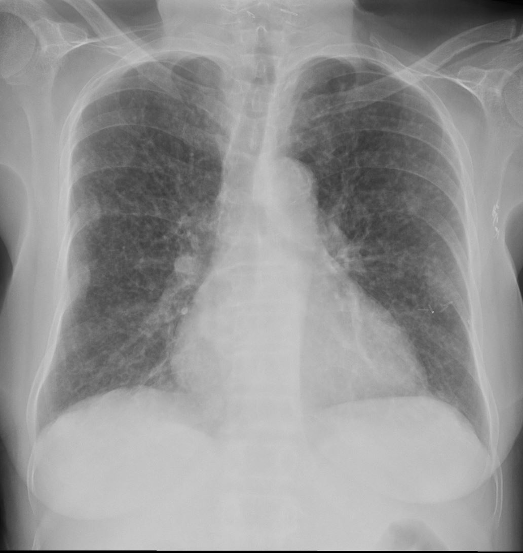
60-year-old immunocompromise female presents with a cough and weight loss CXR shows a diffuse miliary pattern. Final diagnosis was mycobacterium tuberculosis. Associated findings include healed right sided rib fractures and surgical clips in the left axilla
Ashley Davidoff MD TheCommonVein.net 265Lu 136197
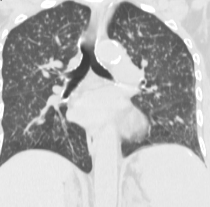
60-year-old female presents with a cough and weight loss. Coronal CT shows miliary nodules throughout both lung fields. The nodules appear to be distributed along the bronchovascular bundles and the lymphatics and are noted in centrilobular, fissural and pleural locations. She responded well to treatment and final diagnosis was mycobacterium tuberculosis.
Ashley Davidoff MD TheCommonVein.net 265Lu 136206
CT Miliary Tuberculosis Centrilobular Nodules Suggesting Arteriolar Small Airway and or Lymphatic Involvement Also Fissural Nodules and Pleural Nodules
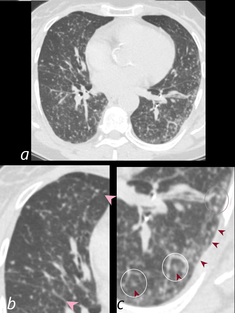
60-year-old immunocompromised female presents with a cough and weight loss. Axial CT shows miliary nodules throughout both lung fields. Some of these nodules are centrilobular (c, maroon arrowheads) and others are fissural based (b, pink arrowheads). In some of the secondary lobules there are 2 centrilobular nodules indicating involvement of the airway and arteriole and or the lymphatics (c white rings). One lobule shows centrilobular and interlobular nodules (c gray ring anteriorly). She responded well to treatment and final diagnosis was mycobacterium tuberculosis.
Ashley Davidoff MD TheCommonVein.net 265Lu 136204cL
Pleural Effusion
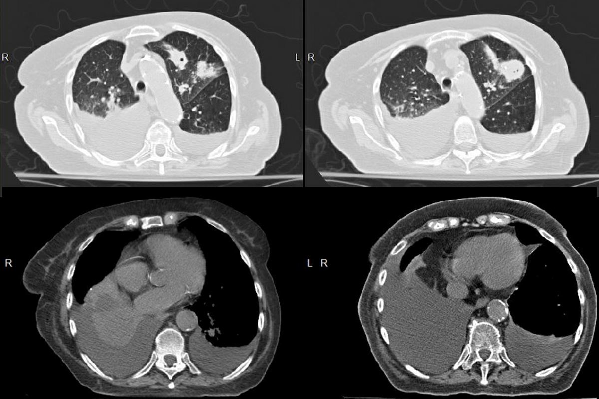
80 year old Russian woman who initially presented with a cavitating LUL nodule that was biopsied and thought to represent sarcoidosis
In December the nodules in the LUL enlarged with an arborising pattern involving the posterior subsegment of the LUL as well as an unchanged RUL ground glass infiltrate
Subsequent diagnosis of TB was made
Initially there was progressive disease in the LUL and lingula with new cavitation in the lingula infiltrate/nodule and extension of the infiltrate in the LUL with a new calcification. These findings were consistent with reactivation TB .
Repeated sputa were positive for acid fast bacilli
More recently new micronodularity was noted in the right lung .
Now 1 month later she presents with a large right pleural effusion and a smaller left effusion
Ashley Davidoff MD The Commonvein.net 31645L02b