
Size
Shape
Position
Bronchopneumonia

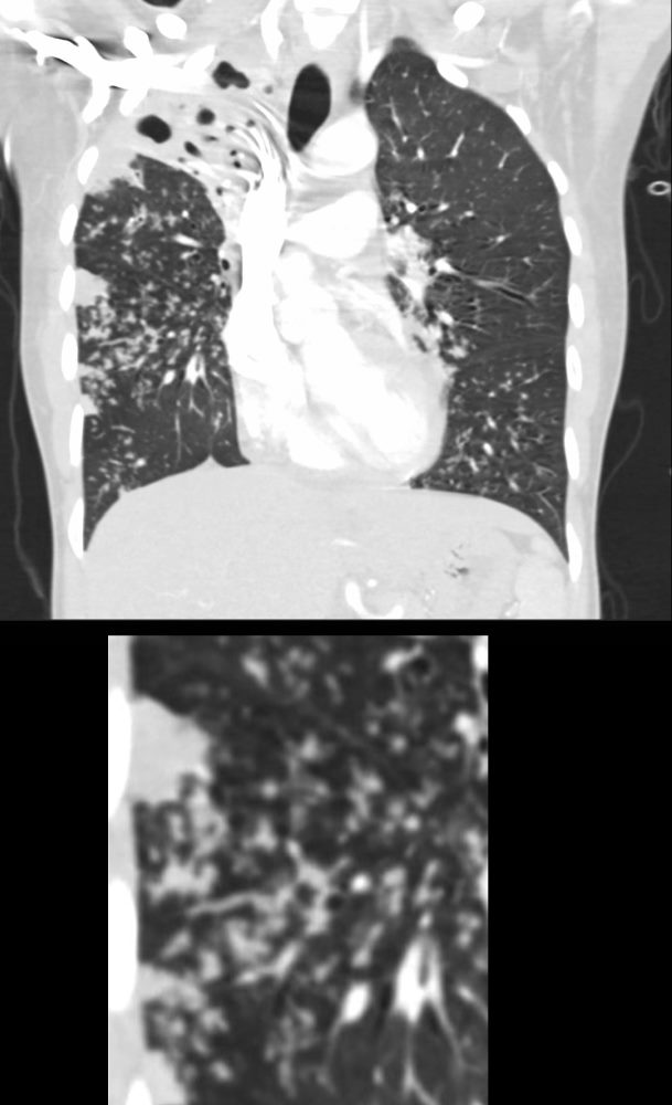
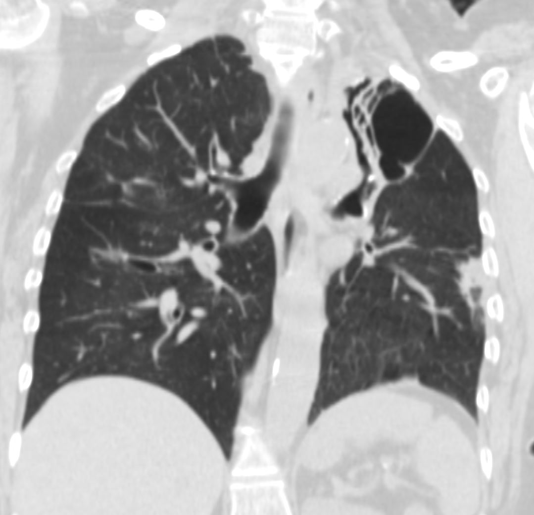
35-year-old woman, presents with dyspnea and fever. Coronal CT shows a bronchocentric pneumonic infiltrate in the superior segment of the left lower lobe and extending into the medial basal segment, (white ring), consistent with a bronchopneumonia. There is an adjacent region of ground glass infiltrate (red ring)
Courtesy Ashley Davidoff MD TheCommonVein.net 289Lu 136560c
Character
Infection Inflammation Malignancy Mechanical/Atelectasis Trauma Metabolic Circulatory- Hemorrhage Immune Infiltrative Idiopathic Iatrogenic Idiopathic
Infection
TB
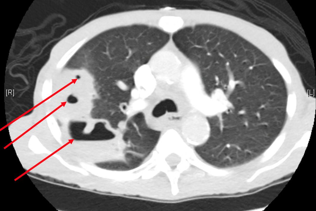
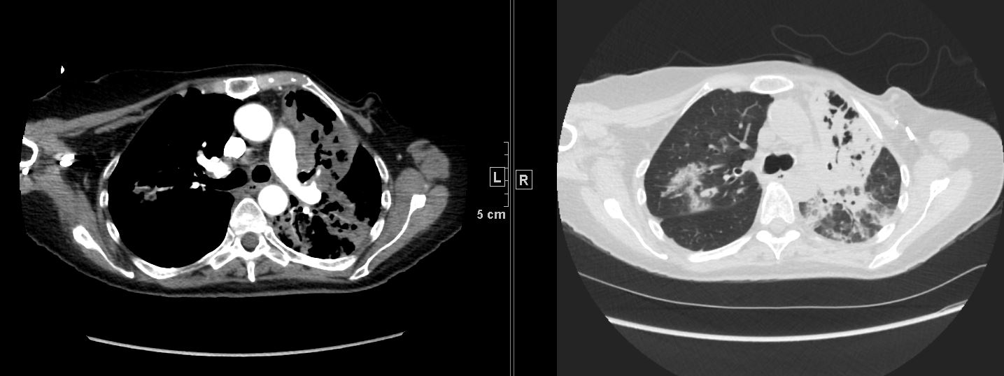
Cavitating TB with Transbronchial Spread
39-year-old immigrant Vietnamese male presents night sweats, fever, and cough. CT in the coronal plane of the chest shows a large cavitating lesion in the right upper lobe, with innumerable micronodules dominantly in the right midlung field, and to lesser extent in the right upper lung field. Some micronodules are probably present in the left lower lobe as well. Close to the largest subsegmental consolidation there is a bronchus which shows thickening of its wall.
Although it has the appearance has a “miliary” pattern, this term is usually referred to the hematogenous spread of the disease
Ashley Davidoff MD TheCommonvein.net 135786c
006Lu
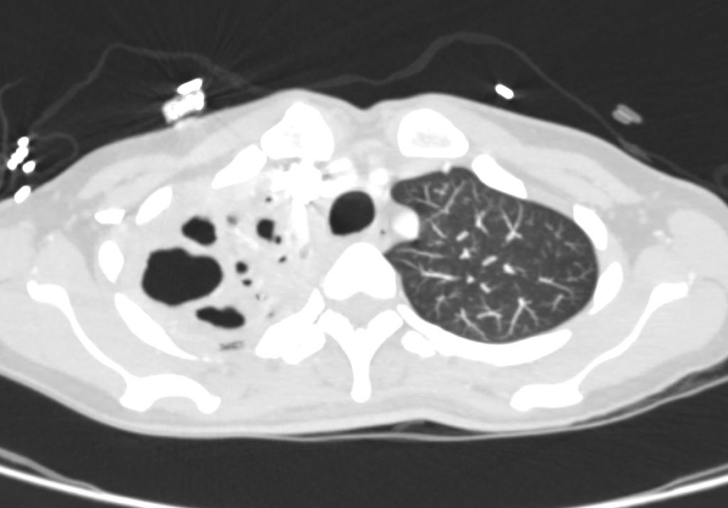
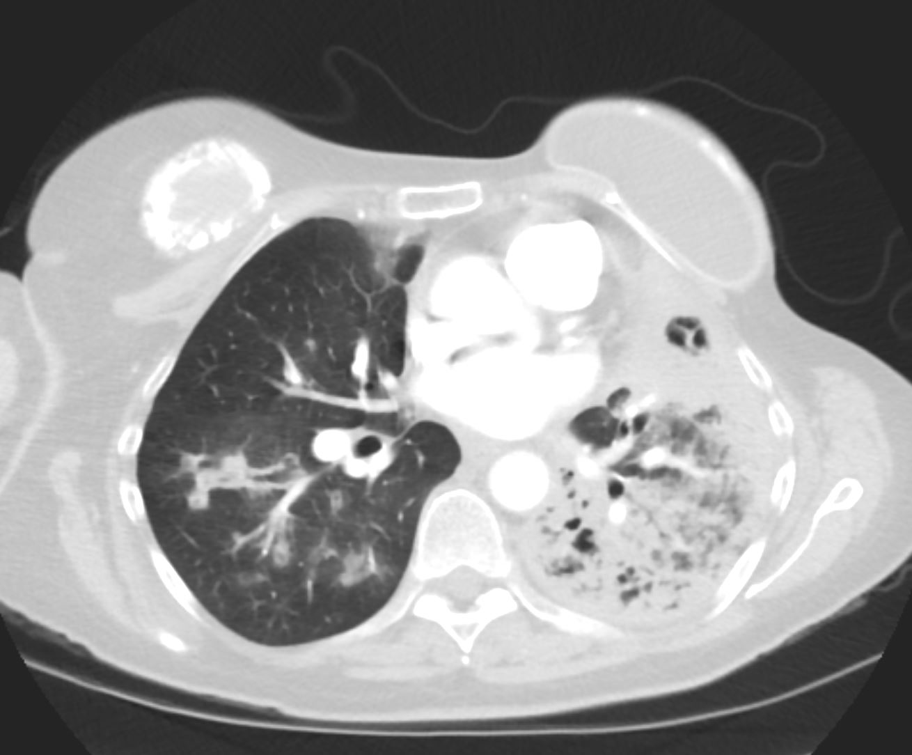
39-year-old immigrant Vietnamese male presents night sweats fever and cough. CT in the axial plane of the chest shows a large cavitating lesion in the right upper apex.
Ashley Davidoff MD TheCommonvein.net 135787 006Lu 006Lu
Courtesy Joseph Cannella, Dr. Christina LeBedis, MD, MS TheCommonVein.net
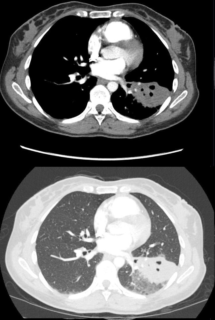
Cavitating Left Lower Lobe Pneumonia with Pseudoaneurysm (PSA)
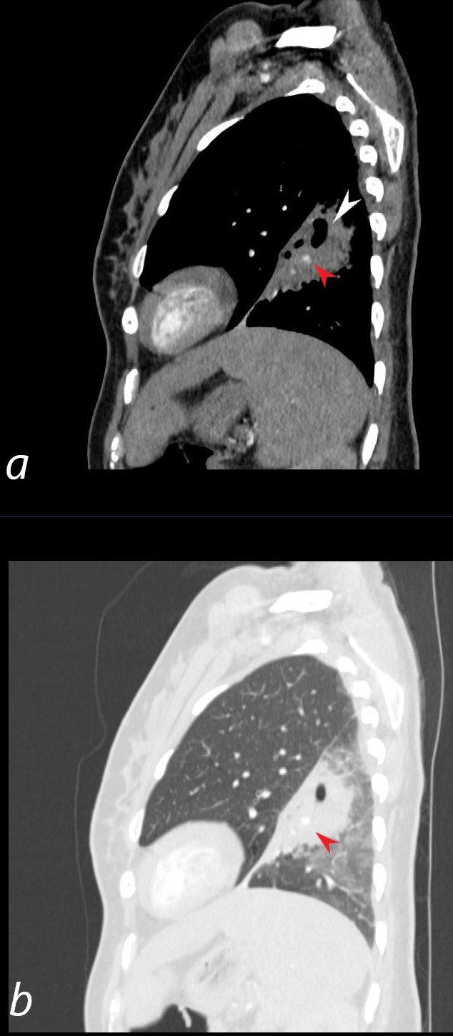
30-year-old female with a history of IVDU presents with a fever and hemoptysis.
CT in the sagittal plane shows a cavitating pneumonia in the anterior segment of the left lower lobe. A focally dilated artery in the consolidation (red arrowheads a, and b) represents a mycotic aneurysm. There are groundglass changes surrounding the consolidation representing either edema or hemorrhage
Ashley Davidoff MD TheCommonVein.net 281Lu 136530cL
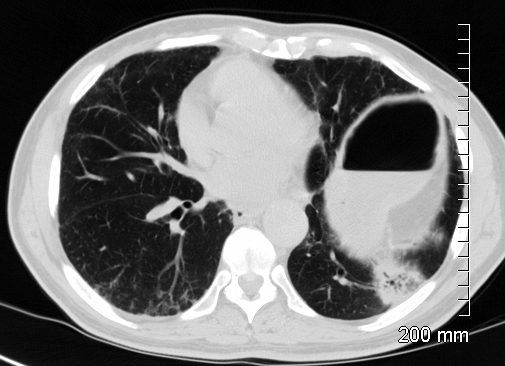
30-year-old female with a history of IVDU presents with a fever and hemoptysis.
CT in the axial plane shows a cavitating pneumonia in the anterior segment of the left lower lobe. There are groundglass changes surrounding the consolidation representing either edema or hemorrhage
Ashley Davidoff MD TheCommonVein.net 281Lu 136524
Inflammation
Neoplasm Mucin Secreting Adenocarcinoma
Ashley Davidoff
TheCommonVein.net
118601
Circulatory Disorders
Immune Disorders
Wegener’s Granulomatosis Granulomatosis with Polyangiitis
70F-year old female presents with hemoptysis and bilateral lower lobe pulmonary infiltrates CT scan shows dense consolidations in the left upper lobe
Ashley Davidoff MD TheCommonVein.net Wegeners-cavitation-010
70F-year old female presents with hemoptysis and bilateral lower lobe pulmonary infiltrates CT scan shows a consolidation in the left lower lobe with early cavitation, and segmental airway thickening and ground glass nodules in the right lower lobe
Ashley Davidoff MD TheCommonVein.net Wegeners-cavitation-014
Ashley Davidoff MD TheCommonVein.net Wegeners-cavitation-028