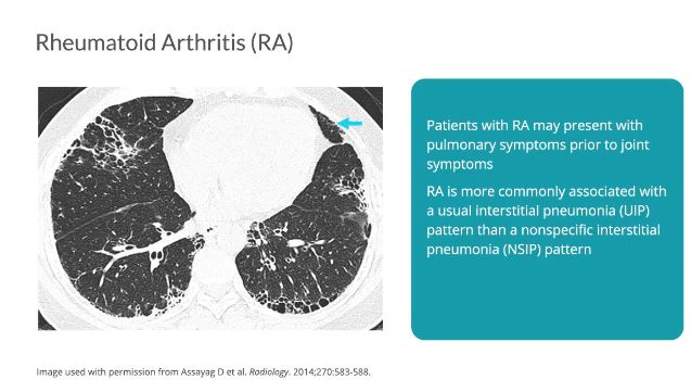
While joint disease is the primary presentation of rheumatoid arthritis, approximately 10-20% of patients present with pulmonary symptoms prior to joint symptoms.19 Pulmonary symptoms associated with RA include interstitial lung disease, pleural thickening or effusions, airway inflammation, pulmonary hypertension, and vasculitis, and confer significant morbidity and mortality.19 RA is more commonly associated with a UIP pattern, distinguishing it from the other connective tissue diseases which often present with NSIP. However, 10-30% of patients with RA will have a radiologic pattern of NSIP.19 Note the subpleural and basilar reticulation, minimal ground glass, traction bronchiectasis, honeycombing, and pleural thickening.
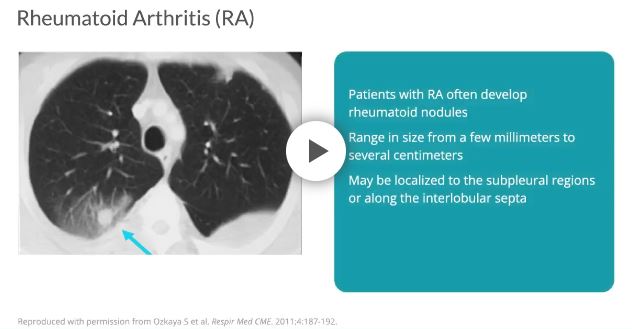
Patients often develop single or multiple rheumatoid nodules in the lung, which can range in size from a few millimeters to several centimeters.20 Rheumatoid nodules are often be located in the subpleural regions or along the interlobular septa.19
Rheumatoid Arthritis (RA)
While joint disease is the primary presentation of rheumatoid arthritis, approximately 10-20% of patients present with pulmonary symptoms prior to joint symptoms.19 Pulmonary symptoms associated with RA include interstitial lung disease, pleural thickening or effusions, airway inflammation, pulmonary hypertension, and vasculitis, and confer significant morbidity and mortality.19 RA is more commonly associated with a UIP pattern, distinguishing it from the other connective tissue diseases which often present with NSIP. However, 10-30% of patients with RA will have a radiologic pattern of NSIP.19 Note the subpleural and basilar reticulation, minimal ground glass, traction bronchiectasis, honeycombing, and pleural thickening.
Patients often develop single or multiple rheumatoid nodules in the lung, which can range in size from a few millimeters to several centimeters.20 Rheumatoid nodules are often be located in the subpleural regions or along the interlobular septa.19

 While joint disease is the primary presentation of rheumatoid arthritis, approximately 10-20% of patients present with pulmonary symptoms prior to joint symptoms.19 Pulmonary symptoms associated with RA include interstitial lung disease, pleural thickening or effusions, airway inflammation, pulmonary hypertension, and vasculitis, and confer significant morbidity and mortality.19 RA is more commonly associated with a UIP pattern, distinguishing it from the other connective tissue diseases which often present with NSIP. However, 10-30% of patients with RA will have a radiologic pattern of NSIP.19 Note the subpleural and basilar reticulation, minimal ground glass, traction bronchiectasis, honeycombing, and pleural thickening.