
67 year old man with prior history of bladder cancer wand now a Non Small Cell Carcinoma of the Right Upper Lobe
Non-Small Cell Lung Cancer (NSLC) with
Lymphangitis Carcinomatosis
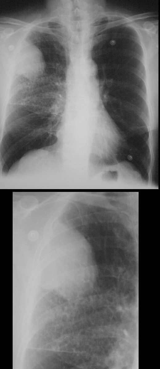
67-year-old man with prior history of bladder cancer. Frontal chest X-ray (CXR) shows a large right upper lobe lung mass. Inferior to the mass, there a polygonal pattern of the intertstitium suggesting lymphangitis carcinomatosis
Ashley Davidoff MD TheCommonVein.net 32270c01
PET scan Positive for LUL Mass and Adrenal Metastasis
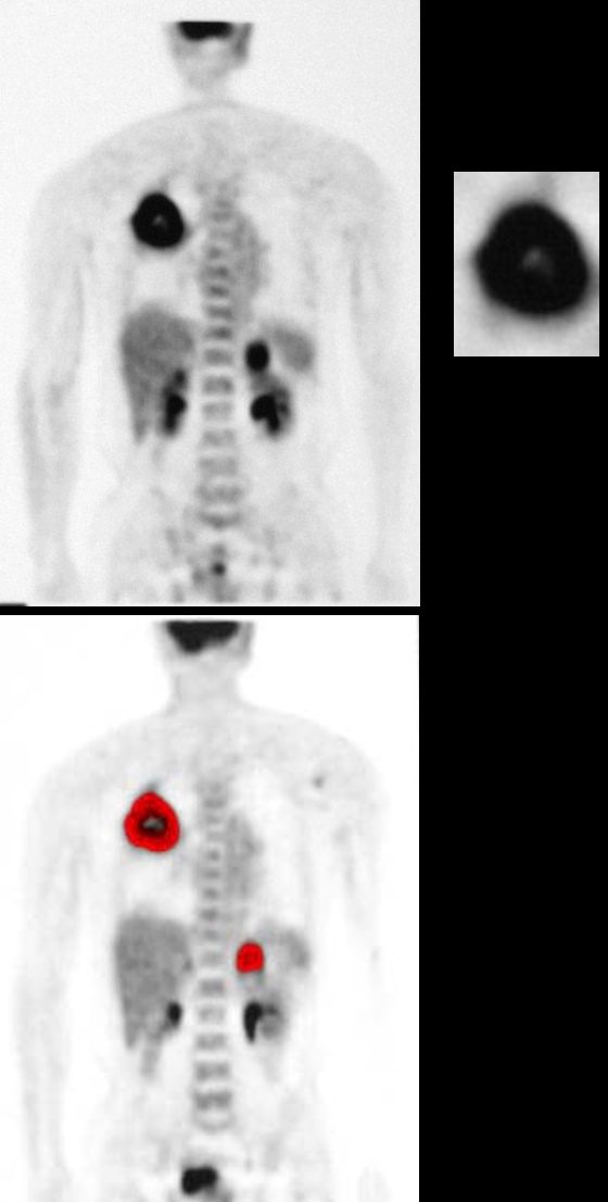
67-year-old man with prior history of bladder cancer. PET scan shows a large PET positive right upper lobe lung mass overlaid in red in the lower panel. The mass has a small central area of a lack of activity (upper panel right) suggesting central necrosis. In addition there is a PET positive left adrenal mass.
Ashley Davidoff MD TheCommonVein.net 32274c
Large RUL Low Density Lung Mass
67-year-old man with prior history of bladder cancer. CT scan in the axial plane shows a large right upper lobe lung mass. The mass has a relatively low density, is mildly heterogeneous and has thickened irregular borders.
Ashley Davidoff MD TheCommonVein.net 32284
Non-Small Cell Lung Cancer (NSLC) with
Lymphangitis Carcinomatosis
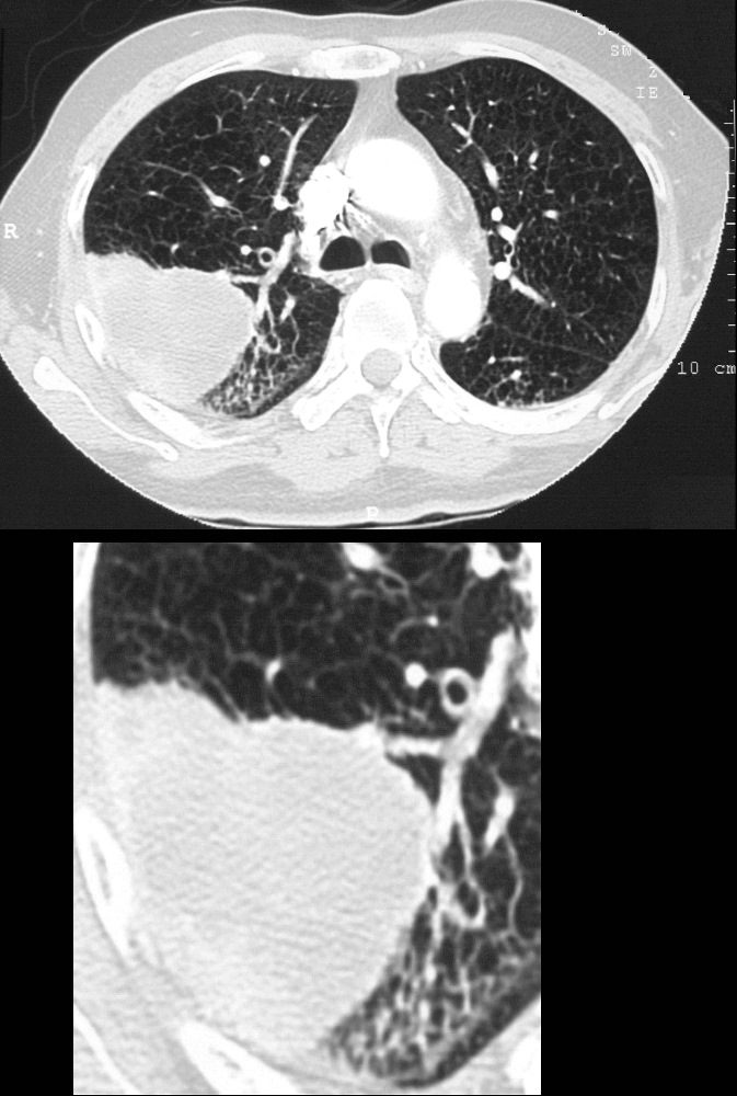
67-year-old man with prior history of bladder cancer. CT scan in the axial plane shows a large right upper lobe lung mass. Posterior to the mass, the interlobular septa are thickened.. These findings suggest lymphangitis carcinomatosis. Peribronchial thickening of the apical segmental airway may be due either to lymphangitis or chronic bronchitis There is background evidence of centrilobular emphysema, dominant in both upper lobes giving an internal comparison of the normal vs abnormal size of the interlobular septa
Ashley Davidoff MD TheCommonVein.net 32285c
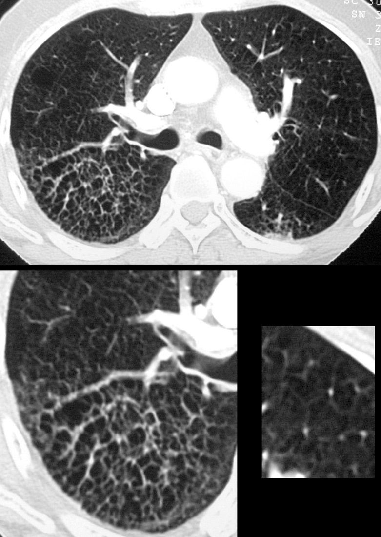
67-year-old man with prior history of bladder cancer. CT scan in the axial plane inferior to the lung mass shows extensive irregular thickening of the interlobular septa magnified in the right lower panel. These findings reflect lymphangitis carcinomatosis. There is background evidence of centrilobular emphysema, dominant in both upper lobes. A magnified view in the right lower panel offers an internal comparison of the abnormal vs the normal size and shape of the interlobular septa
Ashley Davidoff MD TheCommonVein.net 32287c
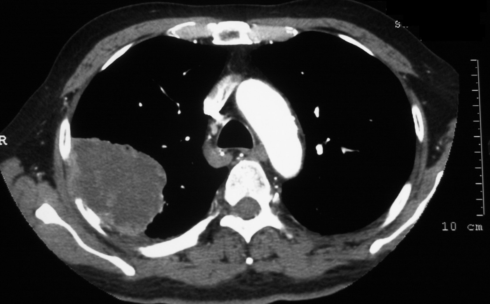
Metastasis to the Left Adrenal Gland
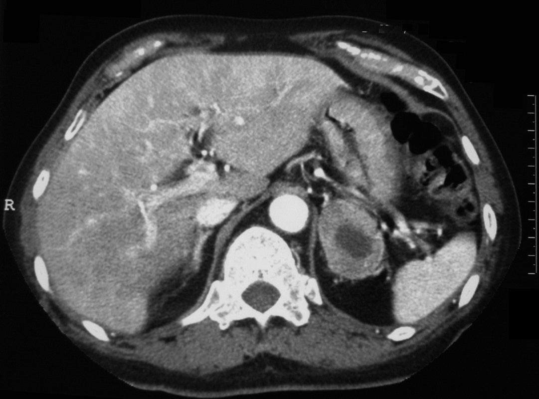
67-year-old man with prior history of bladder cancer. CT scan in the axial of the upper abdomen shows a large metastasis in the left adrenal with central low density. PET scan was positive for this mass and thus a metastatic deposit in confirmed
Ashley Davidoff MD TheCommonVein.net 32290