
Bronchocentric granulomatosis (BCG) is a rare inflammatory lung disease that affects the small airways in the lungs.

Upper lung field distribution
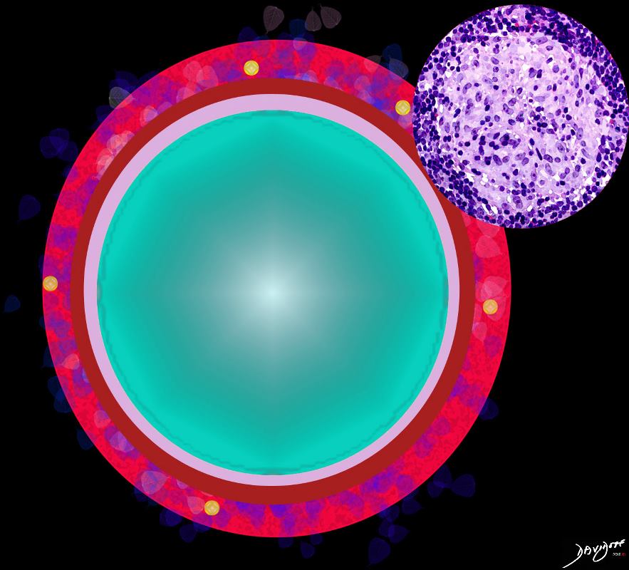
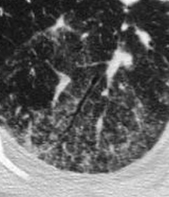
Ashley Davidoff MD TheCommonvein.net lungs-0774
Ashley Davidoff The Common Vein.net
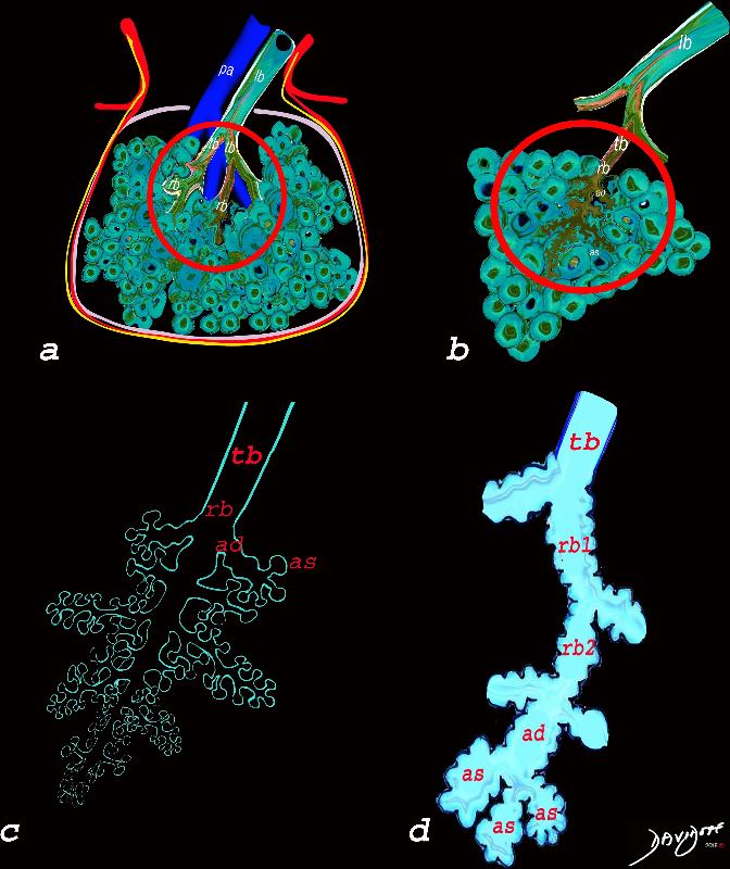
The diagram allows us to understand the the components and the position of the small airways starting in (a) which is a secondary lobule that is fed by a lobular bronchiole(lb) which enters into the secondary lobule and divides into terminal bronchioles (tb) which is the distal part of the conducting airways, and at a diameter of 2mm or less . It divides into the respiratory bronchiole (rb) a transitional airway which then advances into the alveolar ducts(ad) and alveolar sacs (as) Diseases isolated to the small airways do not affect the alveoli and hence there is peripheral sparing Ashley Davidoff MD TheCommonVein.net
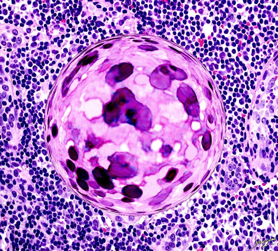
Ashley Davidoff MD The CommonVein.net lungs-0766
Granulomatous inflammation involving a bronchiole. Some of the cells surrounding the lumen are probably degenerating bronchial epithelial cells.
Source Wiki Commons Dr Yale Rosen
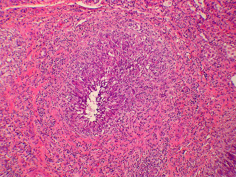
Ashley Davidoff TheCommonVein.net 29288m
- Cause and Predisposing Factors
- BCG is often associated with other conditions, such as
- rheumatoid arthritis,
- systemic lupus erythematosus (SLE), and
- vasculitides.
- The exact cause of BCG is unknown, but it is thought to be an immune-mediated disease
- Symptoms of BCG may include cough, shortness of breath, chest pain, and fever.
- Diagnosis of BCG involves a combination of clinical evaluation, lung function tests, chest imaging and biopsy of lung tissue.
- CT findings
- Thickening of the bronchial walls: BCG is characterized by the formation of granulomas in the walls of the small airways in the lungs, which can cause thickening of the bronchial walls.
- Centrilobular nodules: These are small nodules that are located around the center of the lung lobules. They are a common finding in BCG and may be seen in clusters.
-
- Tree-in-bud appearance: This is a radiologic pattern that can be seen on CT scans of the lungs. It appears as small nodules or branching linear opacities arising from the bronchioles, and is commonly seen in BCG.
- Ground-glass opacities: These are areas of hazy, increased density in the lung tissue that do not completely obscure the underlying lung structures. Ground-glass opacities can be a non-specific finding in many lung diseases, but they are commonly seen in BCG.
- Consolidation: This is an area of lung tissue that appears denser than normal due to the accumulation of fluid, cells, or other materials. Consolidation may be seen in severe cases of BCG.
- Treatment for BCG typically involves immunosuppressive therapy, such as corticosteroids or other medications that suppress the immune system.
- Prognosis for BCG varies depending on the severity of the disease and the presence of other underlying conditions. In some cases, BCG can progress to a more severe form of lung disease called bronchiolitis obliterans.