
47-year-old male presented with a cough
CT Axial Plane – Linear Atelectasis LLL and
Air Fluid Level in the Esophagus
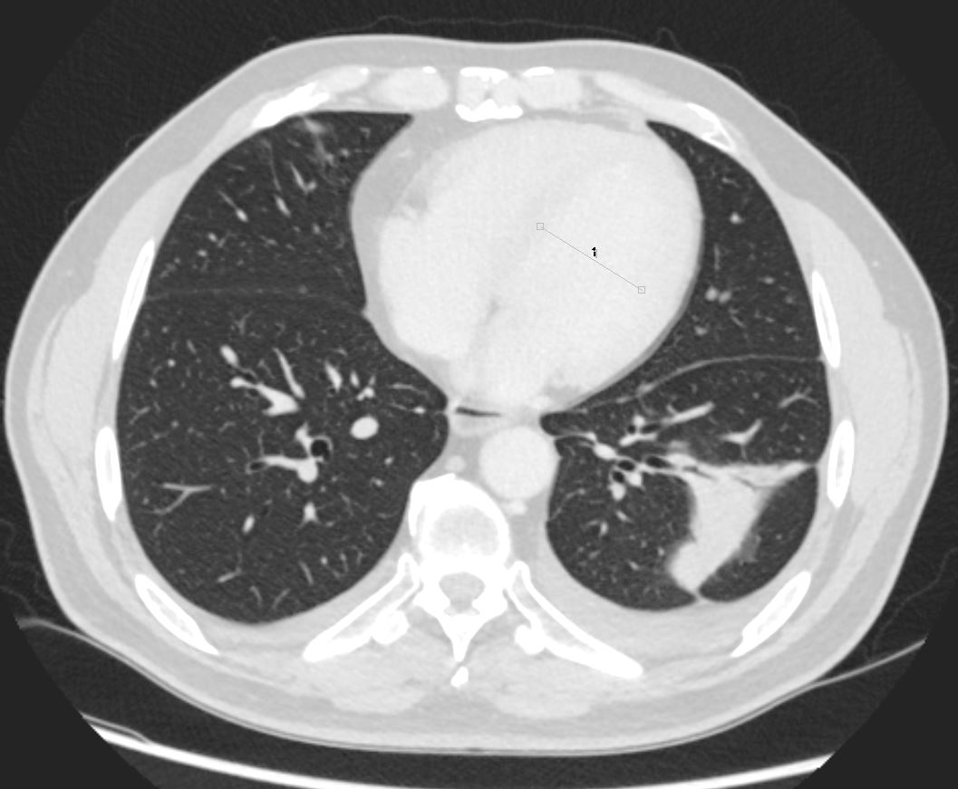
47-year-old male presented with a cough. CT scan in the axial plane shows a wedge-shaped region of subsegmental atelectasis involving the lateral segment of the left lower lobe associated with a small left pleural effusion. A small air-fluid level in a mildly dilated esophagus indicates reflux and raises the possibility of aspiration as a cause for the infiltrate
Ashley Davidoff MD TheCommonVein.net 276Lu 136235
aka discoid atelectasis aka plate-like atelectasis
CT Coronal Plane – Linear Atelectasis LLL
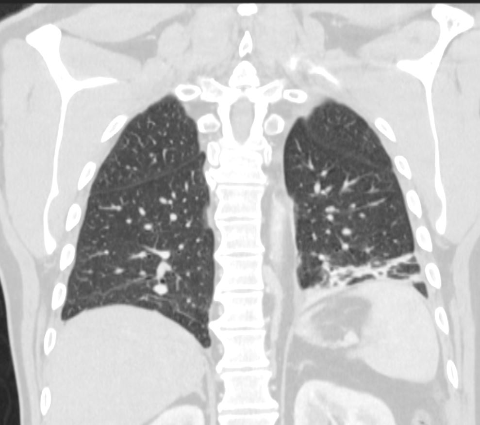
47-year-old male presented with a cough. CT scan in the coronal plane shows a region of subsegmental atelectasis involving a basal segment of the left lower lobe associated with elevation of the left hemidiaphragm indicating volume loss.
Ashley Davidoff MD TheCommonVein.net 276Lu 136236
aka discoid atelectasis aka plate-like atelectasis
3 Months Later
CT Axial Plane – Subsegmental Atelectasis LLL and
Air Fluid Level in the Esophagus

CT scan in the axial plane 3 months later shows persistence of a mild reduction of the size of the wedge-shaped region of subsegmental atelectasis involving the lateral segment of the left lower lobe associated with a persistent small left pleural effusion. A small air-fluid level in a mildly dilated esophagus indicates reflux and raises the possibility of aspiration as a cause for the infiltrate
Ashley Davidoff MD TheCommonVein.net 276Lu 136237
aka discoid atelectasis aka plate-like atelectasis
CT scan in the coronal plane 3 months late
Linear-Discoid-Plate-Like Atelectasis
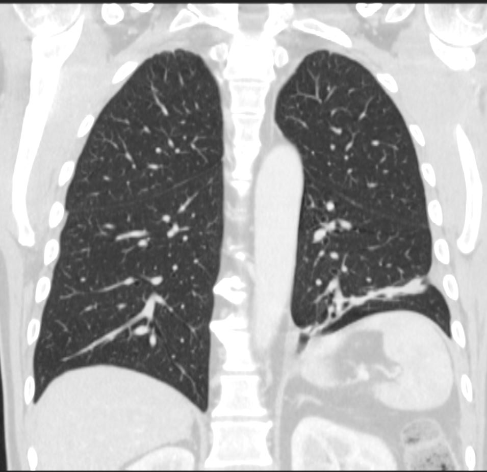
CT scan in the coronal plane 3 months later shows significant improvement of the atelectasis involving a basal segment of the left lower lobe associated with persistent elevation of the left hemidiaphragm indicating volume loss. The atelectasis now has a discoid, linear, or plate-like appearance
Ashley Davidoff MD TheCommonVein.net 276Lu 136238
aka discoid atelectasis aka plate-like atelectasis
CT Linear Atelectasis Over a 3 Month Period
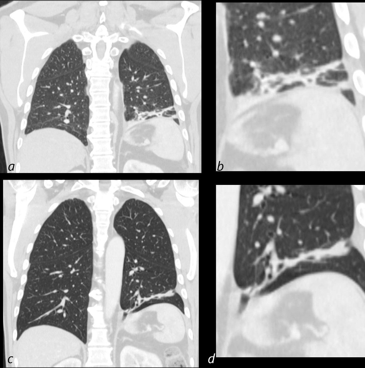
CT scan in the coronal plane (a, magnified in b) shows a region of subsegmental atelectasis involving a basal segment of the left lower lobe associated with elevation of the left hemidiaphragm indicating volume loss. 3 months later (c and d) shows persistence but improvement of the atelectasis involving a basal segment of the left lower lobe associated with persistent elevation of the left hemidiaphragm indicating volume loss. The atelectasis now has a discoid, linear, or plate-like appearance
Ashley Davidoff MD TheCommonVein.net 276Lu 136238cL
aka discoid atelectasis aka plate-like atelectasis