
Parts
Size
Shape
Position
Character
Time Associated Findings
Infection
Inflammation
Malignancy
Mechanical
Atelectasis
Trauma
Metabolic
Circulatory-
Coronary Calcification
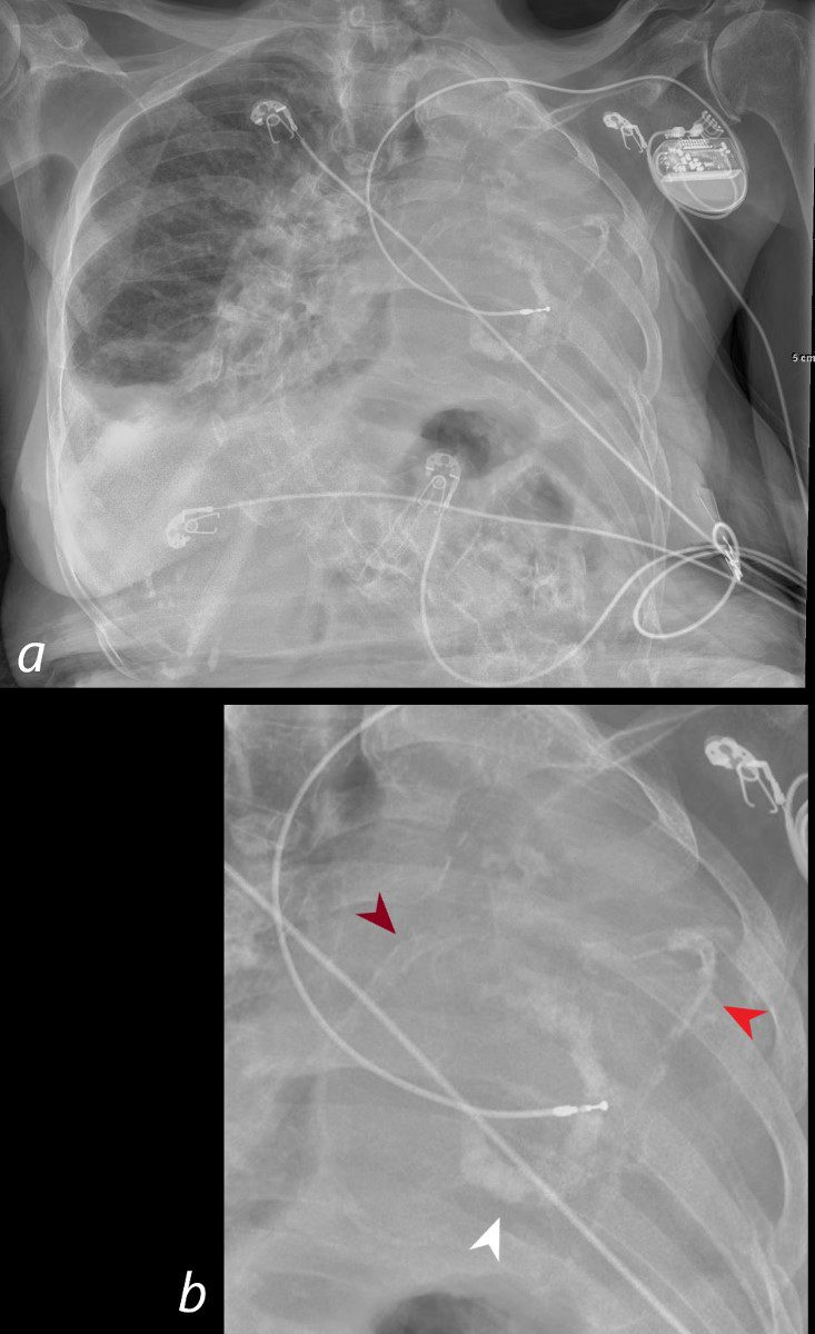
Frontal CXR of a 98-year-old woman showing a left sided white out secondary to a pneumonectomy. The soft tissue structures of the mediastinum have all shifted into the left hemithorax accounting for the white out. The calcified mitral annulus (b, white arrowhead), calcified right coronary artery – RCA (b, maroon arrowhead) and left anterior descending (LAD) – (b, bright red arrowhead) and right ventricle (RV pacemaker lead) confirm the diagnosis of acquired dextrocardia. There is hyperinflation of the right lung which crosses the midline associated with a small right effusion. A significant dextro-thoracic scoliosis with a compensatory levoscoliosis of the lumbar spine is present
Ashley Davidoff MD TheCommonVein.net 269Lu 136234cL