
- Bacterial
- Viral
- CMV

-
PCP – Multicentric Pneumonia
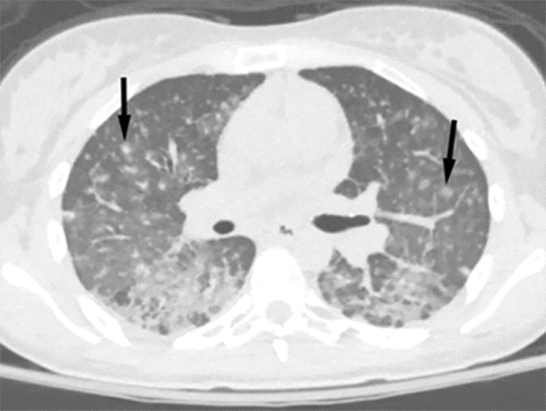
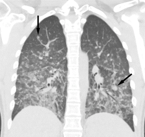
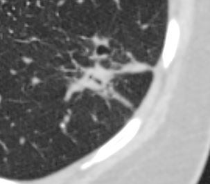
Cytomegalovirus pneumonia. CT scans in a 31-year-old woman with a history of type 1 diabetes mellitus complicated by end-stage renal disease. The patient had previously undergone kidney and pancreas transplant and presented with 2 days of right lower quadrant abdominal pain associated with nausea and vomiting. Upon further work-up, patient was found to have cytomegalovirus viremia. (above) Axial and (be;low) coronal CT images demonstrate diffuse randomly distributed small pulmonary nodules (arrows), many of which are ill-defined and distributed in the secondary pulmonary lobules and perilymphatic regions.
Parekh, M et al Review of the Chest CT Differential Diagnosis of Ground-Glass Opacities in the COVID Era Radiology Vol. 297, No. 3 July 2020COVID 19
-
COVID 19 COVID 19
55-year-old male presents with a fever and a cough.
CXR findings reveal vague peripheral, bibasilar, “ground glass” changes in the lower lung zones.
The CT scan confirms the presence of bilateral, predominantly basilar, nodular, and peripheral mixes ground glass and consolidative opacifications consistent with the diagnosis of COVID 19. Differential diagnosis however includes other viral pneumonias, allergic alveolitis and other multifocal and organizing pneumonias.
Courtesy Kevin Chang MD - Fungal
Ashley Davidoff
TheCommonVein.net
Interval resolution of left lower lobe consolidation 2 months later.
Interval resolution of left lower lobe consolidation 2 months later.
Ashley Davidoff
TheCommonVein.net
-
Links and References
- TCV
- TCV