
Bibasilar Aspiration Pneumonia
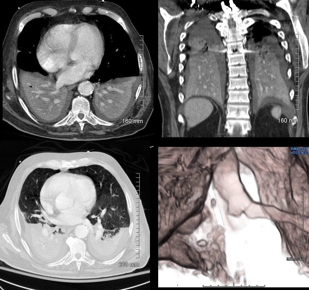
74 year old male alcoholic with bilateral basilar lobar atelectasis caused by bilateral aspiration
CT scan shows airless lower lobes with small bilateral effusions. 3D reconstruction shows total obstruction of the right mainstem bronchus, and patent proximal mainstem bronchus
Ashley Davidoff MD TheCommonVein.net
Aspiration Pneumonia Pulmonary Edema and DAD
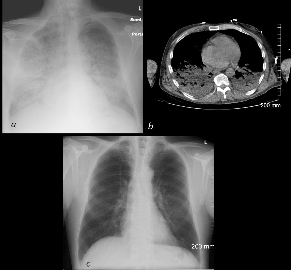
54 year old male alcoholic with seizures presents with diffuse alveolar disease consistent with pulmonary edema (a). CT scan (b) shows bibasilar infiltrates consistent with aspiration.
Follow up CXR 6 months later (c) shows resolution
Ashley Davidoff MD TheCommonVein.net
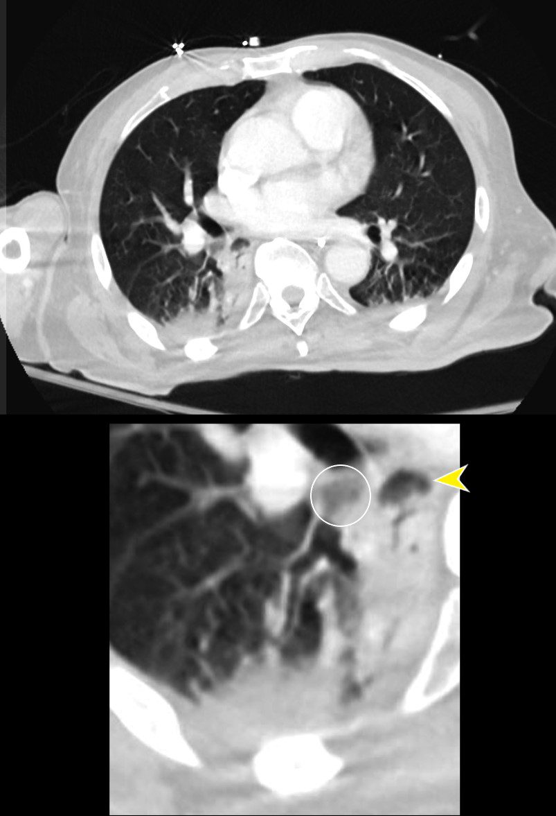
CT Aspirate Occluding the Right Lower Lobe Bronchus
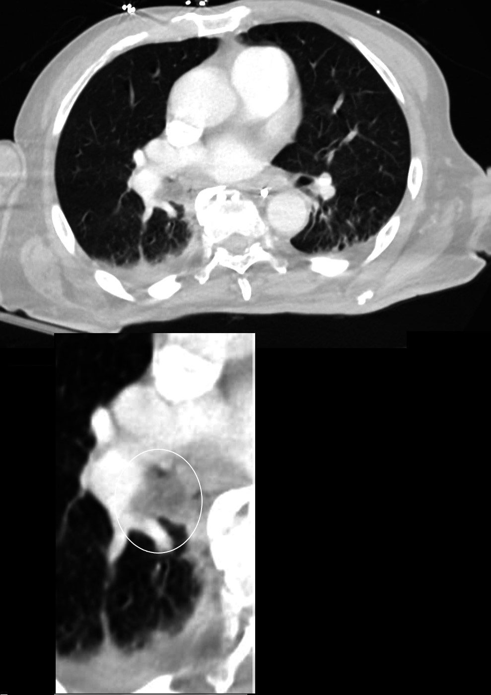
CT of a 72-year-old male with acute dyspnea shows a focal accumulation of low-density aspirate in the right lower lobe (white ring in lower image)
Ashley Davidoff MD TheCommonVein.net 136037c
Aspirate Occluding the Right Lower Lobe Bronchus
CT of a 72-year-old male with acute dyspnea shows a focal accumulation of low-density aspirate in the right lower lobe. Distal to the obstruction the posterior segmental and medial segmental airways are patent, but associated atelectasis is noted in those segments of the right lower lobe. The esophagus is displaced to the right, and appears to contain some aerated content. There is atelectasis of the medial and posterior segments of the right lower lobe secondary to the aspiration
Ashley Davidoff MD TheCommonVein.net 136038
Aspirate Occluding the Right Lower Lobe Bronchus
Medial and Lateral Basal Consolidation
CT of a 72-year-old male with acute dyspnea shows a focal accumulation of low-density aspirate in the right lower lobe (white ring in lower image). Distal to the obstruction the posterior segmental and medial segmental airways are patent, but associated atelectasis is noted in those segments of the right lower lobe. The esophagus is displaced to the right and appears to contain some aerated content.
Ashley Davidoff MD TheCommonVein.net 136038cL
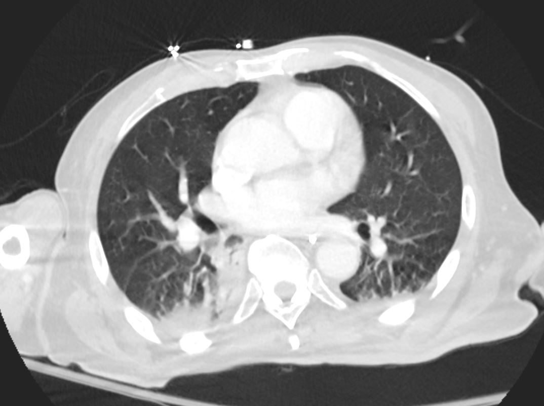
Aspirate Partially Occluding the Right Lower Lobe Bronchus and Extending into the Medial and Posterior Segments with
Associated Atelectasis and Consolidation
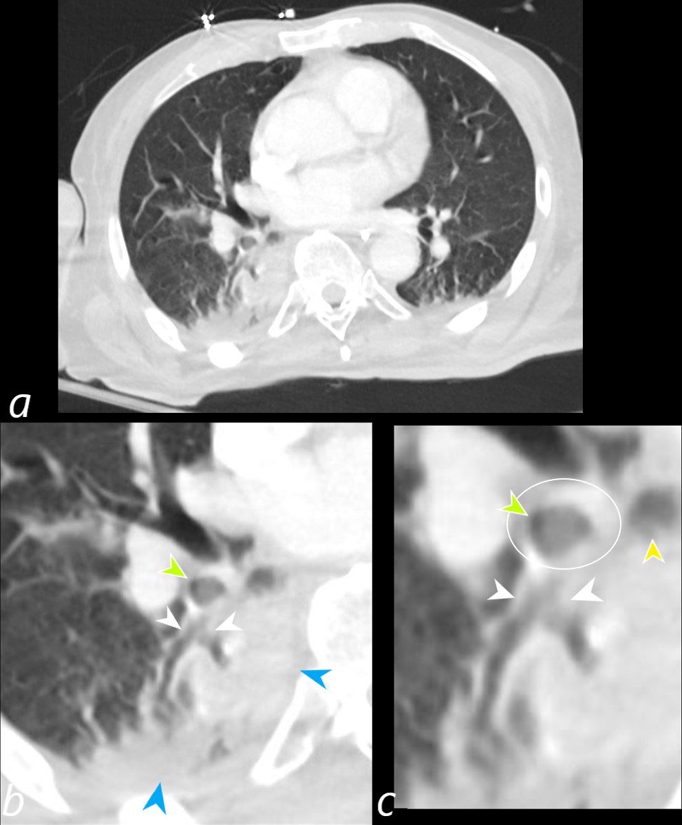
CT of a 72-year-old male with acute dyspnea shows a sub-totally occluded bronchus distal to the more complete obstruction noted in the previous section (green arrowheads b and c, and ringed in white in c). Distally at the branch point of the lower lobe bronchus there is partial filling of the medial and posterior segments (white arrows b and c). Secondary to the aspiration there is post obstructive atelectasis of the medial and posterior segments of the right lower lobe. The esophagus is displaced to the right, and appears to contain some aerated content (yellow arrowhead c).
Ashley Davidoff MD TheCommonVein.net 136041cL