
Asthma Allergic Bronchopulmonary Aspergillosis (ABPA) COPD
CXR – Hyperinflation and LLL infiltrate and Tubular Structures in the Upper Lobes
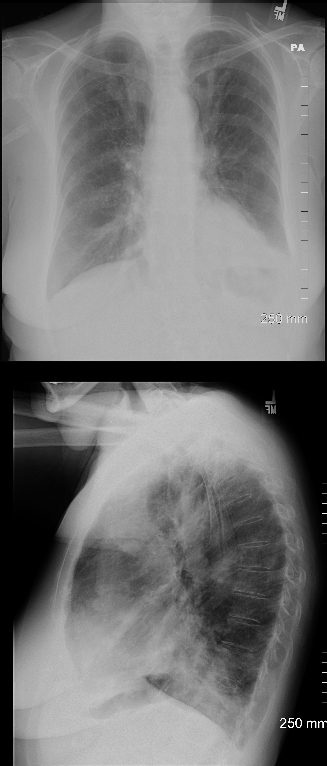
77 year old female with history of asthma, allergic bronchopulmonary aspergillosis (ABPA) and COPD
CXR shows hyperinflation, and consolidation in the left lower lobe silhouetting the left hemidiaphragm, with prominent bronchovascular bundles in the upper lung fields seen both on the PA and the lateral
Ashley Davidoff TheCommonVein.net 227Lu 135131c
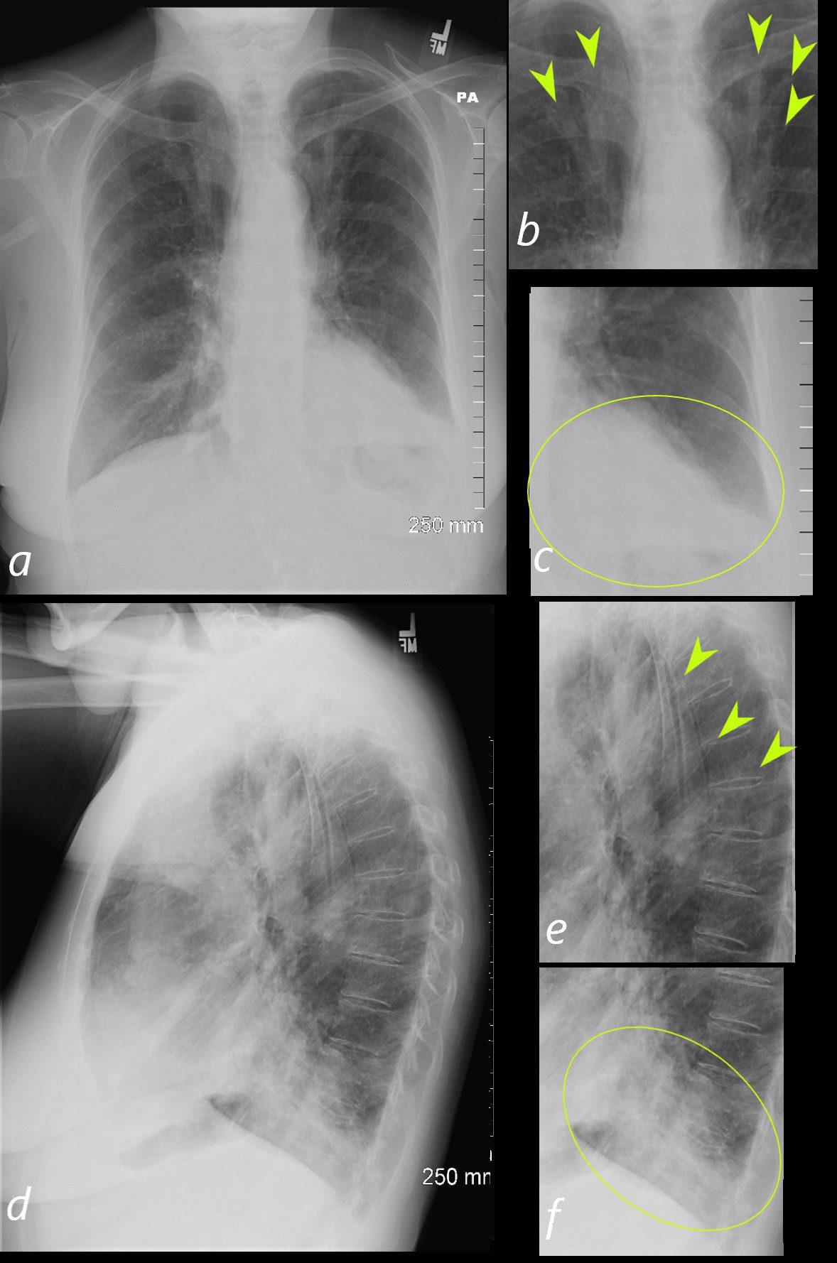
77 year old female with history of asthma, allergic bronchopulmonary aspergillosis (ABPA) and COPD
CXR in the PA projection(a) shows prominent tubular structures in the in the upper lung fields (green arrowheads in b) more prominent than the expected blood vessels seen in the hila. There is an infiltrate in the LLL with silhouetting of the left hemidiaphragm (green oval in c)
On the lateral examination (d) there is a suggestion of hyperinflation, and the tubular structures noted in b, are also appreciated, but are less obvious (green arrowheads in e). The consolidation in the left lower lobe is better appreciated (outlined by the green oval in f)
Ashley Davidoff TheCommonVein.net 227Lu 135161cL
Correlation of the Upper Lobe CXR Findings with the CT

CT – with Noted Tubular Structures in the Upper Lobes
77 year old female with history of asthma, allergic bronchopulmonary aspergillosis (ABPA) and COPD
CXR shows prominent bronchovascular bundles in the upper lung fields (green arrowheads a, and b) . CT shows fluid filled bronchiectatic airways (green arrowheads in image d, which is a magnified image of c) reminiscent of the finger in glove appearance of ABPA)
Ashley Davidoff TheCommonVein.net 227Lu 135161cL03
Correlation of the Lower Lobe CXR Findings with the CT
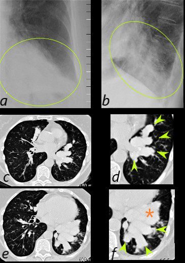
77 year old female with history of asthma, allergic bronchopulmonary aspergillosis (ABPA) and COPD
CXR shows LLL infiltrate in the PA (green oval in a) and lateral views (green oval in d and f) which reflect mucus filled bronchiectatic airways magnified image s of the CT scan of the LLL ) reminiscent of the finger in glove appearance of ABPA. There is a LL infiltrate possibly atelectatic (orange asterisk)
Ashley Davidoff TheCommonVein.net 227Lu 135161cL03
Lower Lobe Bronchiectasis, Finger in Glove and Consolidation
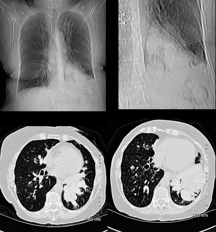
77 year old female with history of asthma, allergic bronchopulmonary aspergillosis (ABPA) and COPD
CT scout (a and magnified in b) shows lobular LLL infiltrate iof the lung Axial images show bilateral bronchiectatic airways . The LLL airways are more affected and are filled with mucus (finger in glove sign left lower image) becoming confluent and consolidative above the left hemidiaphragm right lower image).
Ashley Davidoff TheCommonVein.net 227Lu 135152c
Upper Lobe Bronchiolitis
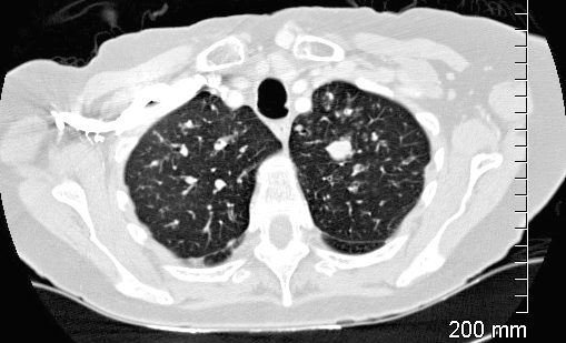
CT of the upper lobes show ground glass micronodules reminiscent of small airway disease
Impacted bronchus is noted in the left apex
Ashley Davidoff TheCommonVein.net 227Lu 135123
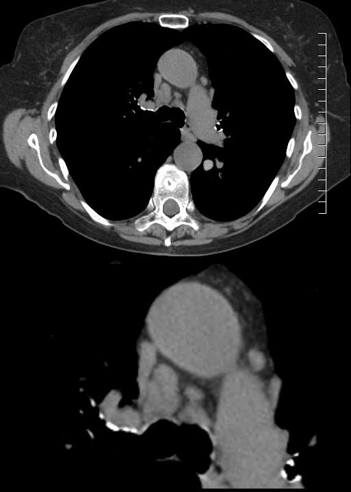
77 year old female with history of asthma, allergic bronchopulmonary aspergillosis (ABPA) and COPD
CT in the axial plane shows minimally enlarged mediastinal lymph nodes
Ashley Davidoff TheCommonVein.net 227Lu 135141c