
Ashley Davidoff
TheCommonVein.net
Source
Signs in Thoracic Imaging
Journal of Thoracic Imaging 21(1):76-90, March 2006.
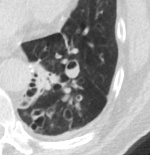
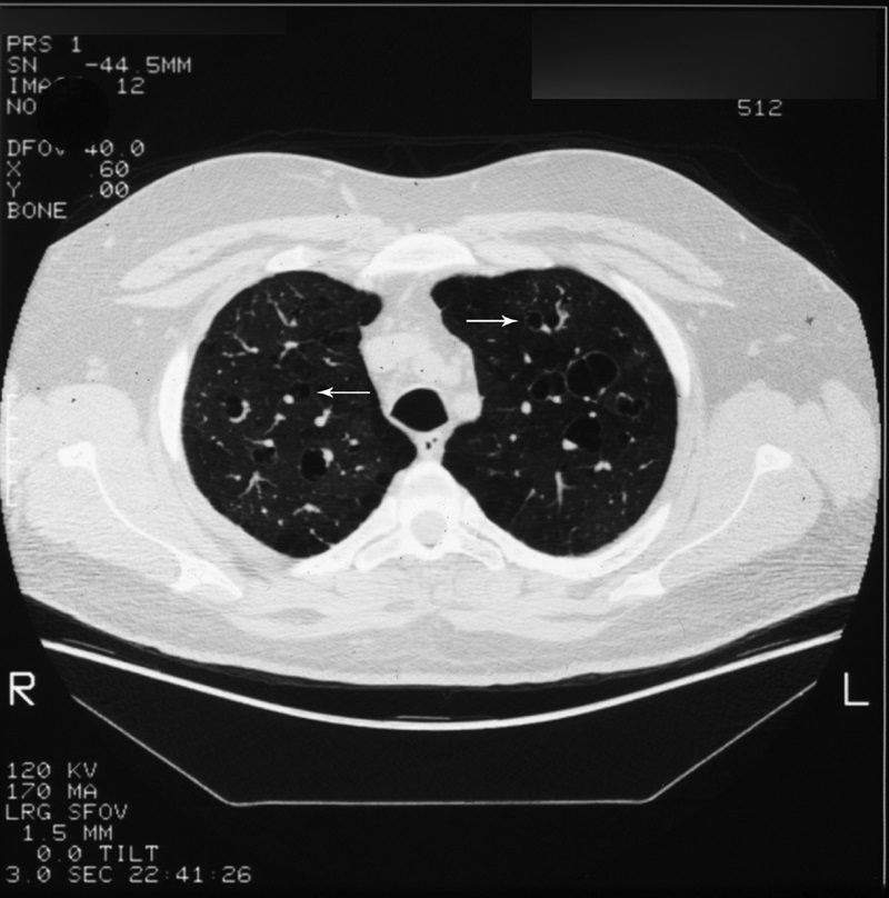
Described on chest CT (Fig. 19, arrows) as a circle of soft-tissue attenuation abutting a lucent ring, this sign depicts the pulmonary artery lying adjacent to a dilated bronchus in cross-section.54–57 The sign is found on CT in patients with bronchiectasis, or irreversible, abnormal bronchial dilatation.56 Accompanying findings, such as peribronchial thickening, lack of bronchial tapering, and visualization of bronchi within 1 cm of the pleura, are all contributing findings confirming the diagnosis. In isolation, an enlarged bronchoarterial ratio may be a spurious finding, as bronchial diameter varies with parameters such as altitude.58 Measurement of the bronchial luminal diameter is made between the inner walls so as not to overestimate in cases with associated peribronchial thickening. Bronchiectasis occurs due to bronchial wall injury, most often due to necrotizing viral or bacterial bronchitis.4,57,58 It can also occur secondary to bronchial obstruction with subsequent mucus production dilating the involved airways, as mentioned previously with the finger-in-glove sign. Finally, in the setting of pulmonary fibrosis or radiation-induced lung injury, traction on the adjacent bronchi will cause dilatation. Other entities, such as cystic fibrosis and Kartagener syndrome, are also associated.4,57