
- Presented 18 months prior
- COPD exacerbation
Patient presenting with multiple months of DOE, noted to hypoxic with minimal exertion - Malignant Neoplasms
Squamous cell carcinoma of lung, stage III, leftClinical Stage IIIB (cT3, cN2, cM0),- Squamous Cell – non-small cell lung carcinoma (squamous) of the Lt lung,
- completed CRT (with weekly carboplatin, paclitaxel regimen), post-CRT Chest CT with partial response, started on durvalumab,
– Durvalumab after CCRT (per PACIFIC trial – N Engl J Med. 2018;379(24):2342); remarkable improvement in PFS (17.2 vs 5.6 months) & median overall survival (12 month survival 83 vs 75%, 24 month survival 66 vs 56%) compared to ChemoRT alone.
-side effects of durvalumab including- pneumonitis, colitis, hepatitis, nephritis, thyroiditis etc.
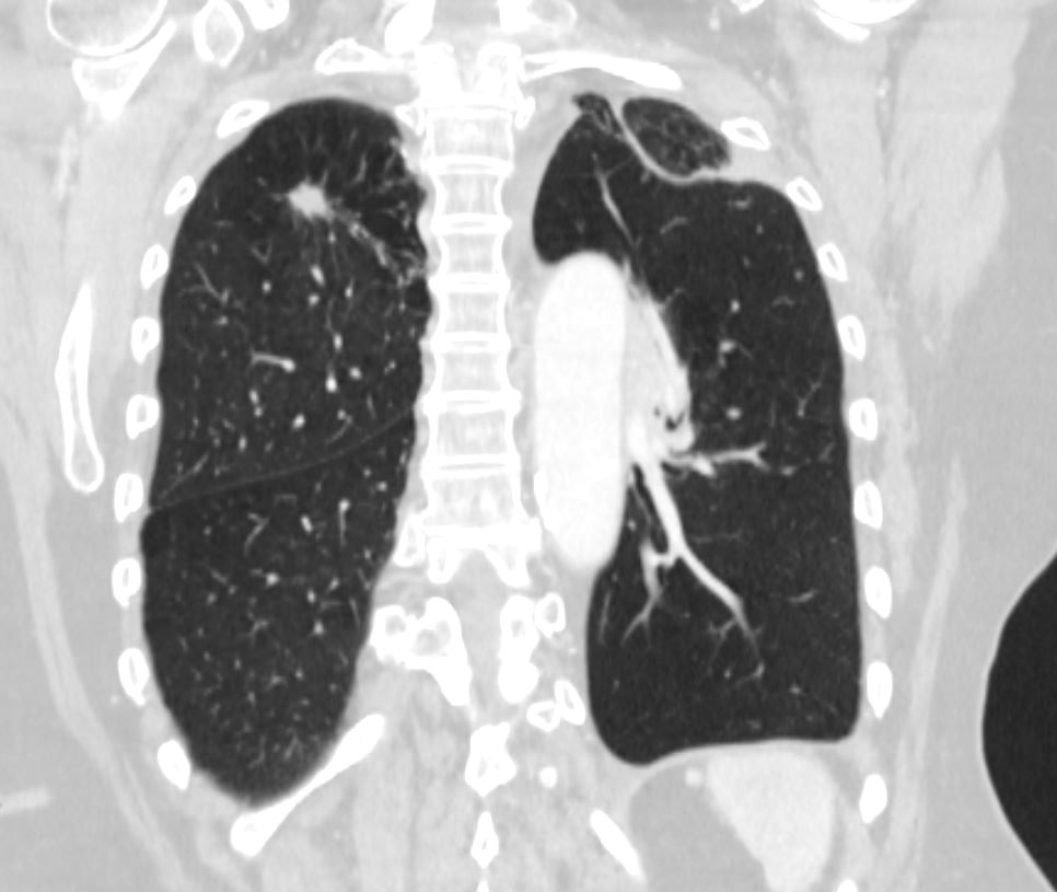
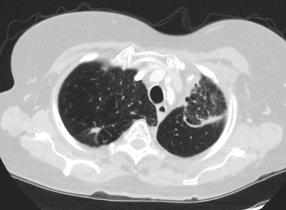
Left Upper Lobe Atelectasis Luftsichel Sign
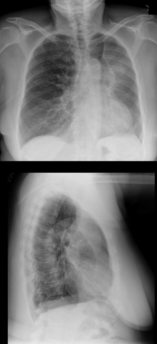
Left Upper Lobe Atelectasis
55-year-old female with central squamous cell carcinoma of the lung with left upper lobe collapse and hyperinflation of the left lower lobe resulting in a Luftsichel sign. The major fissure is displaced superiorly and anteriorly and the hyperinflated left lower lobe is hyperexpanded and fills the apical space resulting in the hyperlucency in the apex.
Ashley Davidoff MD TheCommonVein.net 152Lu 136636c
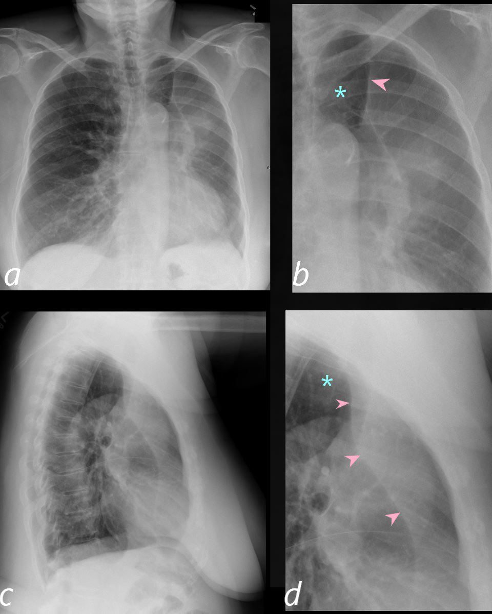
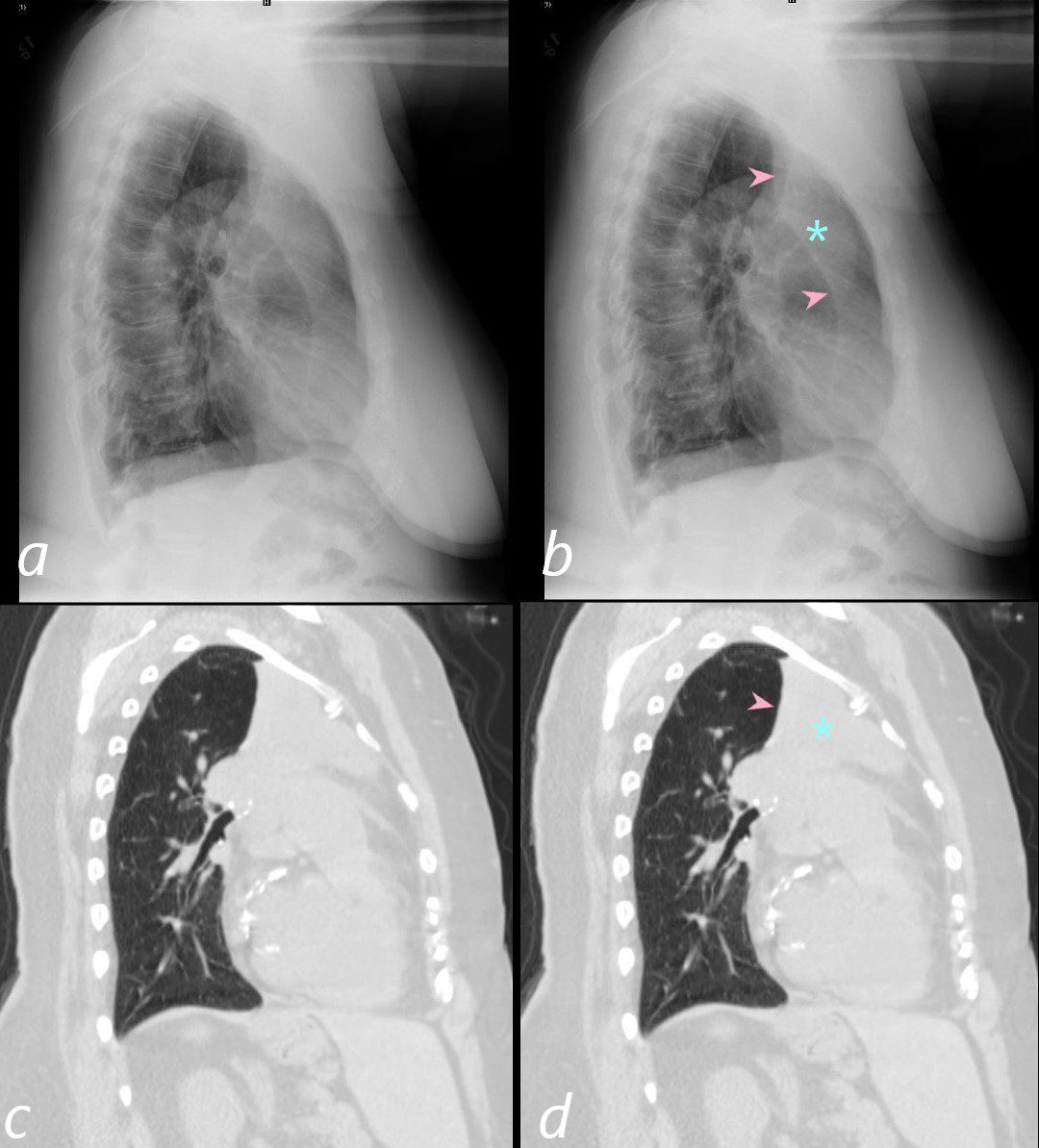
Left Upper Lobe Atelectasis
55-year-old female with central squamous cell carcinoma of the lung with left upper lobe collapse and hyperinflation of the left lower lobe resulting in a Luftsichel sign. The major fissure is displaced superiorly and anteriorly (pink arrowheads in magnified images b and d)
The hyperinflated left lower lobe is hyperexpanded and fills the apical space resulting in the hyperlucency in the apex (teal asterisk b and d).
Ashley Davidoff MD TheCommonVein.net 152Lu 136636c
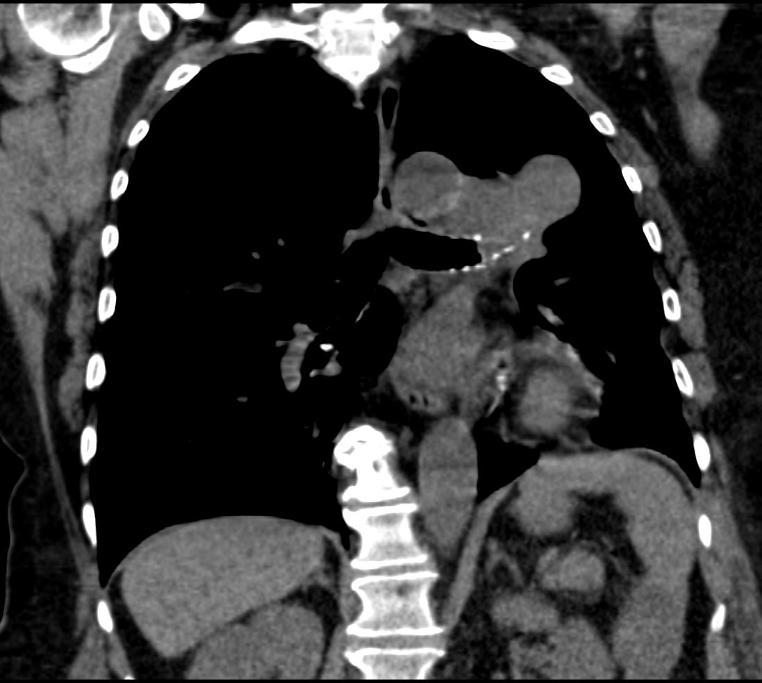
CT scan Coronal View
Left Upper Lobe Atelectasis
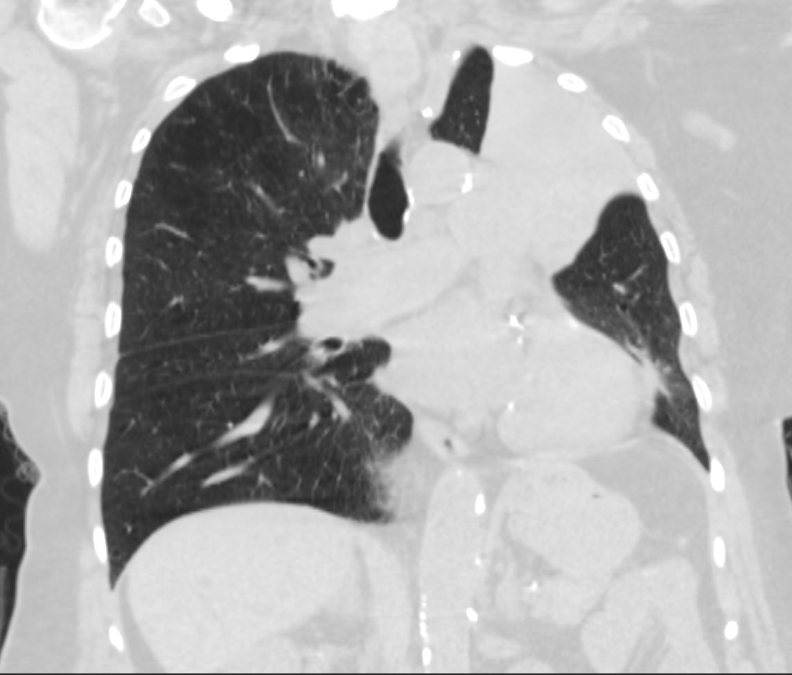
Left Upper Lobe Atelectasis
55-year-old female with central squamous cell carcinoma of the lung with left upper lobe collapse with atelectatic lung collapsed medially and superiorly
Ashley Davidoff MD TheCommonVein.net 152Lu 136642
Mediastinal Shift
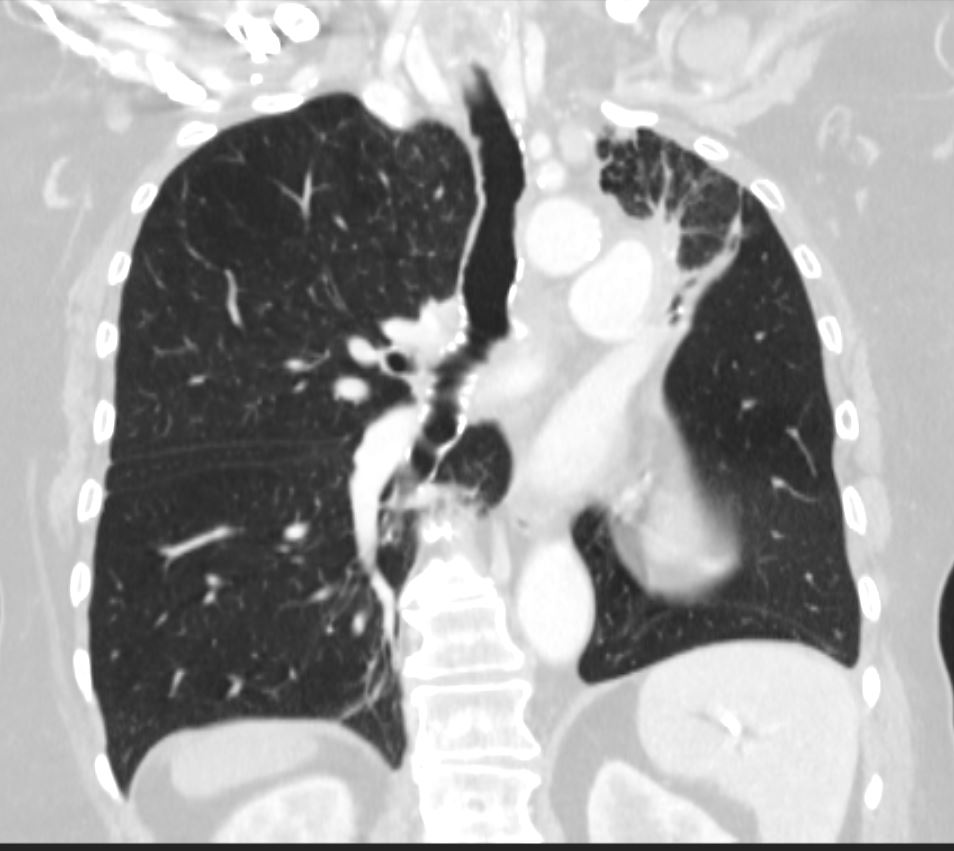
55-year-old female with central squamous cell carcinoma of the lung with left upper lobe collapse and hyperinflation of the left lower lobe resulting in a Luftsichel sign
Noted leftward deviation of the trachea.
Ashley Davidoff MD TheCommonVein.net 152Lu 136648
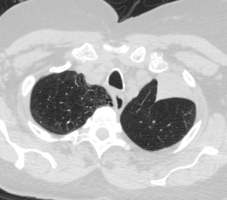
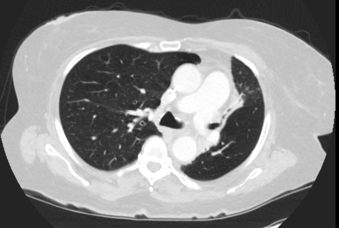
Spiculated Right Upper Lobe Lesion
Left Upper Lobe Atelectasis Hyperlucent Left Lower Lobe
55-year-old female with central squamous cell carcinoma of the lung with left upper lobe collapse and hyperinflation of the left lower lobe resulting in a Luftsichel sign
Noted spiculated lesion in the right upper lobe
Ashley Davidoff MD TheCommonVein.net 152Lu 136649
Atelectatic Lung Collapsed Anteriorly
The Hyperinflated LLL is Seen Occupying the Left Apex
Left Upper Lobe Atelectasis
55-year-old female with central squamous cell carcinoma of the lung with left upper lobe collapse with atelectatic lung collapsed medially and superiorly
The lateral CXR (a and b) shows the displaced major fissure (pink arrowhead) and the atelectatic left upper lobe (teal asterisk) with correlative sagittal CT images
Ashley Davidoff MD TheCommonVein.net 152Lu 136643cL
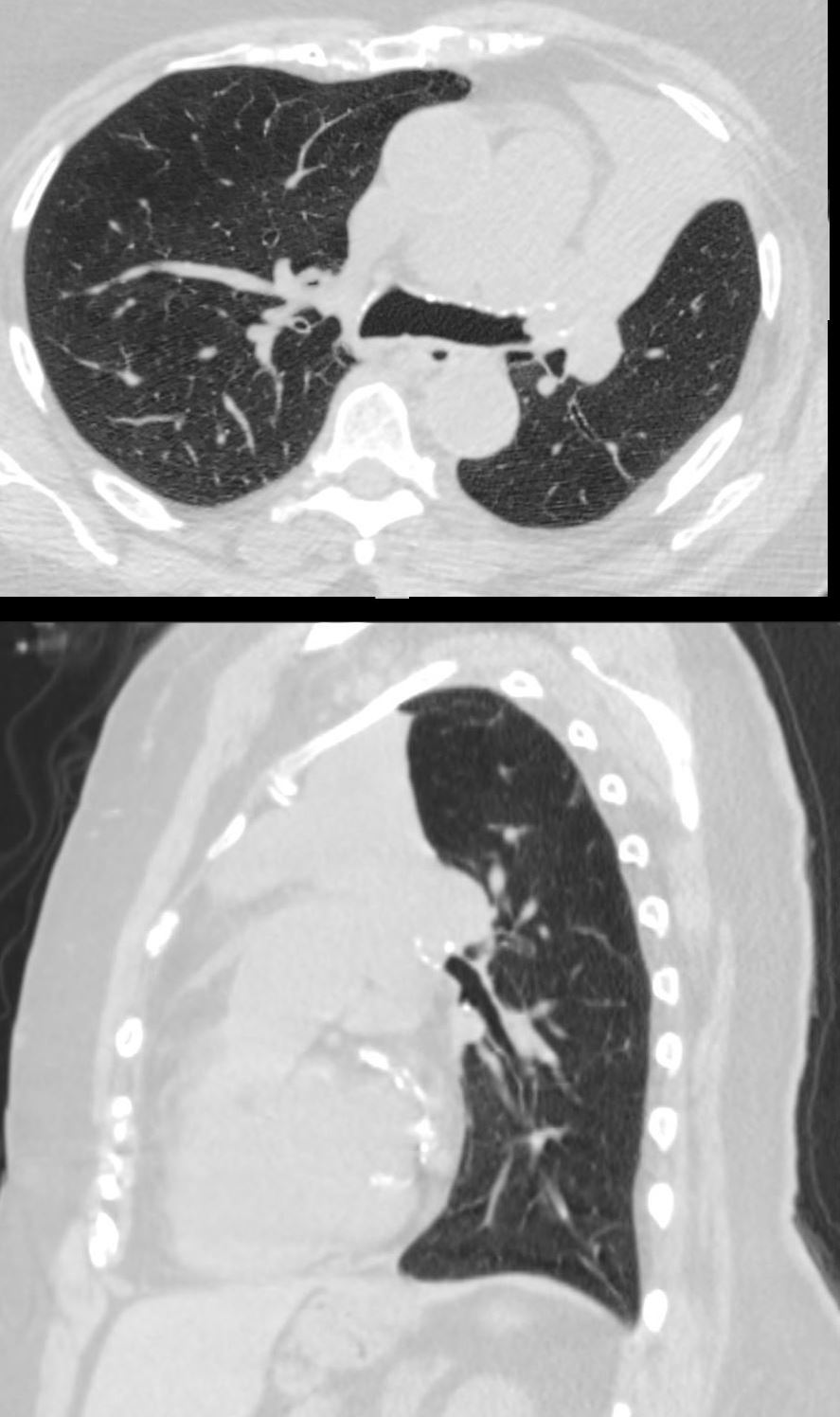
55-year-old female with central squamous cell carcinoma of the lung with left upper lobe collapse in the axial views (above) and sagittal views (below) . There is compensatory hyperinflation of the left lower lobe resulting in cranial migration of the LLL which fills the apex and results in a Luftsichel sign.
Ashley Davidoff MD TheCommonVein.net 152Lu 136639c01
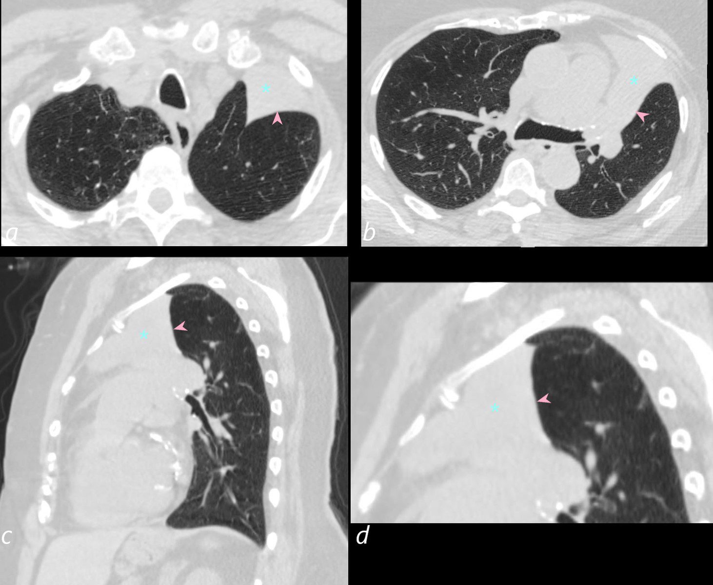
55-year-old female with central squamous cell carcinoma of the lung with left upper lobe collapse (pink asterisks) in the axial views (a,b) and sagittal views (c,d) . There is compensatory hyperinflation of the left lower lobe resulting in cranial migration of the LLL which fills the apex(pink asterisks) and results in a Luftsichel sign.
Ashley Davidoff MD TheCommonVein.net 152Lu 136639cL
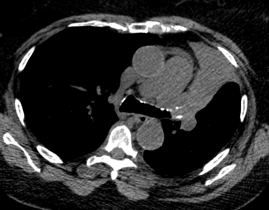
Soft Tissue Density Representing the Tumor is Noted in the
Left Mainstem Bronchus
55-year-old female with central squamous cell carcinoma of the lung with left upper lobe collapse and hyperinflation of the left lower lobe
A soft tissue density representing the tumor is noted in the left mainstem bronchus.
Ashley Davidoff MD TheCommonVein.net 152Lu 136641
Soft Tissue Density Representing the Tumor is Noted in the
Left Mainstem Bronchus
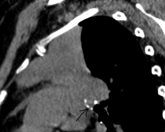
55-year-old female with central squamous cell carcinoma of the lung with left upper lobe collapse with atelectatic lung collapsed anteriorly and superiorly
Soft tissue tumor is noted in the bronchus (arrow) and the atelectatic lung is positioned anteriorly along the anterior chest wall.
Ashley Davidoff MD TheCommonVein.net 152Lu 136644
55-year-old female with central squamous cell carcinoma of the lung with left upper lobe collapse and hyperinflation of the left lower lobe resulting in a Luftsichel sign. The hyperinflated LLL is seen occupying the left apex
Mediastinum is displaced to the left.
Ashley Davidoff MD TheCommonVein.net 152Lu 136640
The Soft Tissue Tumor is noted in the Left Main Stem Bronchus
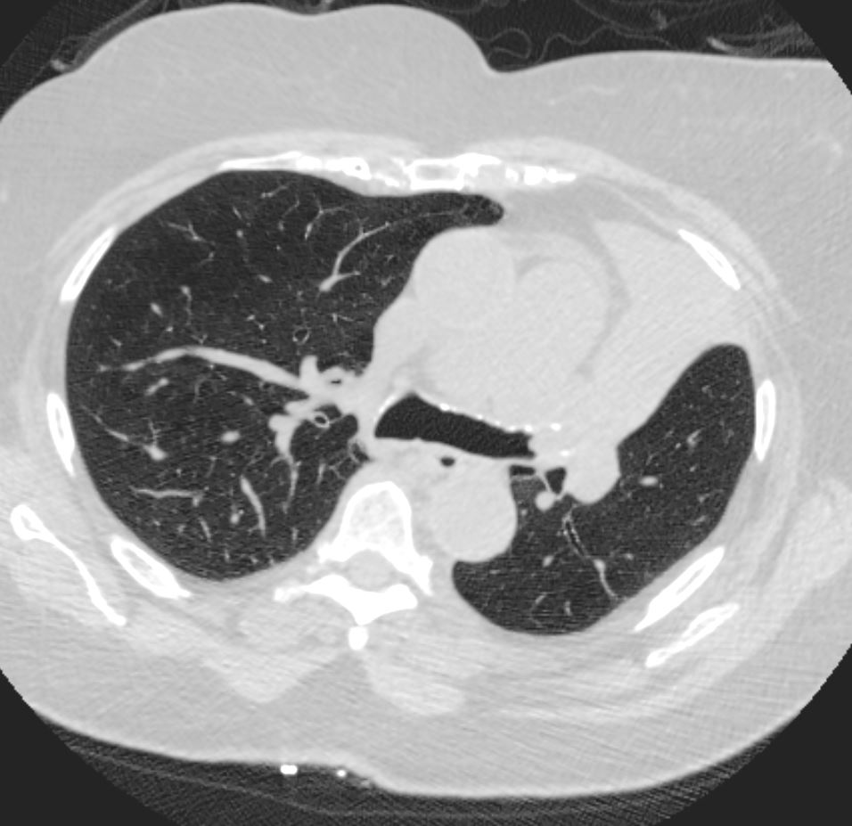
55-year-old female with central squamous cell carcinoma of the lung with left upper lobe collapse and hyperinflation of the left lower lobe resulting in a Luftsichel sign.
The left lung is relatively lucent as a result of hyperinflation . The atelectatic left upper lobe manifests as an anterior soft tissue density along the anterior mediastinum. The soft tissue tumor is noted in the left main stem bronchus
Ashley Davidoff MD TheCommonVein.net 152Lu 136639
55-year-old female with central squamous cell carcinoma of the lung with left upper lobe collapse
The atelectatic left upper lobe manifests as an anterior soft tissue density along the anterior mediastinum and chest wall. A soft tissue density representing the tumor is noted in the left mainstem bronchus.
Ashley Davidoff MD TheCommonVein.net 152Lu
Left Upper Lobe Atelectasis
55-year-old female with central squamous cell carcinoma of the lung with left upper lobe collapse and hyperinflation of the left lower lobe resulting in a Luftsichel sign. Thickening of the major fissure. and some groundglass changes in a small portion of aerated left upper lobe
Noted leftward deviation of the trachea. Noted spiculated lesion in the right upper lobe. Noted spiculated lesion in the right upper lobe
Ashley Davidoff MD TheCommonVein.net 152Lu 136647
Hyperinflated Left Lower Lung Field is Noted Alongside the Atelectatic LUL.
Left Upper Lobe Atelectasis
55-year-old female with central squamous cell carcinoma of the lung with left upper lobe collapse and hyperinflation of the left lower lobe The relatively lucent hyperinflated left lower lung field is noted alongside the atelectatic LUL.
Ashley Davidoff MD TheCommonVein.net 152Lu 136646
Endoscopic view of the Left Main Bronchus showing the
Obstructing Tumor
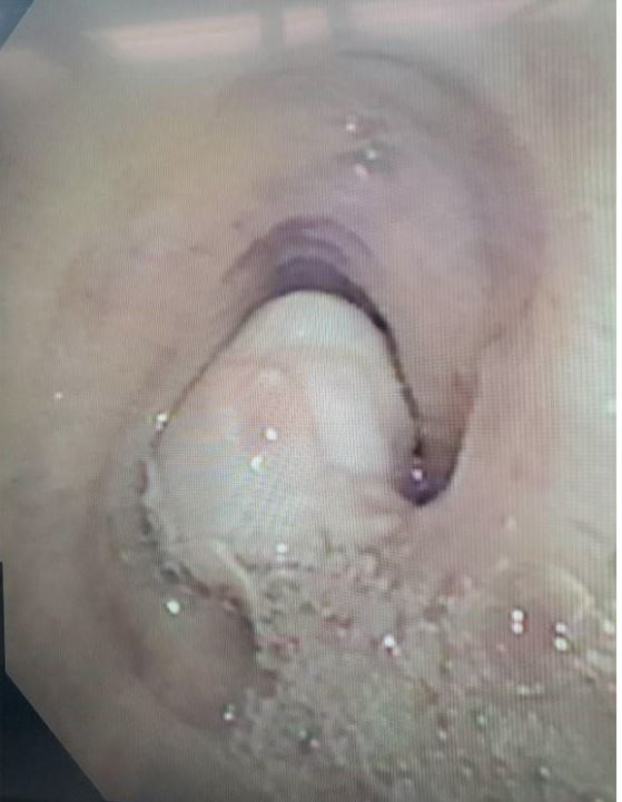
Ashley Davidoff MD TheCommonVein.net 152Lu 136645