
Stage IV Small Cell Lung Carcinoma – Hilum Overlay Sign
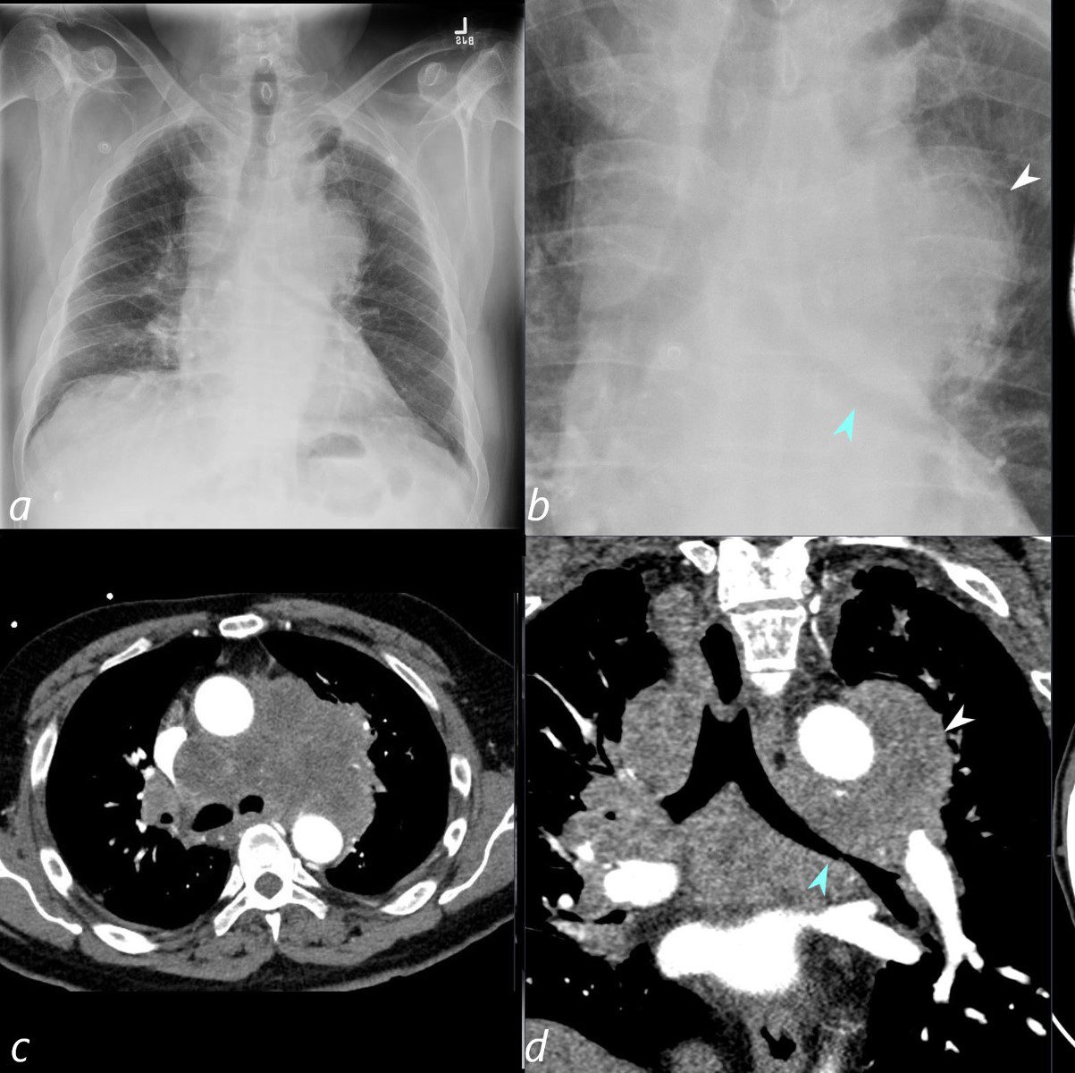
65 year old male presents with a history of chest pain, back pain, myalgias, and night sweats
Frontal CXR shows a left sided mass that silhouettes the aortic knob and the main pulmonary artery (white arrowhead, b) indicating that the mass arises from middle mediastinal tumors, or hilar adenopathy. The mass also compresses and displaces the left mainstem bronchus (teal arrowhead b).
The axial CT (c) shows the extent of the mass and the coronal CT scan (d) confirms the presence of a large mediastinal mass that extends beyond the aortic knob (white arrowhead)
A diagnosis of a large mediastinal small cell carcinoma of the lung was diagnosed
Ashley Davidoff MD TheCommonVein.net 297Lu 136691cL
Compression and Encasement
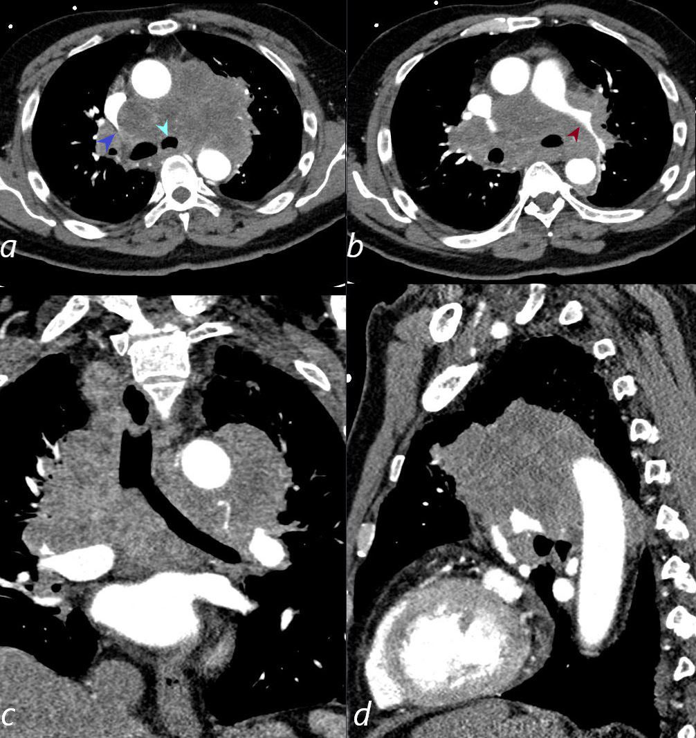
65 year old male presents with a history of chest pain, back pain, myalgias, and night sweats
CT scan in multiple planes shows a left sided mass that extends from the left hilum to the mediastinum and to the ight hilum. The mass compresses the SVC, (blue arrowhead a), narrows the left mainstem bronchus (teal arrowhead, a) and encases the left pulmonary artery., the left mainstem bronchus (teal arrowhead b).
A diagnosis of a large mediastinal small cell carcinoma of the lung was diagnosed
Ashley Davidoff MD TheCommonVein.net 297Lu 136692cL
Extensive Regional Spread
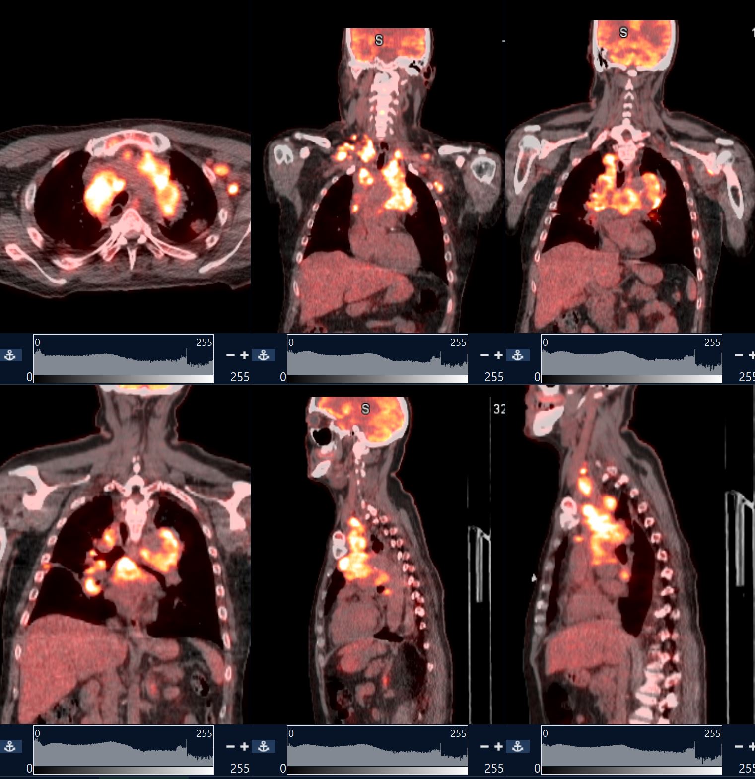
65 year old male presents with a history of chest pain, back pain, myalgias, and night sweats
PET CT scan in multiple planes shows extensive hypermetabolic activity throughout the mediastinum and extending into the right cervical chain, subpectoral region and axilla on the left.
A diagnosis of a large mediastinal small cell carcinoma of the lung with extensive regional metastases was diagnosed
Ashley Davidoff MD TheCommonVein.net 297Lu 136696
Responsive to Treatment
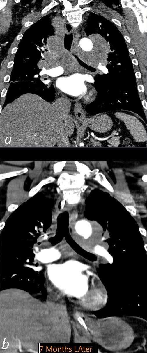
Coronal CT of the chest through the mediastinum at the level of the carina before (a) and 7months following chemotherapy, shows significant reduction of the extensive mediastinal and hilar tumor
Ashley Davidoff MD TheCommonVein.net 297Lu 136698
Endobronchial Spread of Poorly Differentiated Small Cell Carcinoma with Encasement

Courtesy Ashley Davidoff MD.
TheCommonVein.net
32426_02cl

The CT scan of the centrally positioned small cell carcinoma has structural implication as a result of its central location close to large arteries, veins, airways and lymphatics. In this case the centrally placed tumor (dark green in image b) is pushing on the right mainstem bronchus (shown with white arrow) and the lymphatics with peribronchial thickening (image d light green) and extension into the interlobular septa (bright green in d) Subcarinal nodal involvement and left hilar involvement (light green in b)together with small right effusion (a,b) are also noted suggesting advanced disease.
Ashley Davidoff
TheCommonVein.net
87711c01.8s

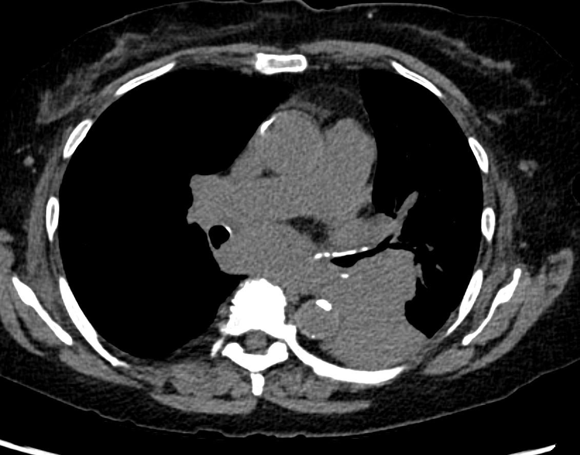
An axial and a sagittal view of a centrally placed carcinoma that lies on the major fissure of the right lung code lung mass fissure likely carcinoma central probability of it being a squamous cell carcinoma or a small carcinoma is high because of its central location.
keywords lung mass nodule major fissure cancer carcinoma central CTscan
Ashley Davidoff
TheCommonVein.net
75675c01.8s
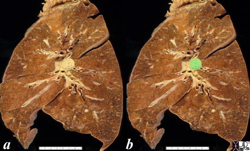
The anatomical specimen shows a 2cms central lesion near the hilum of the left lung causing extrinsic impression on the central bronchovascular bundle. The relatively small size and central location suggests a squamous cell carcinoma though a non small cell carcinoma is also possible
Ashley Davidoff
TheCommonVein.net
32323c02.8s
Small Cell Lung Carcinoma with Early Atelectasis
Diagnosis Small Cell Lung Carcinoma with Early Atelectasis
Ashley Davidoff MD TheCommonVein.net
Diagnosis Small Cell Lung Carcinoma with Early Atelectasis
Ashley Davidoff MD TheCommonVein.net
1 Week Later Almost Complete White Out with Near Total Left Lung Collapse

Diagnosis Small Cell Lung Carcinoma
CXR shows almost complete “white out” of the left lung caused by a large hilar mas Diagnosis Small Cell Lung Carcinoma
Ashley Davidoff MD TheCommonVein.net