
Percutaneous Biopsy
Percutaneous biopsy of the lung offers the ability to characterize lung lesions with very few contraindications. Complications include pneumothorax (10-49%) and hemoptysis (8-20%) which generally resolve without treatment (Gohari).
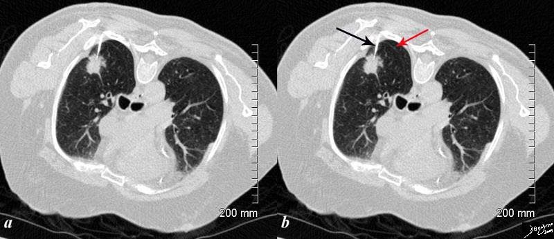
The CT is from an 81 year old man with remote history of smoking who presents with an asymptomatic lung mass. The lesion was PET positive, without nodal activity suggesting that it was either an infection, inflammatory lesion or a primary lung cancer. A biopsy revealed adenocarcinoma of the lung. In the image the patient has been placed in the prone projection. The needle (arrow is shown) within the spiculated lesion. The radiologist needed to avoid the major fissure which is seen as a fine curvilinear structure posteriorly (red arrow), negotiate the the the scapula and two closely spaced ribs. Without CT guidance this procedure would have been almost impossible to biopsy.
Courtesy Ashley Davidoff MD
TheCommonVein.net
85221c01.8s
Small Pneumothorax

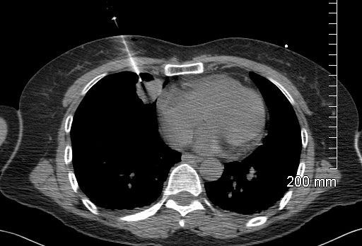
The CT through a mass in the LUL shows focal pleural calcification overlaid in green (2,4,) in this patient with asbestos related disease. A pneumothorax followed the biopsy revealing that the pleural plaques are positioned on the parietal pleura.(3,4) A small amount of air introduced from the xylocaine injection outlines the parietal pleura.(5,6 – green)
Ashley Davidoff
TheCommonVein.net
32017c.jpg
Ashley Davidoff
TheCommonVein.net
134384
Transbronchial Biopsy
Transbronchial evaluation of central lung cancers. When the lesions are visible washings have a yield of 79%, brushings 92%, and forceps 93%. For peripheral lesions that are <2cms yield is 20% and when they are >2cms yield is 50-80%. It is a safe procedure sometimes complicated by pneumothorax (10%), and less commonly hemorrhage (.2%) laryngospasm (.13%).