
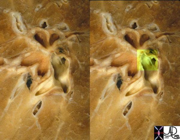
Web Across the Pulmonary Artery
Chronic PE
This is an image of a web across the pulmonary artery, (overlaid in green in the second image) residual from a previous pulmonary embolus.
Courtesy Ashley Davidoff MD. and Jeffrey Peirce 32300c
CTPA Pulmonary Embolus 15 Years Ago
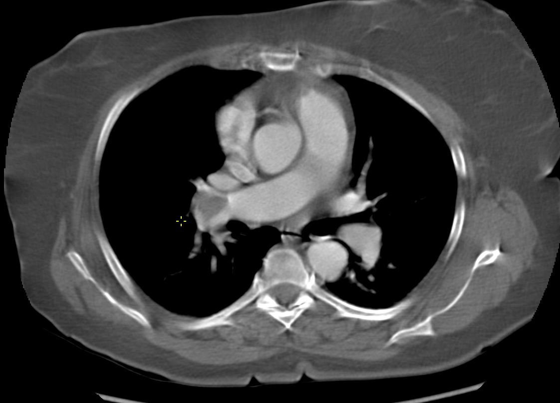
CTPA 15 years ago shows a large thrombotic embolus in the right main pulmonary artery extending into the middle lobe artery
Ashley Davidoff MD TheCommonVein.net 136172
Pulmonary Embolus 15 Years Later
Obstructive Dystrophic Calcification
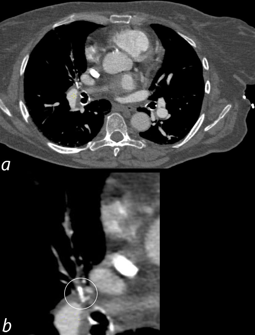
CTPA 15 years following an acute pulmonary embolus reveals an obstructive dystrophic calcification at the origin of the middle lobe pulmonary artery (b ringed in white). There is a suggestion of paucity of contrast in the middle love
Ashley Davidoff MD TheCommonVein.net 136173cL
Obstructive Dystrophic Calcification
Attenuated Middle Lobe Artery
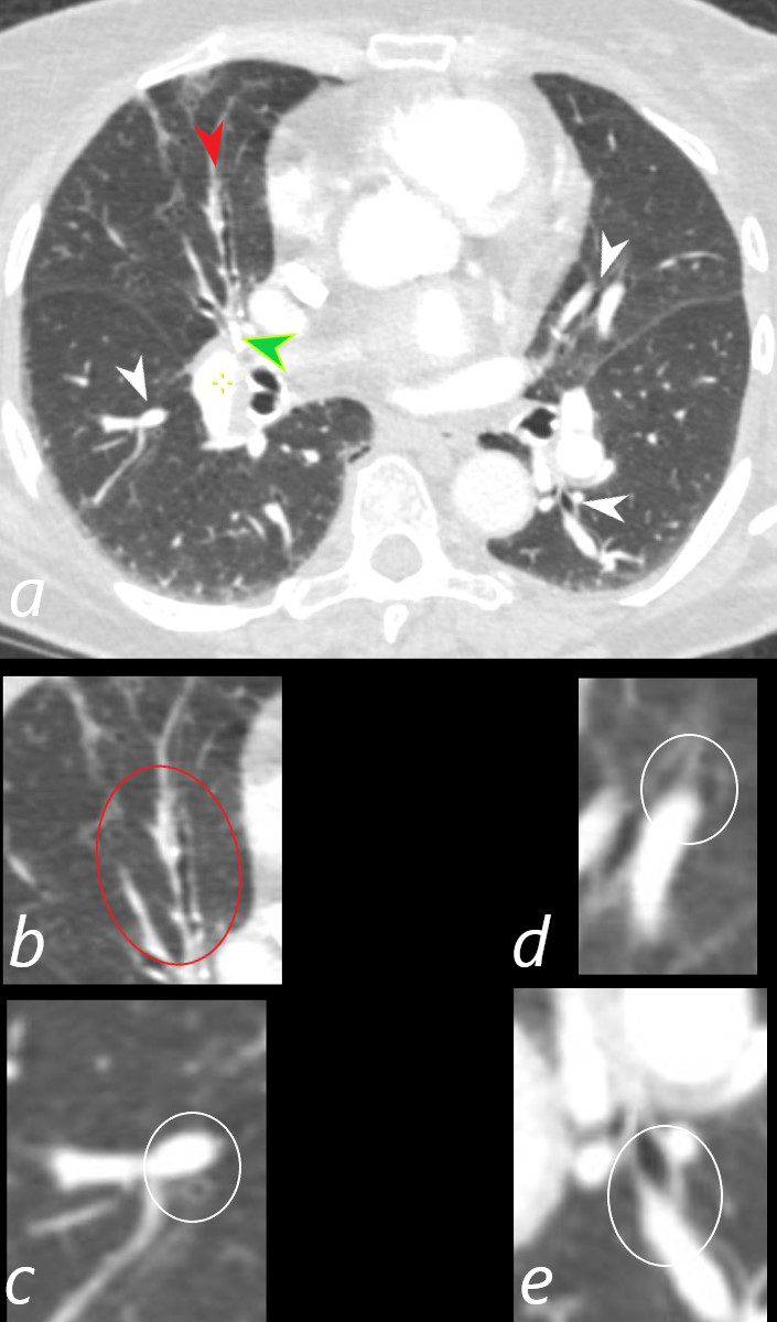
CTPA 15 years following an acute pulmonary embolus, reveals an obstructive dystrophic calcification at the origin of the middle lobe pulmonary artery (a, green arrowhead) The middle lobe arteries distal to the calcification are attenuated due to proximal obstruction (a, red arrowhead and b red ring) . The normal sized arteries are shown in images ringed in images c, d and e demonstrating the normal ratio of arterial to bronchiole size. There is only mild scarring and ground glass change in the middle despite the apparent decrease in size of the artery.
Ashley Davidoff MD TheCommonVein.net 136175cL01