
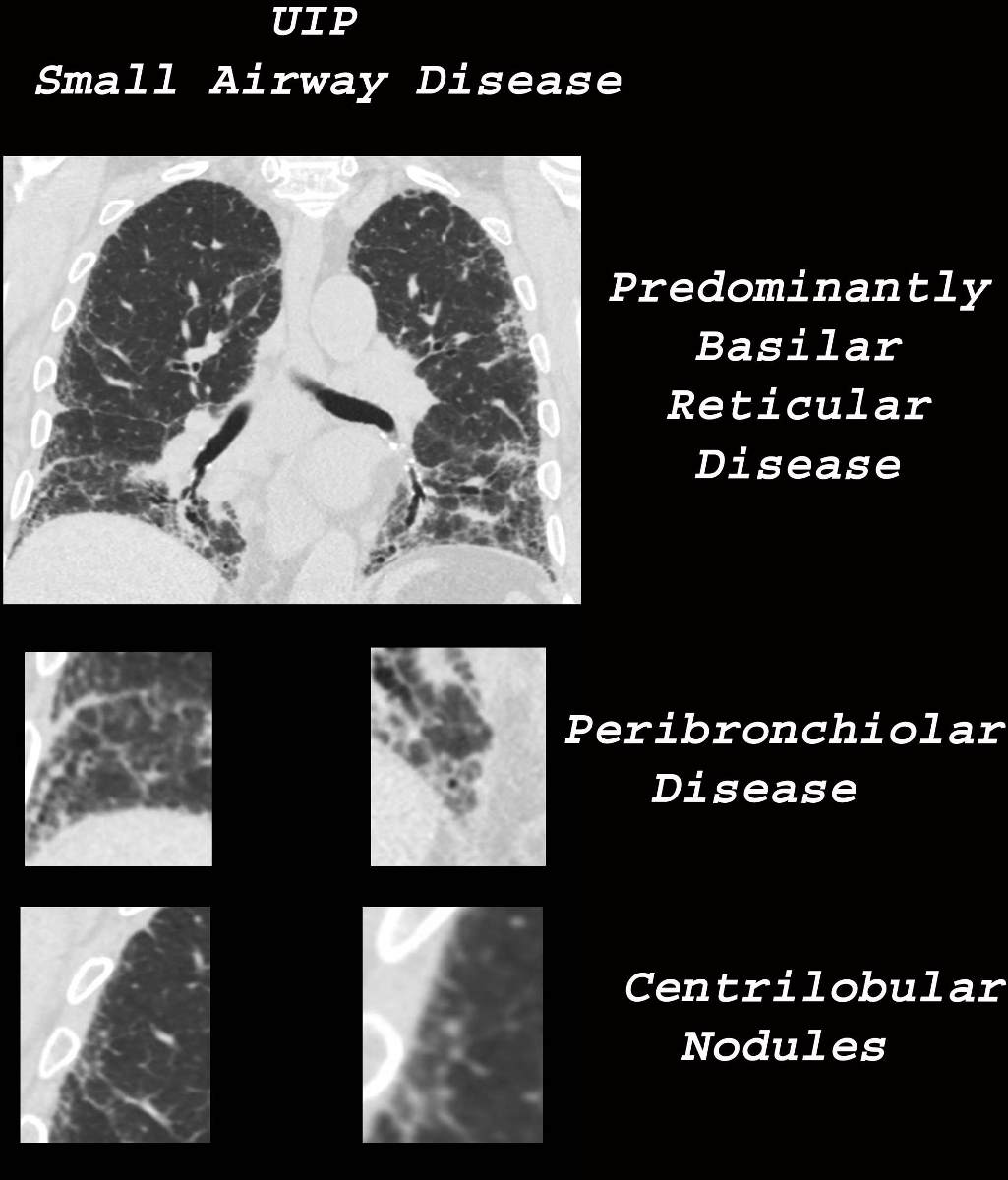
Reticular Disease
Basilar Peripheral Disease
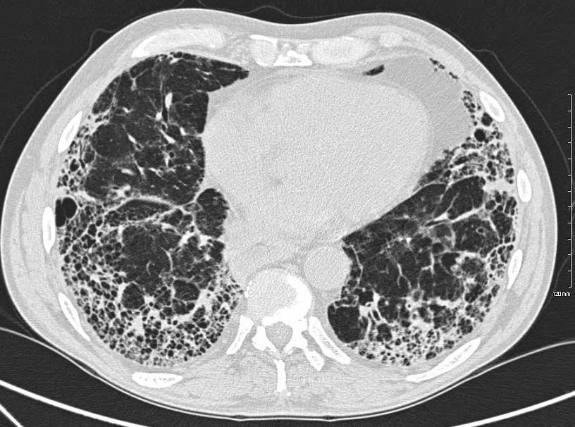
Ashley Davidoff MD TheCommonVein.net uip-002c-CT
CT UIP and Honeycomb Lung in an84 year old Female
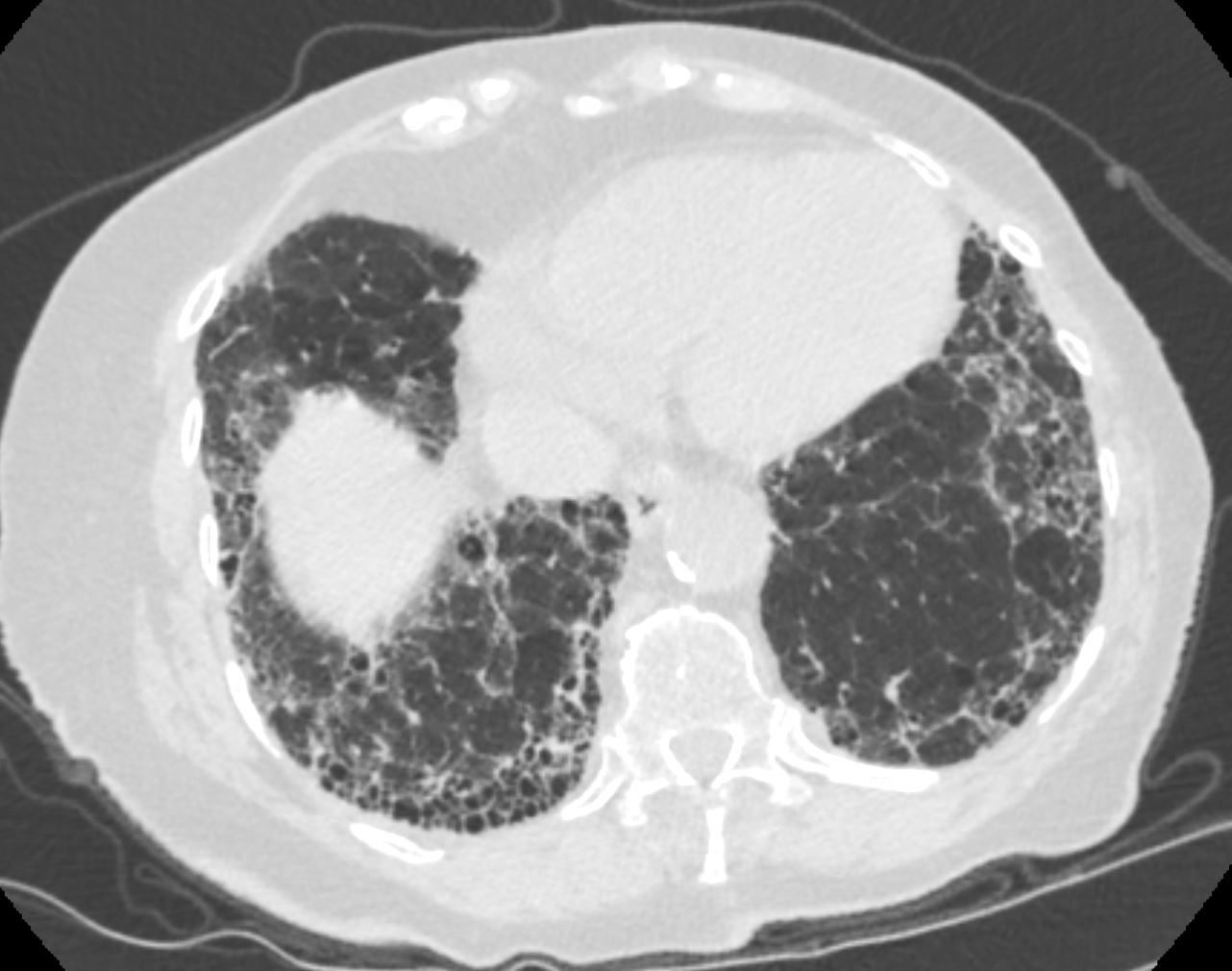
CT scan in the axial plane through the lung bases of an 84 year old female with UIP showing the typical changes of honeycomb lung in the periphery of both lung bases, and in this instant more prominent at the right base
Ashley Davidoff MD TheCommonVein.net 136453
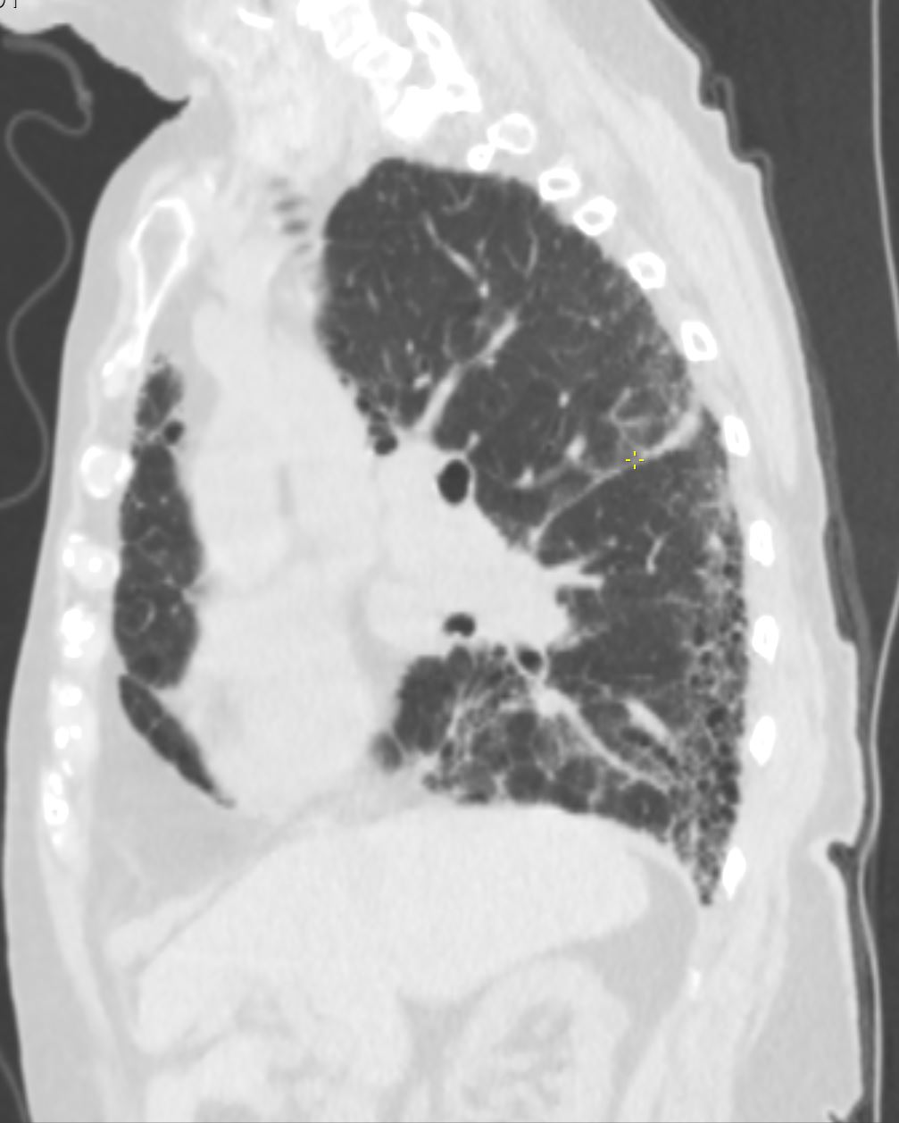
CT scan in the sagittal plane through the right lung of an 84 year old female with UIP showing the typical changes of honeycomb lung in the periphery posteriorly.
Ashley Davidoff MD TheCommonVein.net 136454
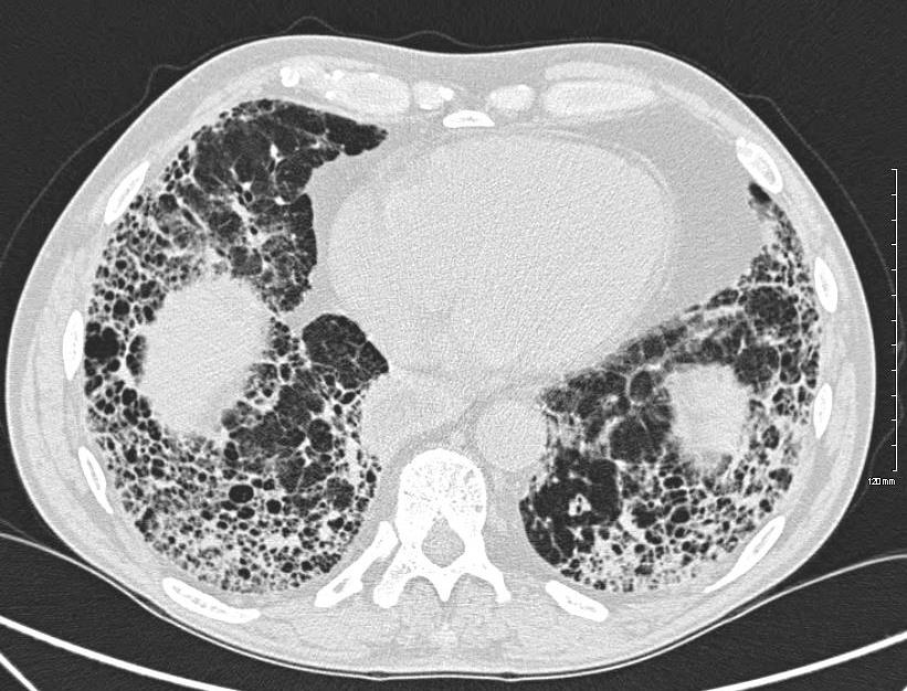
58 year old male wit hfamily history of IPF Features on the CT reveal a dominant pattern of extensive honeycombing in the lower lobes
Ashley Davidoff MD thecommonvein.net 134902-lungs UIP
Ashley Davidoff MD thecommonvein.net 134901-lungs UIP