
Pooja Sikka MD Ashley Davidoff MD
Problem
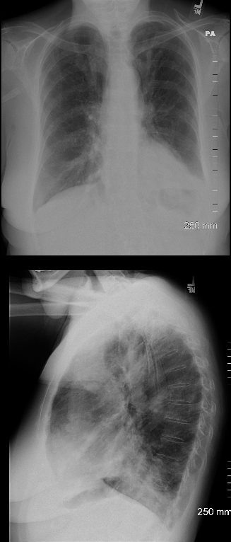
77 year old female with history of asthma, allergic bronchopulmonary aspergillosis (ABPA) and COPD
CXR shows hyperinflation, and consolidation in the left lower lobe silhouetting the left hemidiaphragm, with prominent bronchovascular bundles in the upper lung fields seen both on the PA and the lateral Diagnosis: Asthma Allergic Bronchopulmonary Aspergillosis (ABPA) COPD
Ashley Davidoff TheCommonVein.net
Source
Signs in Thoracic Imaging
Journal of Thoracic Imaging 21(1):76-90, March 2006.
Solution Next
Back to “Image First” Case List
Links and References