
Total Lung Collapse
Total Left Lung Collapse
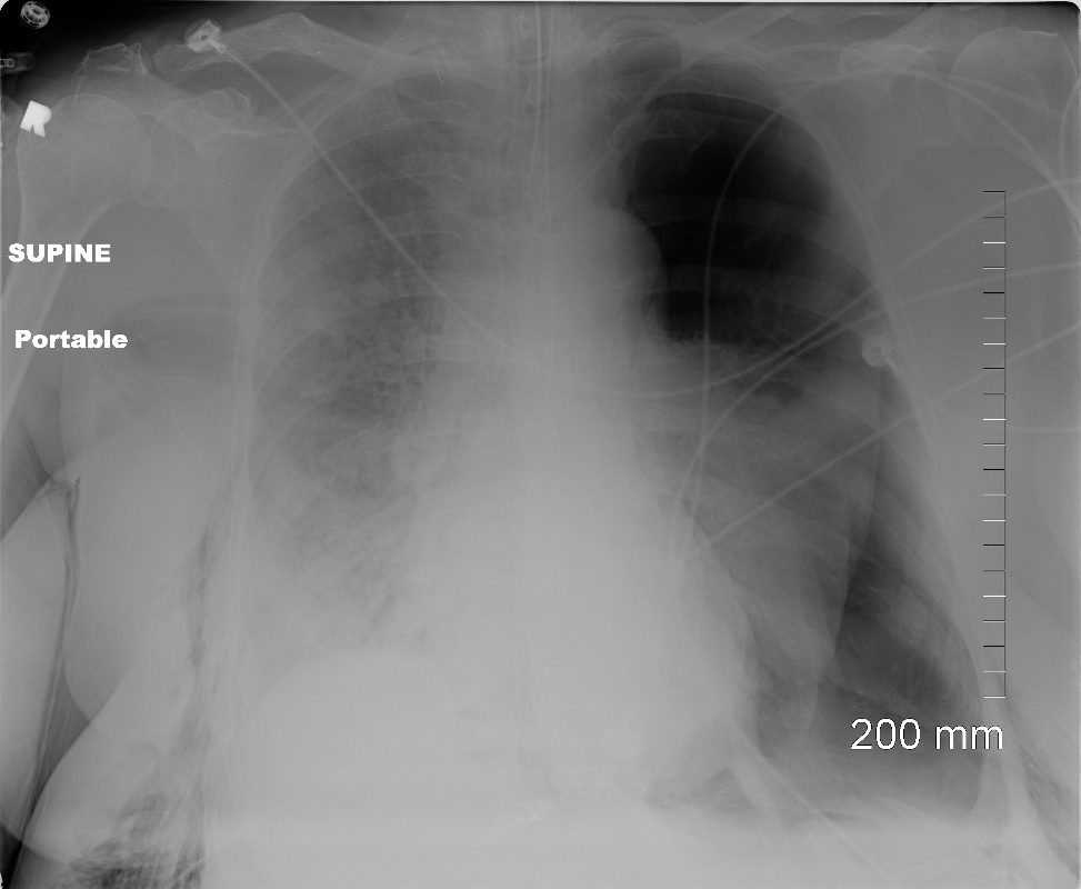
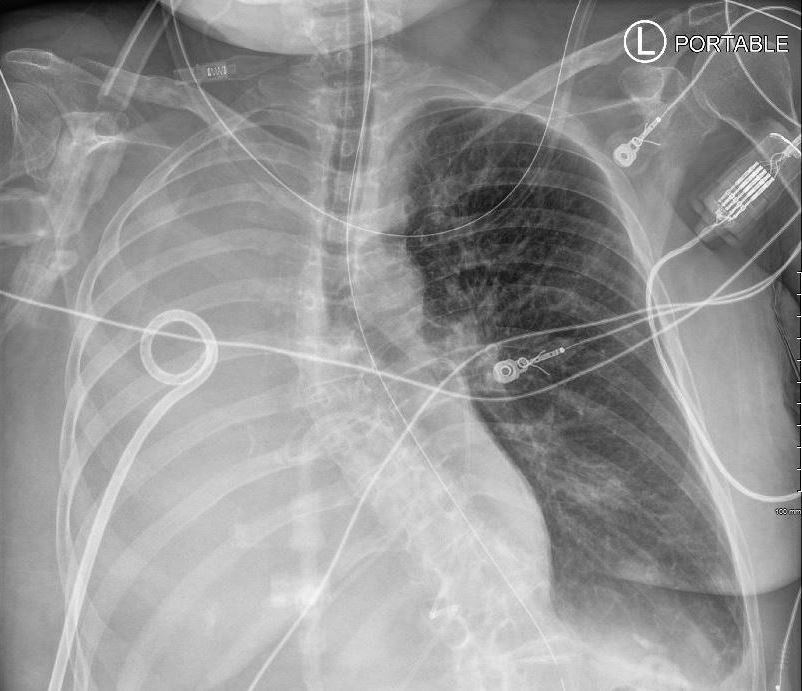
Tension Pneumothorax
Ashley Davidoff MD TheCommonVein.net
77949
49 year old male with a cough presents for a Chest Xray which showed a tension pneumothorax. Chest tube was placed emergently in the radiology department.
Ashley Davidoff MD TheCommonVein.net
117300c
Total Right Lung Collapse
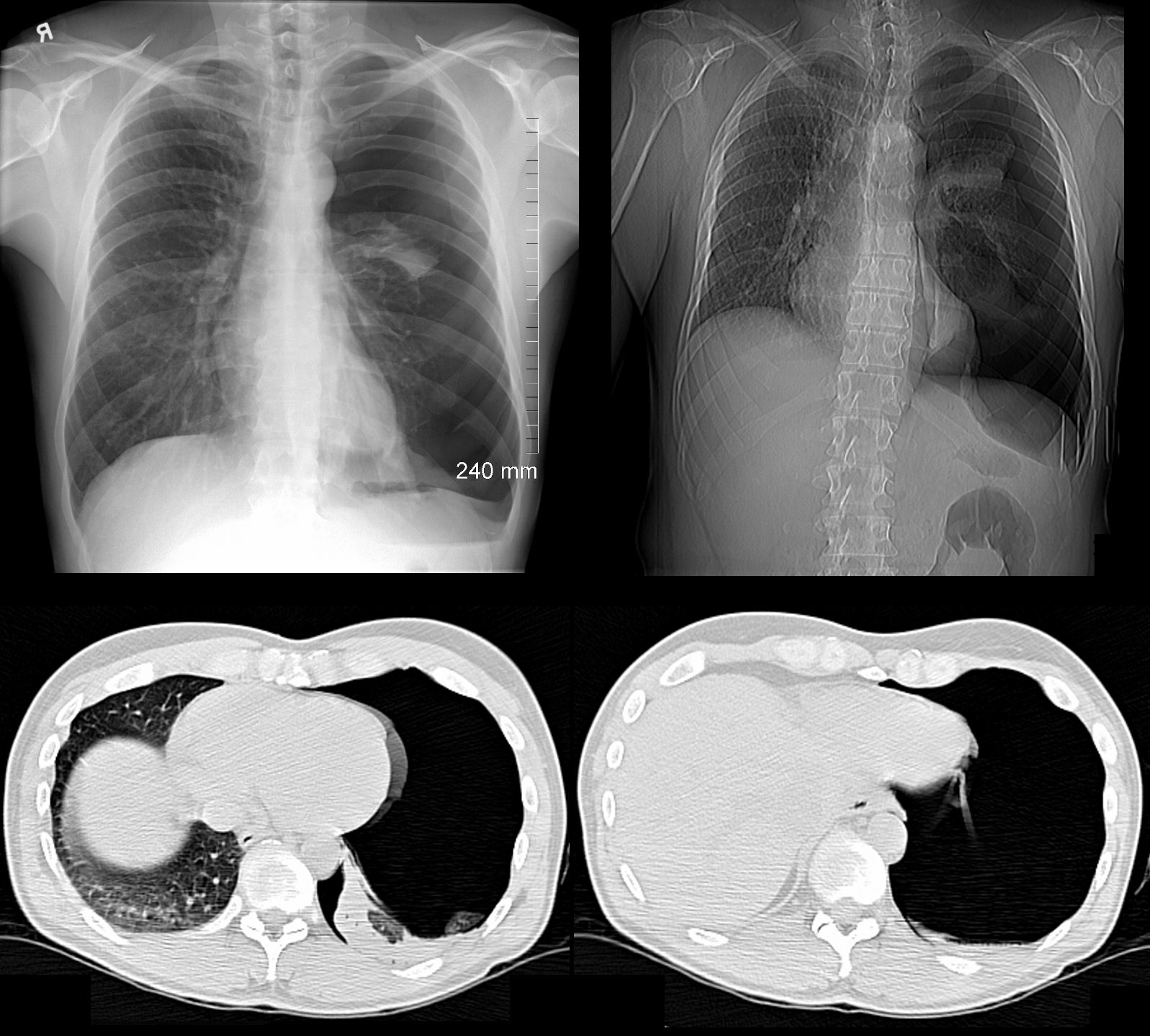
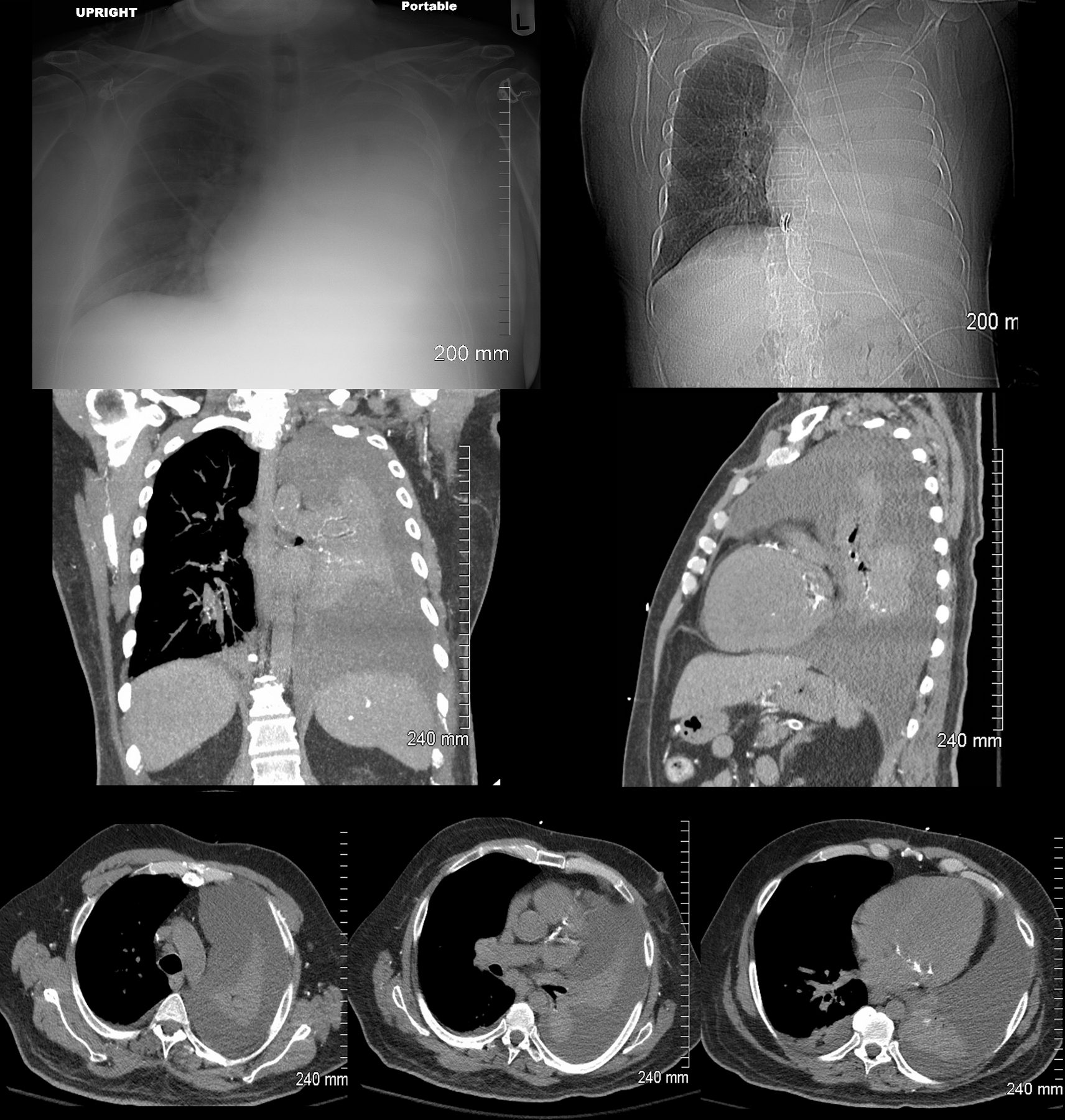
Tension Hydrothorax
from large pleural effusion and probable hemothorax under tension with atelectasis of the right lung
85-year-old female with a history of lung cancer, presents with dyspnea and hypotension. CXR shows white out of the right hemithorax with pressure effect characterised by narrowing of the distal trachea cardio-mediastinal shift to the left and atelectasis in the left lower lobe.
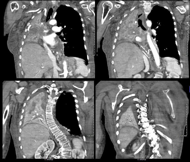
Ashley Davidoff MD TheCommonVein.netSee 106Lu 118468
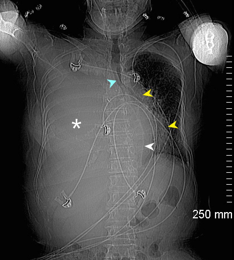
85-year-old female with a history of lung cancer, presents with a dyspnea and hypotension. Scout film prior to the CT scan shows “white out” of the right hemithorax (white asterisk) with pressure effect characterised by narrowing of the trachea (blue arrow) mediastinal shift yellow arrows) and herniation into the left chest characterised by a leftward shift of the azygo-esophageal junction line (white arrow).
Ashley Davidoff MD TheCommonVein.ne 106Lu 118463L
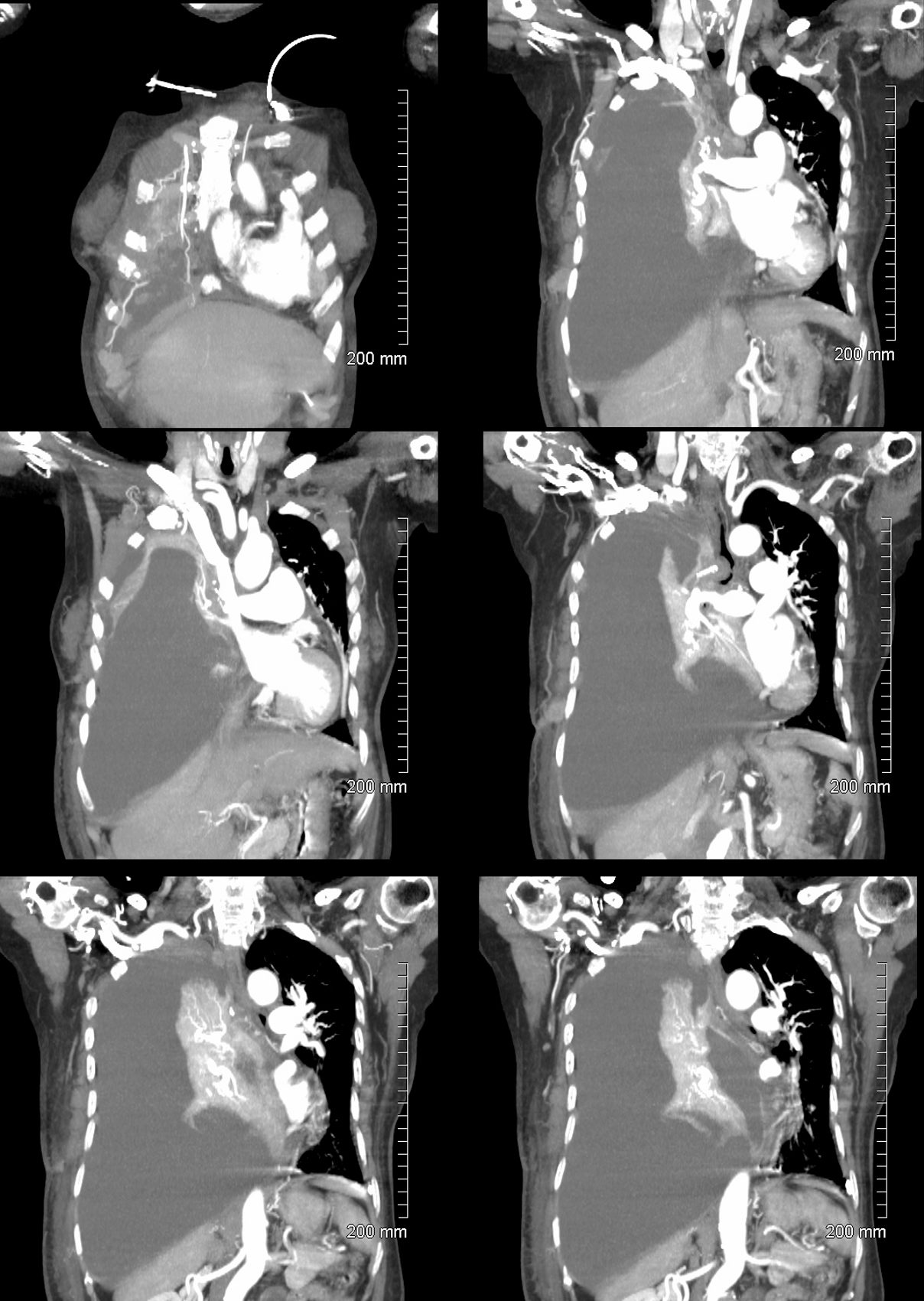
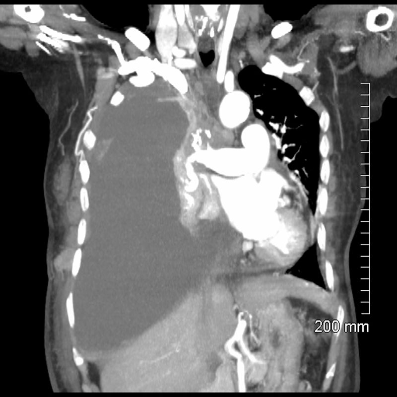
85-year-old female with a history of lung cancer, presents with a dyspnea and hypotension. CT scan shows a large right pleural effusion under pressure, with mediastinal shift to the right. In addition, there is compression of the heart with back up of venous return due the pressure effect on the heart and vascular structures. Among the structures showing venous distension are the SVC (blue arrowhead,a) right sided upper limb veins (blue arrowhead b) and the left upper pulmonary veins (red arrowhead, b. The effusion in the right pleural cavity with atelectatic lung herniates into the left hemithorax, (white arrowhead, c). There is a dense sediment in the pleural fluid (red arrowhead, d) suggesting blood in the pleural cavity. The left atrium is compressed (maroon arrowhead, d)
Ashley Davidoff MD TheCommonVein.net106Lu 118467c

85-year-old female with a history of lung cancer, presents with a dyspnea and hypotension. Reconstruction of the CT scan in the coronal plane, shows a large right pleural effusion under pressure with herniation into the left chest (white asterisk e,and f) , with mediastinal shift to the left (yellow arrowhead b, c, d). In addition, there is compression of the heart with back up of venous return due the pressure effect on the heart and vascular structures. Among the structures showing venous distension are the SVC (blue arrowhead, c) right sided upper limb veins (blue arrowhead d) and the left upper pulmonary veins (red arrowhead, d and f). The density of the systemic venous abd arterial systems is similar, but vascular structures as noted by the green arrowhead in a could represent venous collaterals.
Ashley Davidoff MD TheCommonVein.ne 106Lu 118467cL
85-year-old female with a history of lung cancer, presents with a dyspnea and hypotension. CT scan shows a large right pleural effusion under pressure, with mediastinal shift to the left. In addition, there is compression of the heart with back up of venous return due the pressure effect on the heart and vascular structures. The effusion in the right pleural cavity with atelectatic lung herniates into the left hemithorax.
Ashley Davidoff MD TheCommonVein.net 106Lu 118467
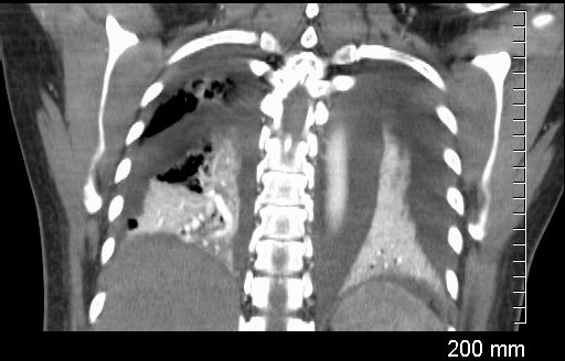
Coronal CT through the lungs show bilateral pleural effusions with compressive atelectasis
Ashley Davidoff MD TheCommonvein.net
238Lu
Compressive Atelectasis and Complex Pleural Effusions
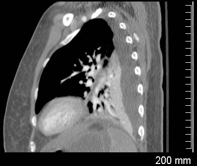
Sagittal CT through the left lung shows undulations of the posterior surface of the left lung, and the suggesting differing pressures on the lung parenchyma by the effusions and indicating complexity and loculation.
Ashley Davidoff MD TheCommonVein.net
238Lu
Whole Lung Atelectasis Due to Obstruction
59F shows total white out caused by collapse of right lung with an
occluded right main step bronchus associated with a
large right sided effusion. The occlusion is likely due to proximal cancer. A pigtail drain has been placed to drain the effusion
Ashley Davidoff MD TheCommonVein.net 104 Lu
59F shows total collapse of left lung with an
occluded right main step bronchus(top right image)associated with a
right sided effusion. The occlusion is likely due to proximal cancer
Ashley Davidoff MD TheCommonVein.net 104 Lu
Total Lung Compressive Atelectasis
Effusion
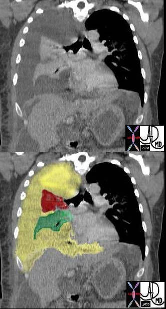
In this case there a large right sided pleural effusion (yellow) with secondary atelectasis of the right lung. (red and green) This coronal CT of the chest at the level of the left ventricle shows a large right pleural effusion which lies between the visceral and parietal pleura. Once the effusion is large enough to weaken the capillary forces that hold the parietal and visceral pleura together, it fail, and the lung collapses which is what is noted on this image – ie total lung collapse because of loss of cohesive adhesive forces.
Courtesy of: Ashley Davidoff, M.D. TheCommonvein.net 42558c
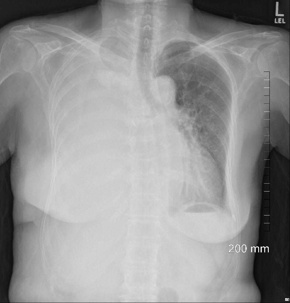
White Out of the CXR with Passive Compressive Atelectasis of the Left Lung
48 year-old male presents with a dyspnea. CXR shows a total white out of the left chest with pulmonary congestion. CT scan shows a large left pleural effusion with total atelectasis of the left lung. Incidental note is made of premature calcific coronary artery disease.
Ashley Davidoff MD TheCommonVein.net