
A 36 year-old female with known history significant for HFrEF from postpartum cardiomyopathy (8 years prior to presentation) with ICD complicated by recurrent PE, renal infarcts, & congestive hepatopathy presents to the ED with abdominal pain.
35-year-old female with a 8 year history of post- partum cardiomyopathy presents with of chest pain.
CXR – Post Partum Cardiomyopathy
External Defibrillator
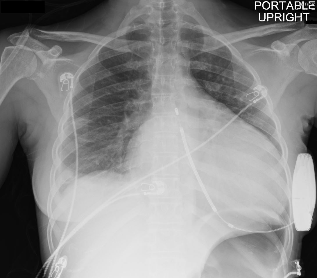
35-year-old female with a 8 year history of post- partum cardiomyopathy presents with of chest pain. Frontal CXR shows global cardiomegaly, blunting of the right costophrenic angle with a suggestion of a subsegmental infiltrate in the right costophrenic angle, and a region of linear atelectasis in the right mid lung field. A small loculated right effusion is present. An external defibrillator is noted. No definite CHF
Ashley Davidoff MD TheCommonVein.net 258Lu 136164
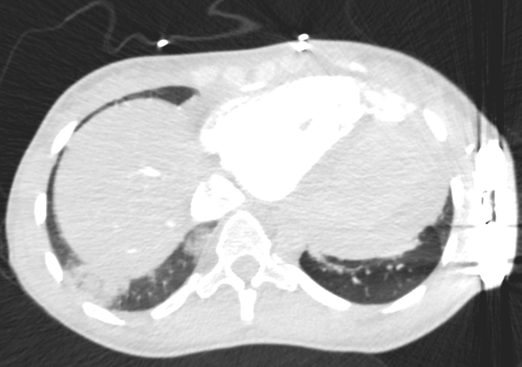
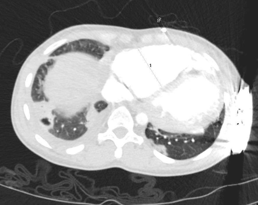
CT – Post Partum Cardiomyopathy with Pulmonary Emboli to Right Lower Lobe
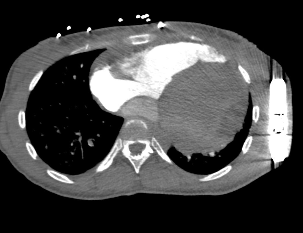
35-year-old female with an 8-year history of post- partum cardiomyopathy presents with a history of chest pain. CT of chest with contrast in an axial projection, at the level of the heart, shows an enlarged left ventricle. The right lower lobe segmental arteries show filling defects and absence of contrast compared to the left lower lobe arteries. An external defibrillator is present.
Ashley Davidoff MD TheCommonVein.net 258Lu 136165
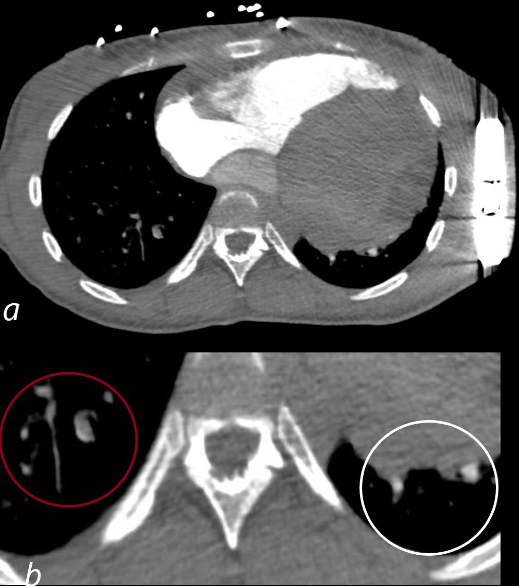
35-year-old female with an 8-year history of post- partum cardiomyopathy presents with a history of chest pain. CT of the chest with contrast in an axial projection, at the level of the heart, shows an enlarged left ventricle. The right lower lobe segmental arteries show filling defects and absence of contrast (maroon circle in b), compared to the left lower lobe arteries (white circle b). An external defibrillator is present.
Ashley Davidoff MD TheCommonVein.net 258Lu 136165cL
Right Lower Lobe Wedge Shaped
Ground Glass Hemorrhagic Infarct
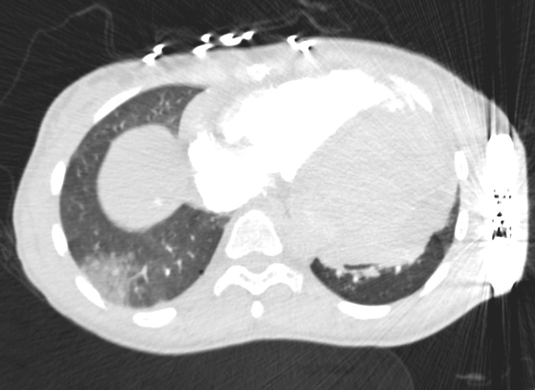
35-year-old female with an 8-year history of post- partum cardiomyopathy presents with a history of chest pain. CT of the chest with contrast in an axial projection, at the level of the heart, shows a subsegmental wedge shaped ground glass infarct in the posterior or lateral region of the right lower lobe likely reflecting a hemorrhagic infarct. An external defibrillator is present.
Ashley Davidoff MD TheCommonVein.net 258Lu 136169a
Posterior Recess
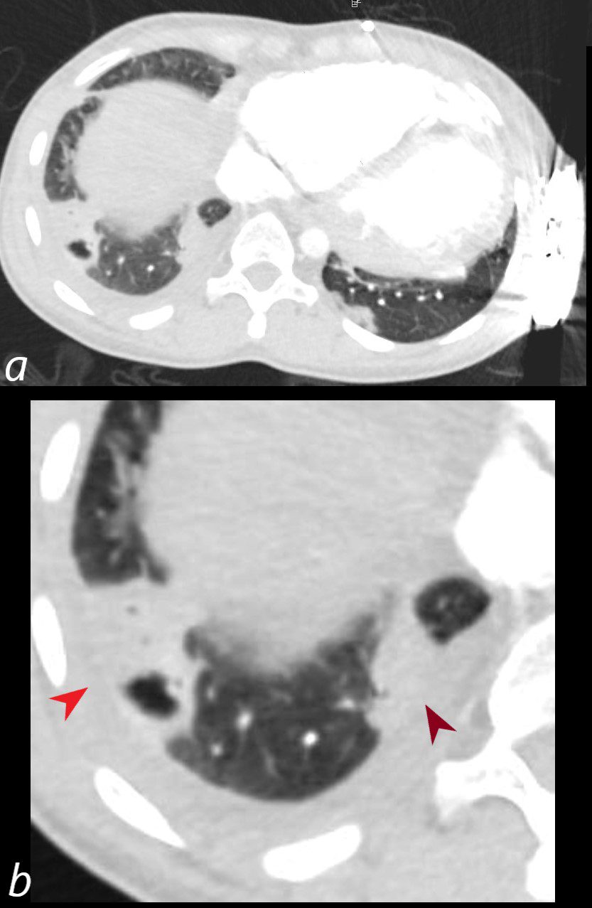
Ground Glass Hemorrhagic Infarcts with Necrosis
35-year-old female with an 8-year history of post- partum cardiomyopathy presents with a history of chest pain. CT of the chest with contrast in an axial projection, at the level of the heart, shows a subsegmental wedge shaped ground glass infarct with suggestion of necrosis in the posterior or lateral region of the right lower lobe. A second similar but less complex subsegmental region of ground glass opacity, also suspicious for infarction is noted medially. The right ventricle is enlarged. An external defibrillator is present.
Ashley Davidoff MD TheCommonVein.net 258Lu 136169b
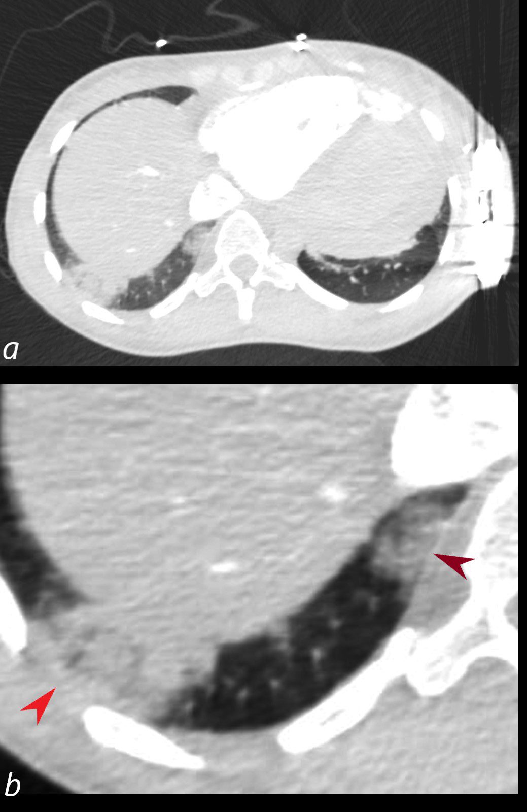
35-year-old female with an 8-year history of post- partum cardiomyopathy presents with a history of chest pain. CT of the chest with contrast in an axial projection, at the level of the heart, shows a subsegmental wedge shaped ground glass infarct with suggestion of necrosis in the posterior or lateral region of the right lower lobe (b red arrow). A second similar but less complex subsegmental region of ground glass opacity, also suspicious for infarction is noted medially (b, maroon arrowhead). The right ventricle is enlarged. An external defibrillator is present.
Ashley Davidoff MD TheCommonVein.net 258Lu 136169cL
Tricuspid Regurgitation
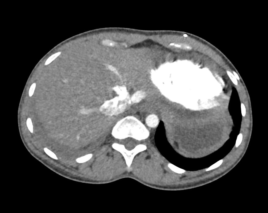
35-year-old female with an 8-year history of post- partum cardiomyopathy presents with a history of chest pain. CT of the chest with contrast in an axial projection, at the level of the liver in the arterial phase, shows reflux of contrast into the hepatic veins indicating tricuspid regurgitation.
Ashley Davidoff MD TheCommonVein.net 258Lu 136170
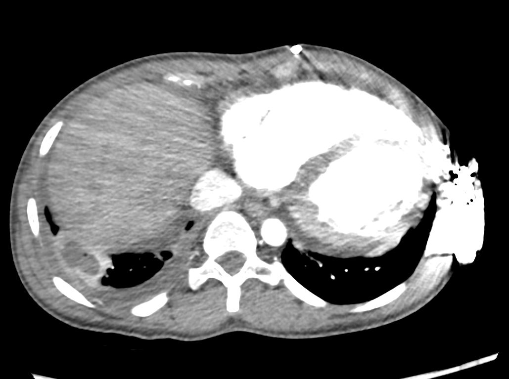
Nutmeg Liver
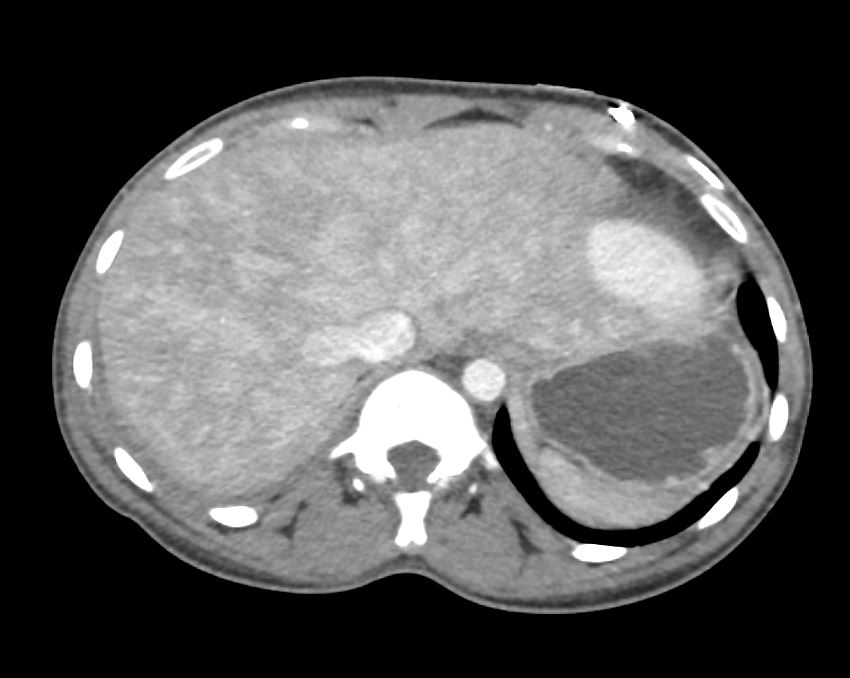
35-year-old female with an 8-year history of post- partum cardiomyopathy presents with a history of chest pain. CT of the chest with contrast in an axial projection, at the level of the liver in the portal venous phase, shows heterogeneous nodular enhancement of the liver consistent with nutmeg liver due to chronic hepatic congestion
Ashley Davidoff MD TheCommonVein.net 258Lu 136171
10 days Later
Necrotizing Pulmonary Infarction with Cavitation s/p Pulmonary Embolus
35-year-old female with an 8-year history of post- partum cardiomyopathy presents with a history of ongoing chest pain 10 days following acute pulmonary infarction. CT of the chest with contrast in an axial projection, at the level of the heart shows an enlarged right ventricle, cavitation of the previously identified posterolateral infarction, a persistent wedge shaped subsegmental infarct medially, a small right pleural effusion, and subsegmental atelectasis in the left lower lobe.
Ashley Davidoff MD TheCommonVein.net 258Lu 136173
35-year-old female with an 8-year history of post- partum cardiomyopathy presents with a history of ongoing chest pain 10 days following acute pulmonary infarction. CT of the chest with contrast in an axial projection, at the level of the heart shows an enlarged right ventricle, cavitation of the previously identified posterolateral infarction (b, red arrowhead), a persistent wedge shaped subsegmental infarct medially (b maroon arrowhead), a small right pleural effusion, and subsegmental atelectasis in the left lower lobe.
Ashley Davidoff MD TheCommonVein.net 258Lu 136173cL
1 Week Later Presents with a Fever
New Abscess in Region of Prior Infarction
35-year-old female with an 8-year history of post- partum cardiomyopathy presents with a history of ongoing chest pain and fever 1 week following new cavitation of a pulmonary infarction. CT of the chest with contrast in an axial projection, at the level of the heart shows an enlarged right ventricle, evolution of the posterolateral infarction into an abscess with a bubble of air, enhancing wall, a small right pleural effusion with suggestion of pleural enhancement in the left lower lobe.
Ashley Davidoff MD TheCommonVein.net 258Lu 136174