
Stage IV Small Cell Lung Carcinoma – Hilum Overlay Sign
65 year old male presents with a history of chest pain, back pain, myalgias, and night sweats
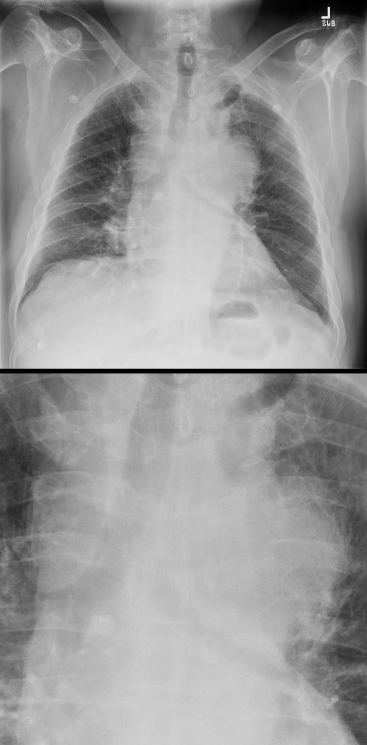
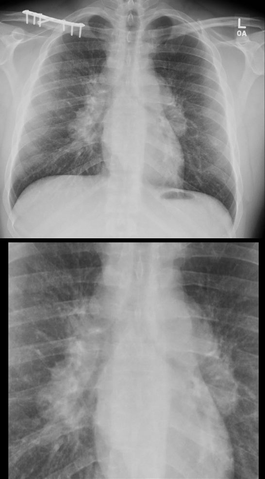
Frontal CXR shows a left sided mass that silhouettes the aortic knob and the main pulmonary artery indicating that the mass arises from middle mediastinal tumors, hilar adenopathy, or a large pericardial effusion – hilum overlay sign.
A diagnosis of a large mediastinal small cell carcinoma of the lung was diagnosed
Ashley Davidoff MD TheCommonVein.net 297Lu 136690c
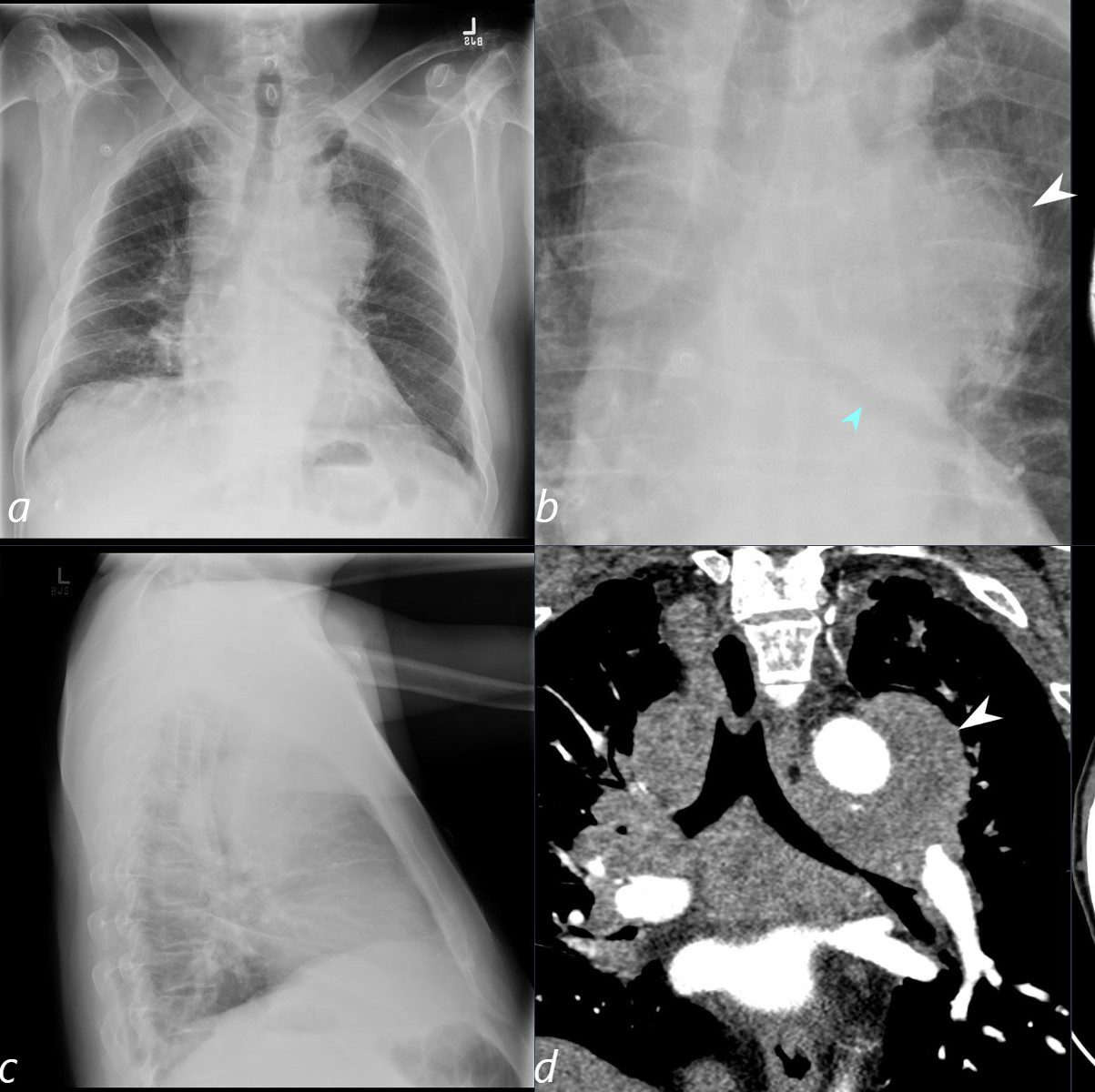
65 year old male presents with a history of chest pain, back pain, myalgias, and night sweats
Frontal CXR shows a left sided mass that silhouettes the aortic knob and the main pulmonary artery (white arrowhead, b) indicating that the mass arises from middle mediastinal tumors, hilar adenopathy, or a large pericardial effusion. The mass also compresses and displaces the left mainstem (teal arrowhead b) and confirmed in d.
The coronal CT scan (d) confirms the presence of a large mediastinal mass that extends beyond the aortic knob (white arrowhead)
A diagnosis of a large mediastinal small cell carcinoma of the lung was diagnosed
Ashley Davidoff MD TheCommonVein.net 297Lu 136690cL
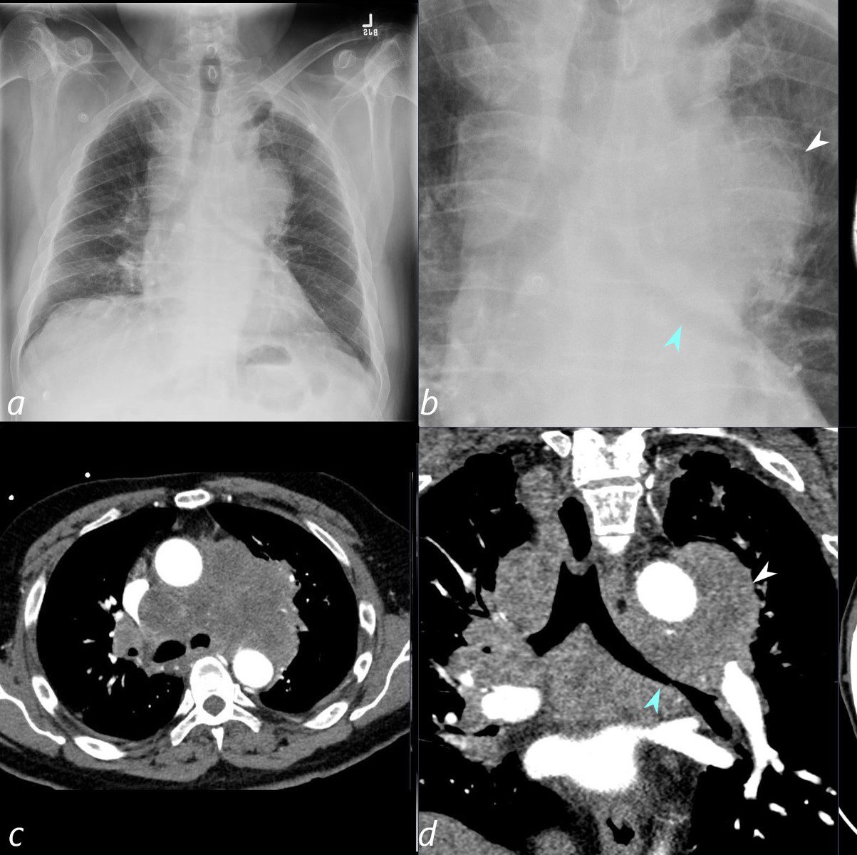
65 year old male presents with a history of chest pain, back pain, myalgias, and night sweats
Frontal CXR shows a left sided mass that silhouettes the aortic knob and the main pulmonary artery (white arrowhead, b) indicating that the mass arises from middle mediastinal tumors, or hilar adenopathy. The mass also compresses and displaces the left mainstem bronchus (teal arrowhead b).
The axial CT (c) shows th extent of the mass and the coronal CT scan (d) confirms the presence of a large mediastinal mass that extends beyond the aortic knob (white arrowhead)
A diagnosis of a large mediastinal small cell carcinoma of the lung was diagnosed
Ashley Davidoff MD TheCommonVein.net 297Lu 136691cL
Source
Signs in Thoracic Imaging
Journal of Thoracic Imaging 21(1):76-90, March 2006.
In evaluating densities projecting over the hila on PA chest radiographs, it is important to evaluate whether the opacity blends with the normal hilar shadow of the proximal pulmonary arteries and obscures clear visualization or if the mass and hilar structures are overlapping but distinct from one another. If the hilar vessels are sharply delineated, then it can be assumed that the overlying mass is anterior (Fig. 15) or posterior to the centrally located vascular structures and the intervening aerated lung enables crisp visualization of the pulmonary vessels.12,13 However, when the mass is inseparable from the proximal pulmonary arteries, it is assumed that the structures are immediately adjacent to one another, with the lack of interposing air obscuring the margins of the normal hilar structures. This sign was originally described by Benjamin Felson.12,13
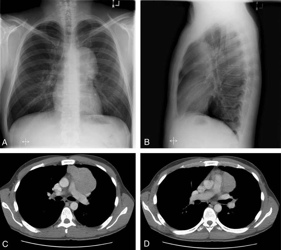
29-year-old male presents with night sweats
CXR in the PA projection shows bilateral hilar enlargement consistent with hilar adenopathy. Wedge biopsy and culture revealed a diagnosis of MAC (mycobacterium avium complex) The silhouetting of the pulmonary vessels indicates that the soft tissue density arises from the hilum and this case from mediastinal lymph nodes (hilum overlay sign)
Ashley Davidoff MD TheCommonvein.net 135804c 247Lu