
Vessels
Arteriole, Bronchiole Venule Lymphatics
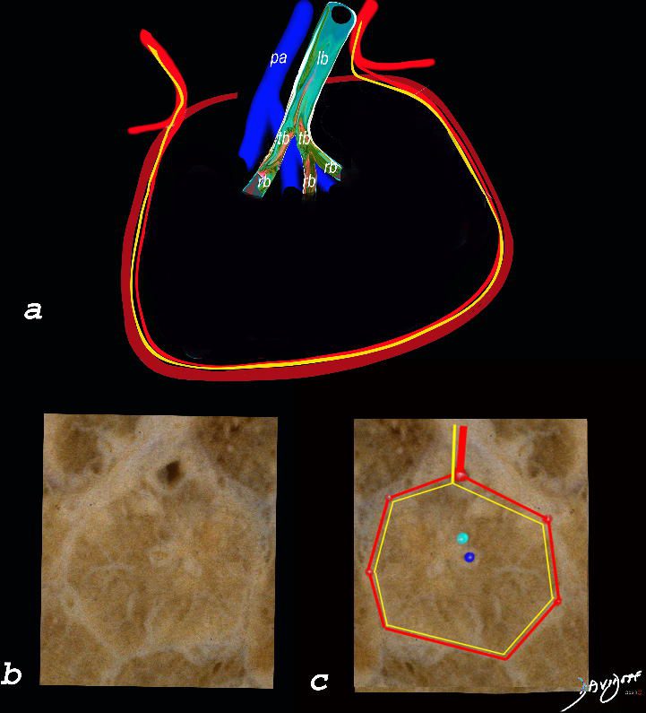
The top image (a) shows an anatomic drawing of a secondary lobule of the lung subtended by a lobular bronchiole (lb) and arteriole (pa). The interlobular septum contains the venule (red) lymphatic (yellow) and septum (maroon)
The anatomical specimen of the lung (b) shows normal intralobular parenchyma while image c shows the centrilobular arteriole (navy blue) and centrilobular bronchiole (teal) and interlobular venule (red) and lymphatics (yellow) The interlobular septum is slightly thickened
Ashley Davidoff TheCommonVein.net
Moderate CHF with Interstitial Edema
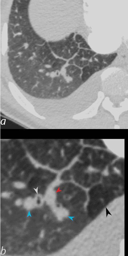
50-year-old female with diabetes, chronic renal failure and congestive heart failure. CT in the axial plane through the right posterior recess, shows thickened interlobular septa at the right base, congested arterioles (light blue arrowheads, b), alongside the bronchioles, peribronchial cuffing (white arrowheads, b), a congested pulmonary venule in the interlobular septum (red arrowhead arrowheads, b), ground glass changes and a secondary lobule demonstrating mosaic attenuation (black arrowhead arrowheads, b). The IVC is dilated and a small complex effusion is present.
Ashley Davidoff MD TheCommonvein.net 135783cL 193Lu
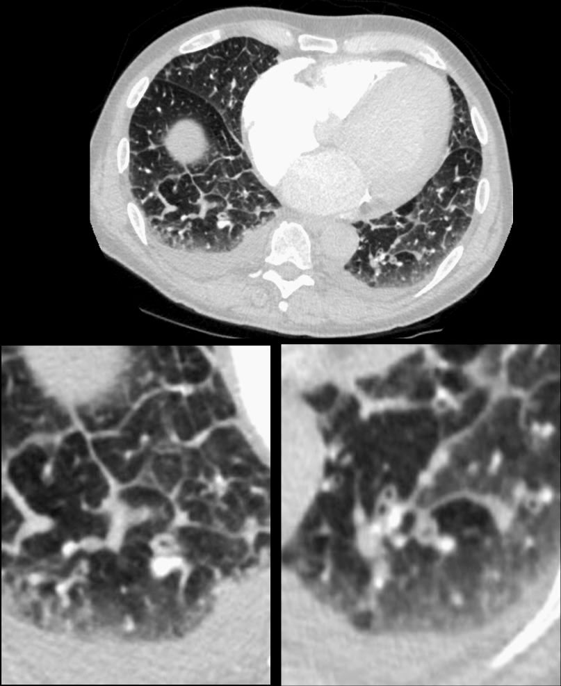
74-year-old man presents with dyspnea and orthopnea. CT shows thickening of the interlobular septa (Kerley B lines), peribronchial cuffing, and enlargement of the lobular arteriole in the right lower lobe. There is a suggestion of vasoconstriction of the arteriole as it enters the secondary lobule. ground glass changes in the some of the secondary lobules on the left and perhaps mosaic attenuation vs normal secondary lobule at the right base are noted. Additionally, there are small bilateral effusions right greater than left. The mild irregular shape of the effusions suggests that they are partially loculated. These findings indicate moderate congestive heart failure with interstitial edema.
Ashley Davidoff MD TheCommonVein.net 135775c01
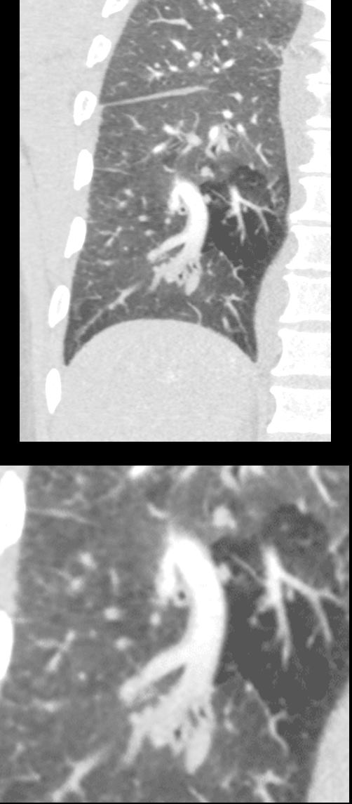
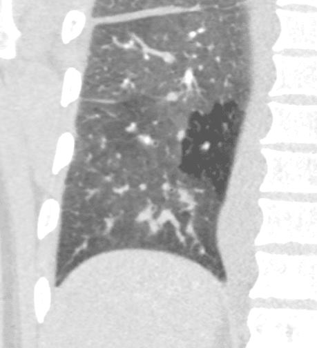
50-year-old female with diabetes, chronic renal failure with congestive heart failure. CT in the coronal plane shows diffuse ground glass changes, Kerley B lines, edema in the fissure, peribronchial cuffing, enlargement of the pulmonary artery, and mosaic attenuation
Ashley Davidoff MD TheCommonvein.net 135778c 193Lu
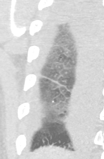
50 year old female with diabetes, chronic renal failure with congestive heart failure. CT in the coronal plane shows diffuse ground glass changes, Kerley B lines, edema in the fissure, and mosaic attenuation
Ashley Davidoff MD TheCommonvein.net 135779c 193Lu
50-year-old female with diabetes, chronic renal failure and congestive heart failure. CT in the coronal plane through the posterior aspect of the chest, shows diffuse ground glass changes, thickening of the interlobular septa, thickening of the fissure and centrilobular nodules reflecting arteriolar congestion.
Ashley Davidoff MD TheCommonvein.net 135780 193Lu
Angiocentric Disease
-
-
- CHF
- Pulmonary Hypertension
- Vasculitis
- Talicosis
- Hemorrhage
- Hemosiderosis
- Metastatic Calcification
-
Links and References
- TCV