
Infection
ABPA
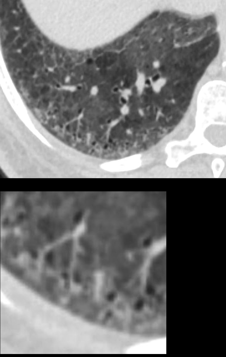
CT Allergic Bronchopulmonary Aspergillosis (ABPA)
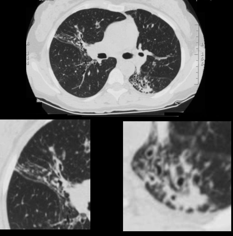
48 year old female with a history of asthma presents with productive cough. CT scan 18 months prior shows multicentric foci of bronchial wall thickening , in the segmental and subsegmental airways, in the middle lobe with crowding of the airways in the RML indicating atelectasis. (upper panel magnified lower left). In the LLL there is bronchiolectasis with thickened airways and a focal subsegmental consolidation (lower panel right) . Flexible bronchoscopy revealed mucus plugs and aspergillus was isolated.
Ashley Davidoff MD TheCommonVein.net
Known Upper Lobe Latent TB
Basilar Bronchiolectasis Probable Childhood Infection
COPD Latent TB Basilar Bronchiolectasis
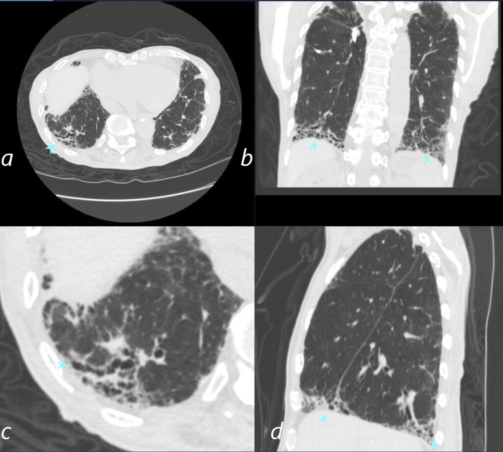
80- year-old non-smoker with childhood history of treated TB, presents with a chronic cough
CT scan in the axial plane (a, magnified in b) shows a wedge shaped conglomerate of dillated bronchioles (blue arrowheads. The coronal image (b) and sagittal image b and c) again show the wedge shaped regions of bibasilar bronchiolectasis
Ashley Davidoff TheCommonVein.net292Lu 136631cL
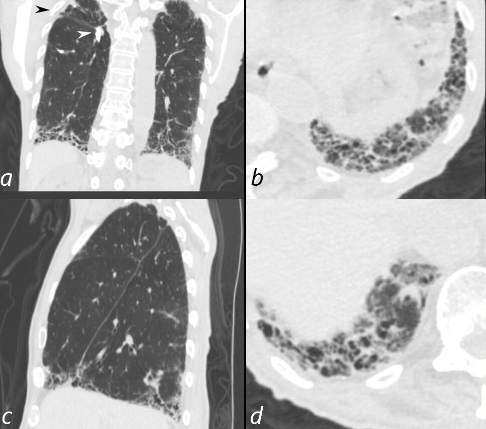
80- year-old non-smoker with childhood history of treated TB, presents with a chronic cough
CT scan in the coronal plane (a) shows volume loss in the right upper lobe (black arrowhead) and a subtending calcified broncholith (white arrowhead, a).
Basilar peripheral bronchiolectasis is demonstrated in the coronal (a) and sagittal planes (b) , and magnified in the axial planes at the left posterior recess (c) and right posterior recess (d)
Ashley Davidoff MD TheCommonVein.net 292Lu 136632cL
Inflammation
ABPA
CT Allergic Bronchopulmonary Aspergillosis (ABPA)
48 year old female with a history of asthma presents with productive cough. CT scan 18 months prior shows multicentric foci of bronchial wall thickening , in the segmental and subsegmental airways, in the middle lobe with crowding of the airways in the RML indicating atelectasis. (upper panel magnified lower left). In the LLL there is bronchiolectasis with thickened airways and a focal subsegmental consolidation (lower panel right) . Flexible bronchoscopy revealed mucus plugs and aspergillus was isolated.
Ashley Davidoff MD TheCommonVein.net
DIP
51-year-old female smoker with a history of COPD asthma and pulmonary hypertension presents with progressive dyspnea. Axial CT through the lower lung fields shows patchy ground glass changes in the middle lobe inferior ligula and lower lobes and some regions of mosaicism. Focal regions of interlobular septal thickening are noted left lower lobe and evidence of thick walled bronchiolectasis in the right lower lobe (lower panel). Pathology confirmed a diagnosis of DIP
Ashley Davidoff MD TheCommonVein.net 252Lu 135972c
Bronchiolectasis Mosaicism and the Secondary Lobule
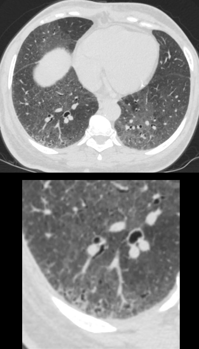
51-year-old female smoker with a history of COPD asthma and pulmonary hypertension presents with progressive dyspnea. Axial CT through the right posterior recess shows patchy ground glass changes with some regions of mosaicism. The bronchovascular bundle subtending 2 secondary lobules is highlighted in the lower panel. The centrilobular arteriole and ectatic bronchiole are magnified
Pathology confirmed a diagnosis of DIP
Ashley Davidoff MD TheCommonVein.net 252Lu 135981c
Malignancy Mechanical/Atelectasis Trauma Metabolic Circulatory- Hemorrhage Immune Infiltrative Idiopathic Iatrogenic Idiopathic