
65 yo F with PMHx of HTN, HLD, T2DM, OSA, hypothyroidism, cataracts/glaucoma, and depression who presents to ILD clinic for evaluation of pulmonary nodules.
Recent history to note:
5 years ago
New cough in for which she had a CXR showing multiple nodular opacities in the R hemithorax. Subsequent CT chest showed innumerable bilateral pulmonary nodules ranging between 4-8 mm many of which were bronchocentric Noted mediastinal LAD.
PET/CT showed that most of the nodules demonstrated low level FDG avidity though there was an intense FDG uptake of the pleural-based opacity of the medial segment of the RLL. Subsequent surgical lung biopsy showed focal fibrosis and foreign body giant cell reaction, no evidence of malignancy.
Subsequent biopsy showed necrotizing-granuloma
Ashley Davidoff MD TheCommonVein.net
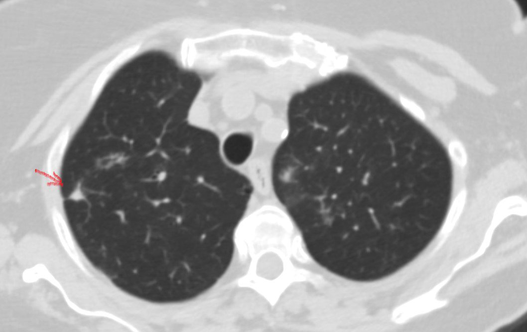
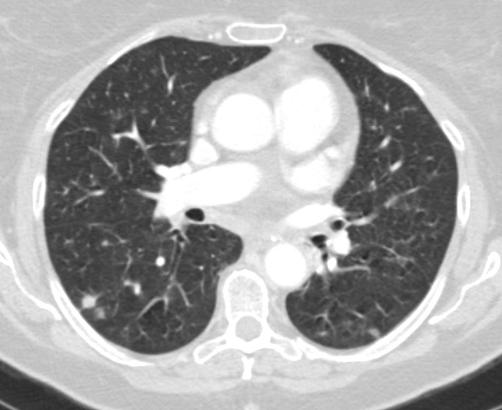
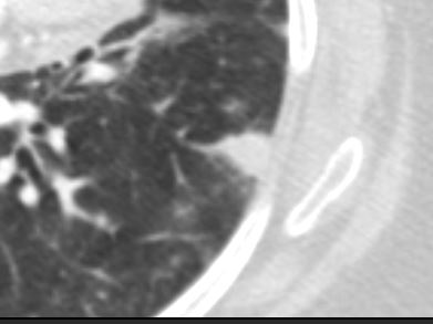
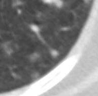
Bronchocentric Nodule Left Upper Lobe
65 F presents 6 years ago with both solid nodules and bronchocentric nodules
Ashley Davidoff MD TheCommonVein.net
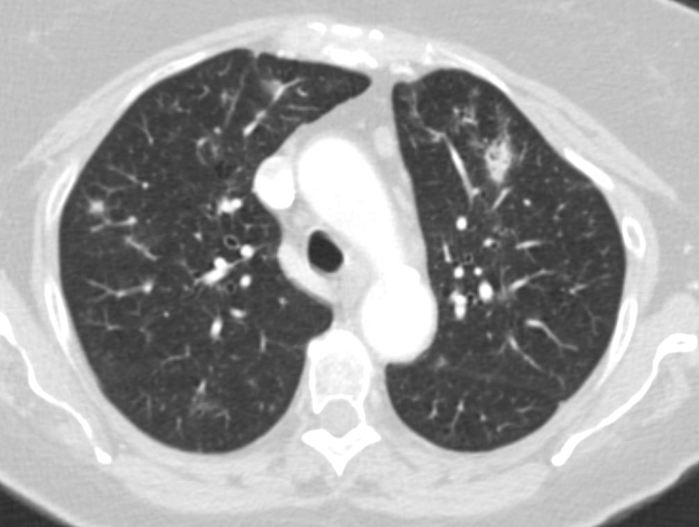
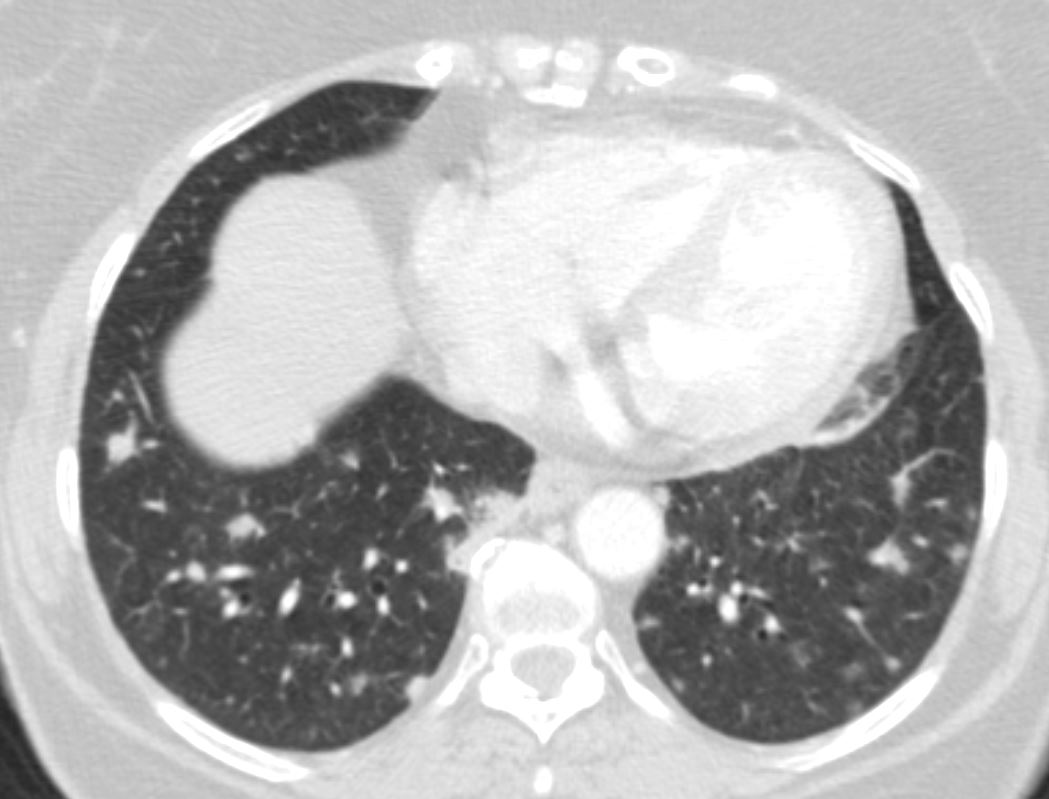
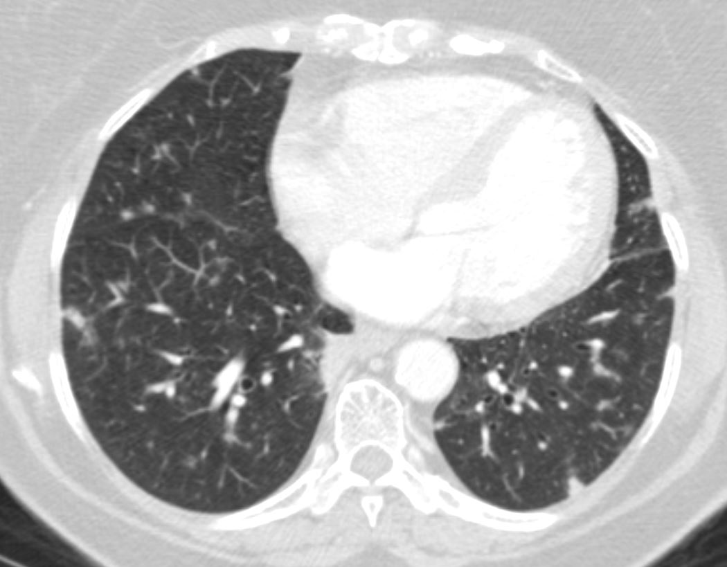
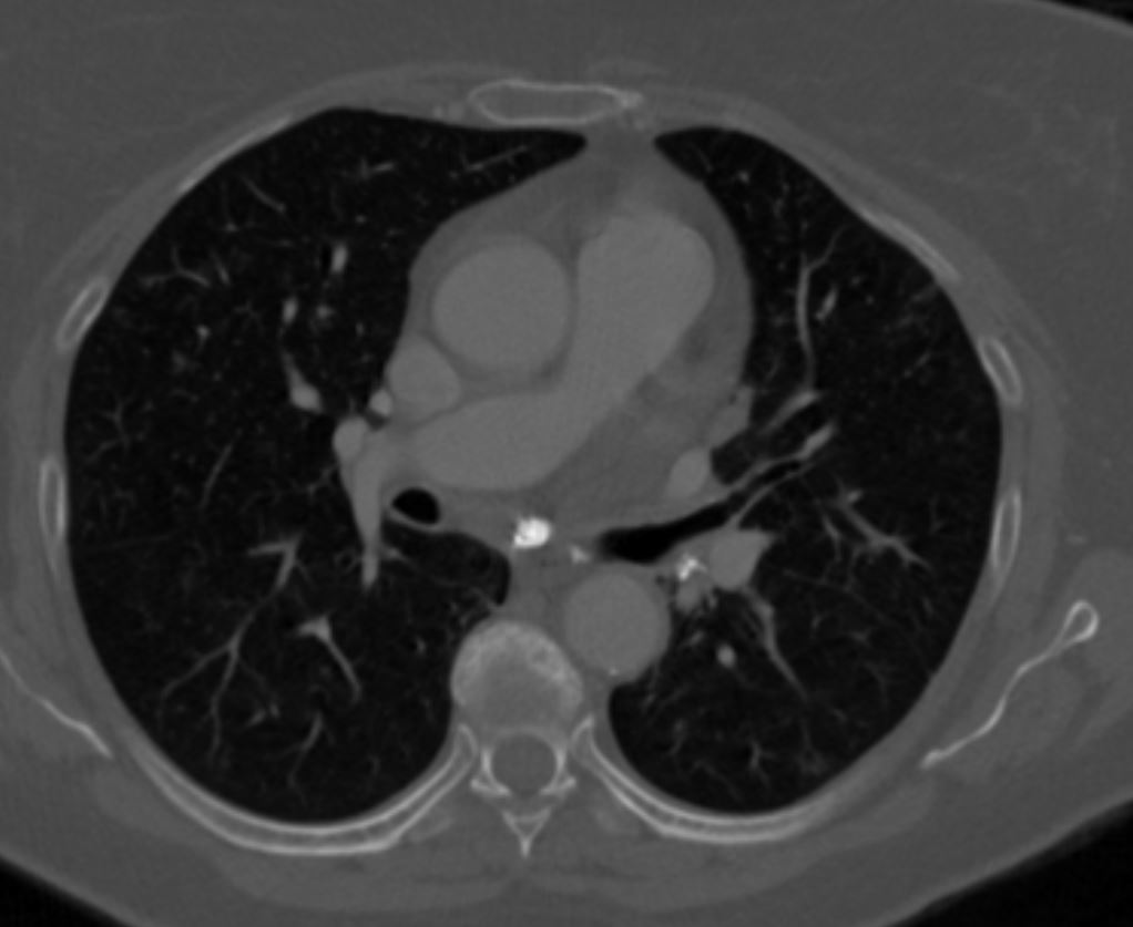
Bronchocentric Nodules and Suggestion of Micronodules
65 F presents 6 years ago with both solid nodules and bronchocentric nodules
Ashley Davidoff MD TheCommonVein.net
65 F presents 6 years ago with both solid nodules and bronchocentric nodules
Ashley Davidoff MD TheCommonVein.net
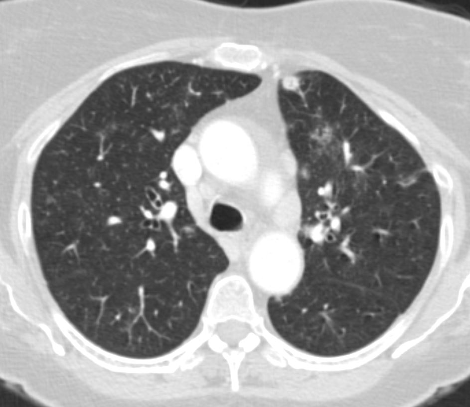
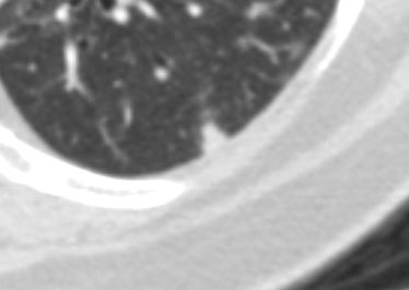
Solid Nodules
Nodules showed waxing and waning and subsequent biopsy showed necrotizing-granuloma
Ashley Davidoff MD TheCommonVein.net
Nodules showed waxing and waning and subsequent biopsy showed necrotizing-granuloma
Ashley Davidoff MD TheCommonVein.net
Nodules showed waxing and waning and subsequent biopsy showed necrotizing-granuloma
Ashley Davidoff MD TheCommonVein.net
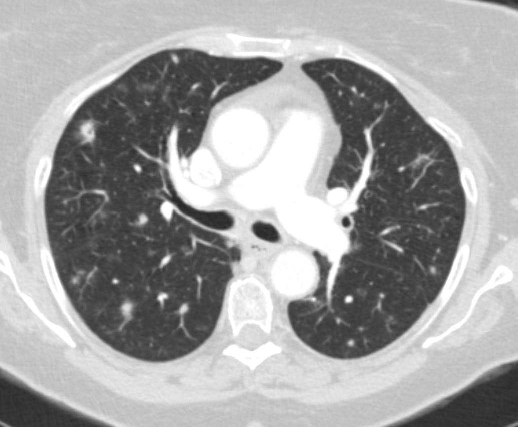
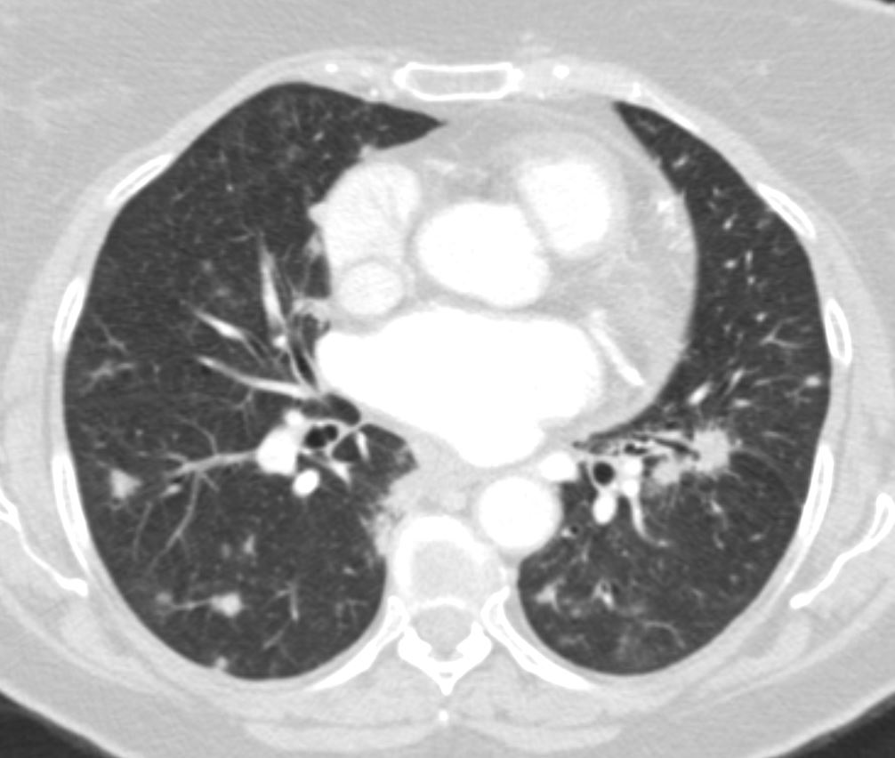
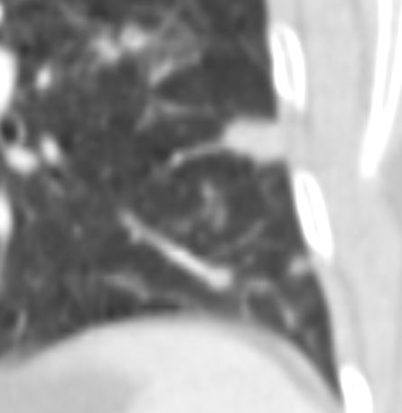
Wedge Shaped Defects Along the Broncho-vascular Bundle
65 F presents 6 years ago with both solid nodules and bronchocentric nodules There is a larger nodule sis conglomerate nodule around the left lower lobe segmental airways
Nodules showed waxing and waning and subsequent biopsy showed necrotizing-granuloma
Ashley Davidoff MD TheCommonVein.net
65 F presents 6 years ago with both solid nodules and bronchocentric nodules There is a larger nodule sis conglomerate nodule around the left lower lobe segmental airways
Nodules showed waxing and waning and subsequent biopsy showed necrotizing-granuloma
Ashley Davidoff MD TheCommonVein.net
65 F presents 6 years ago with both solid nodules and bronchocentric nodules There is a larger nodule sis conglomerate nodule around the left lower lobe segmental airways
Nodules showed waxing and waning and subsequent biopsy showed necrotizing-granuloma
Ashley Davidoff MD TheCommonVein.net
65 F presents 6 years ago with both solid nodules and bronchocentric nodules There is a larger nodule sis conglomerate nodule around the left lower lobe segmental airways
Nodules showed waxing and waning and subsequent biopsy showed necrotizing-granuloma
Ashley Davidoff MD TheCommonVein.net
65 F presents 6 years ago with both solid nodules and bronchocentric nodules There is a larger nodule sis conglomerate nodule around the left lower lobe segmental airways
Nodules showed waxing and waning and subsequent biopsy showed necrotizing-granuloma
Ashley Davidoff MD TheCommonVein.net
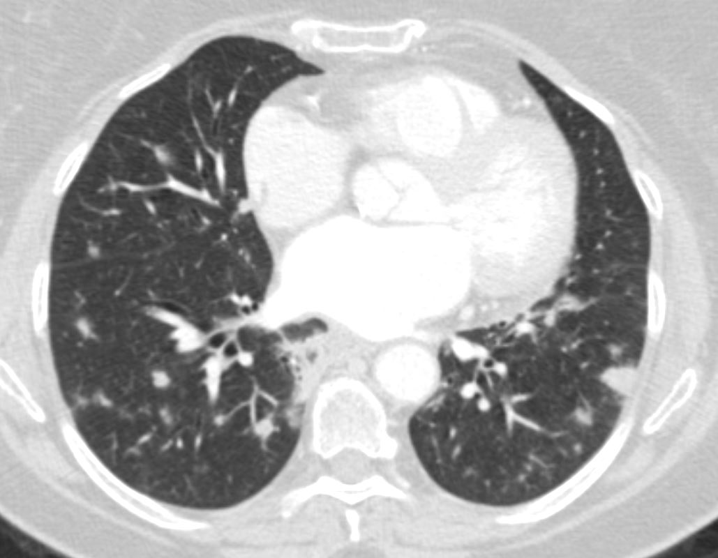
Solid Nodules Along the Small Vessels of the Bronchovascular Bundle in the Left Lower Lobe
65 F presents 6 years ago with both solid nodules and bronchocentric nodules This nodule is solid
Nodules showed waxing and waning and subsequent biopsy showed necrotizing-granuloma
Ashley Davidoff MD TheCommonVein.net
65 F presents 6 years ago with both solid nodules and bronchocentric nodules This nodule is solid
Nodules showed waxing and waning and subsequent biopsy showed necrotizing-granuloma
Ashley Davidoff MD TheCommonVein.net
65 F presents 6 years ago with both solid nodules and bronchocentric nodules There is a larger nodule sis conglomerate nodule around the left lower lobe segmental airways
Nodules showed waxing and waning and subsequent biopsy showed necrotizing-granuloma
Ashley Davidoff MD TheCommonVein.net
65 F presents 6 years ago with both solid nodules and bronchocentric nodules There is a larger nodule sis conglomerate nodule around the left lower lobe segmental airways
Nodules showed waxing and waning and subsequent biopsy showed necrotizing-granuloma
Ashley Davidoff MD TheCommonVein.net
4 years ago
Surveillance imaging from showed stable scattered solid spiculated and GG nodules measuring up to 6 mm decreased from 6 years ago
2 years ago she reported having weight loss to her PCP prompting her to check CT chest. P
CT 1 yearago
showed interval growth of a 1.5 cm spiculated RUL nodule (previously measured 6 x 5 mm in 4 years prior) which was FDG-avid on subsequent PET/CT
concerning for malignancy. She was sent for IR biopsy (1 year ago ) which showed necrotizing granuloma without culture growth and no evidence of malignancy.