
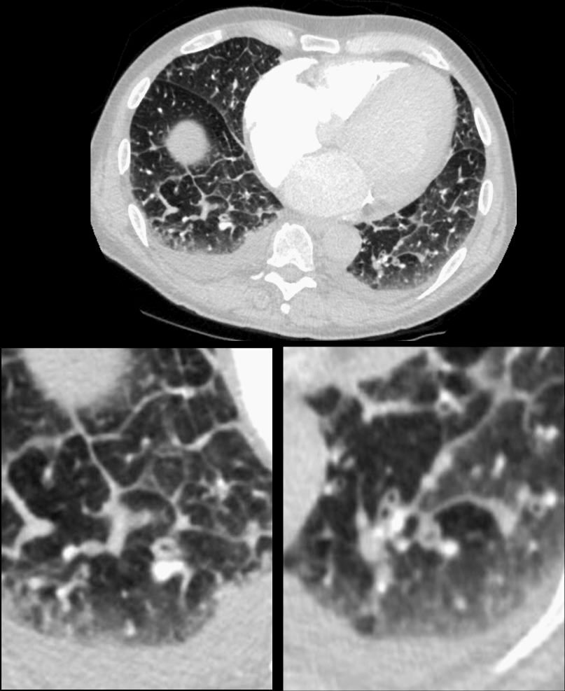
3 months later the patient presented with chest pain and a cough. CT of the chest in the axial plane shows new bilateral lower lobar regions of irregular interlobular septal thickening noted in the right lower lobe a, magnified in b). Ringed in image c and d are 2 side by side secondary lobules with irregular septal thickening centrilobular nodules and other intralobular nodules likely reflecting lymphatic involvement.
Given the changes in the right upper lobe these findings likely reflect lymphangitis carcinomatosa
Ashley Davidoff MD TheCommonVein.net 013Lu 136062cL
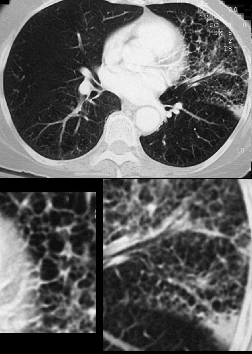
72 year old female showing reticular changes at the lung bases characterised by irregular thickening of the interlobular septa geometric distortion of the secondary lobules changes Ashley Davidoff TheCommonVein.net
136228
Reticulation ILD Geometric Distortion of the Secondary Lobules
72 year old female showing reticular changes at the lung bases characterised by irregular thickening of the interlobular septa geometric distortion of the secondary lobules changes Ashley Davidoff TheCommonVein.net
136229
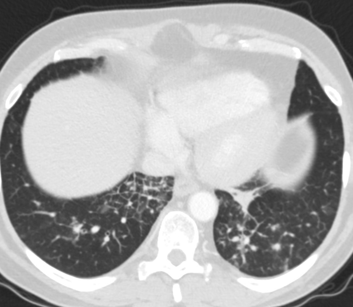
70-year-old female presents with a cough, fever and leukocytosis. CT in the axial plane shows extensive lymphangitis characterized by thickening of the interlobular septa in the inferior aspect of the upper lobe below the necrotizing pneumonia.
Ashley Davidoff MD TheCommonVein.net 260Lu 31631c
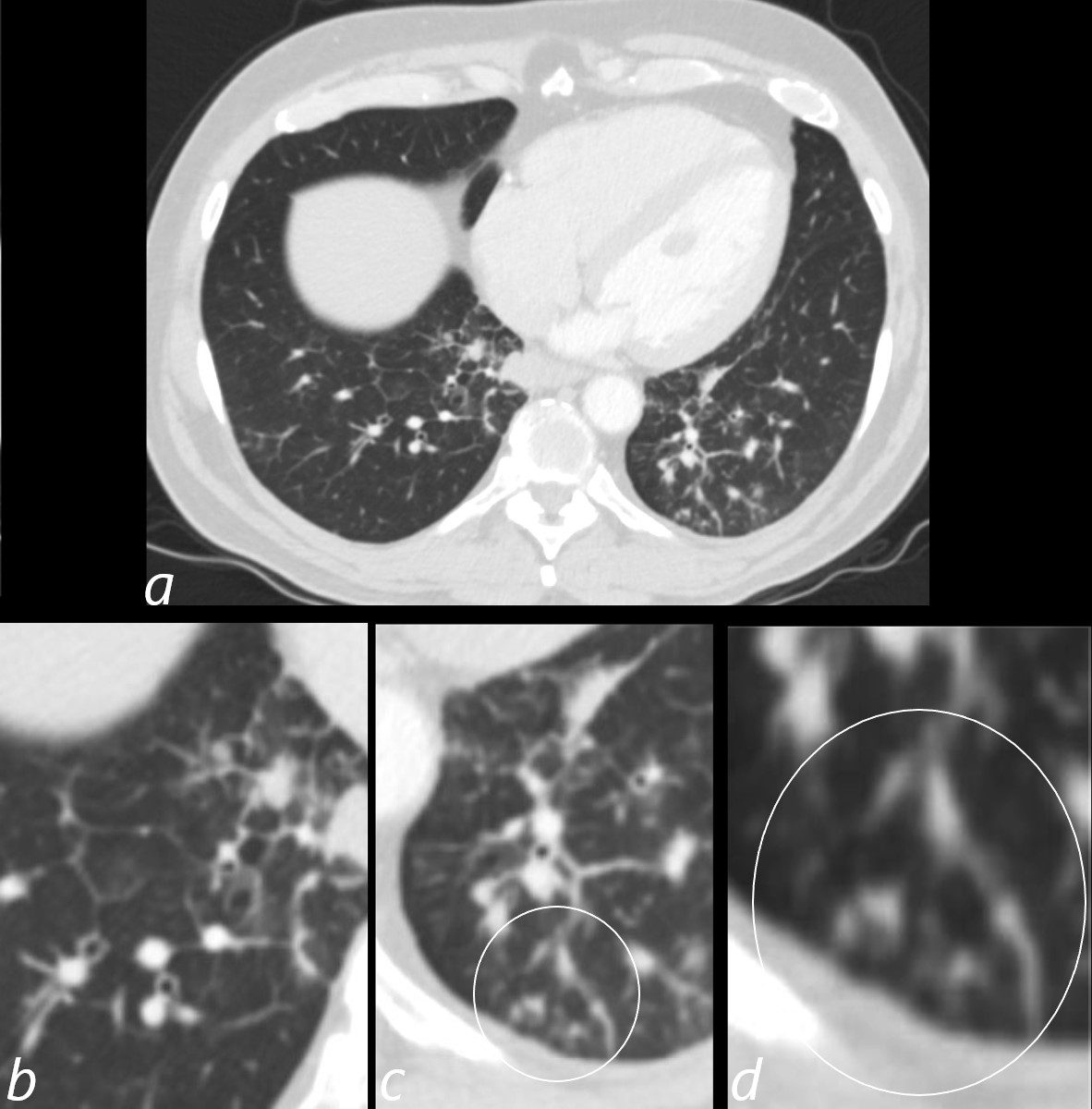
3 months later the patient presented with chest pain and a cough. CT of the chest in the axial plane shows new bilateral lower lobar regions of irregular interlobular septal thickening noted in the right lower lobe a, magnified in b). Ringed in image c and d are 2 side by side secondary lobules with irregular septal thickening centrilobular nodules and other intralobular nodules likely reflecting lymphatic involvement.
Given the changes in the right upper lobe these findings likely reflect lymphangitis carcinomatosa
Ashley Davidoff MD TheCommonVein.net 013Lu 136062cL
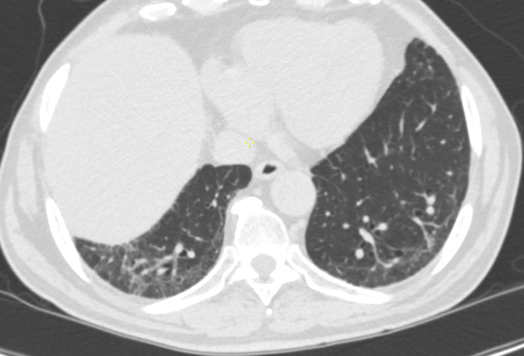
3 months later the patient presented with chest pain and a cough. CT of the chest in the axial plane shows new bilateral lower lobar regions of irregular interlobular septal thickening bilaterally more prominent on the right with nodular changes at the left base.
Given the changes in the right upper lobe these findings likely reflect lymphangitis carcinomatosa
Ashley Davidoff MD TheCommonVein.net 013Lu 136063
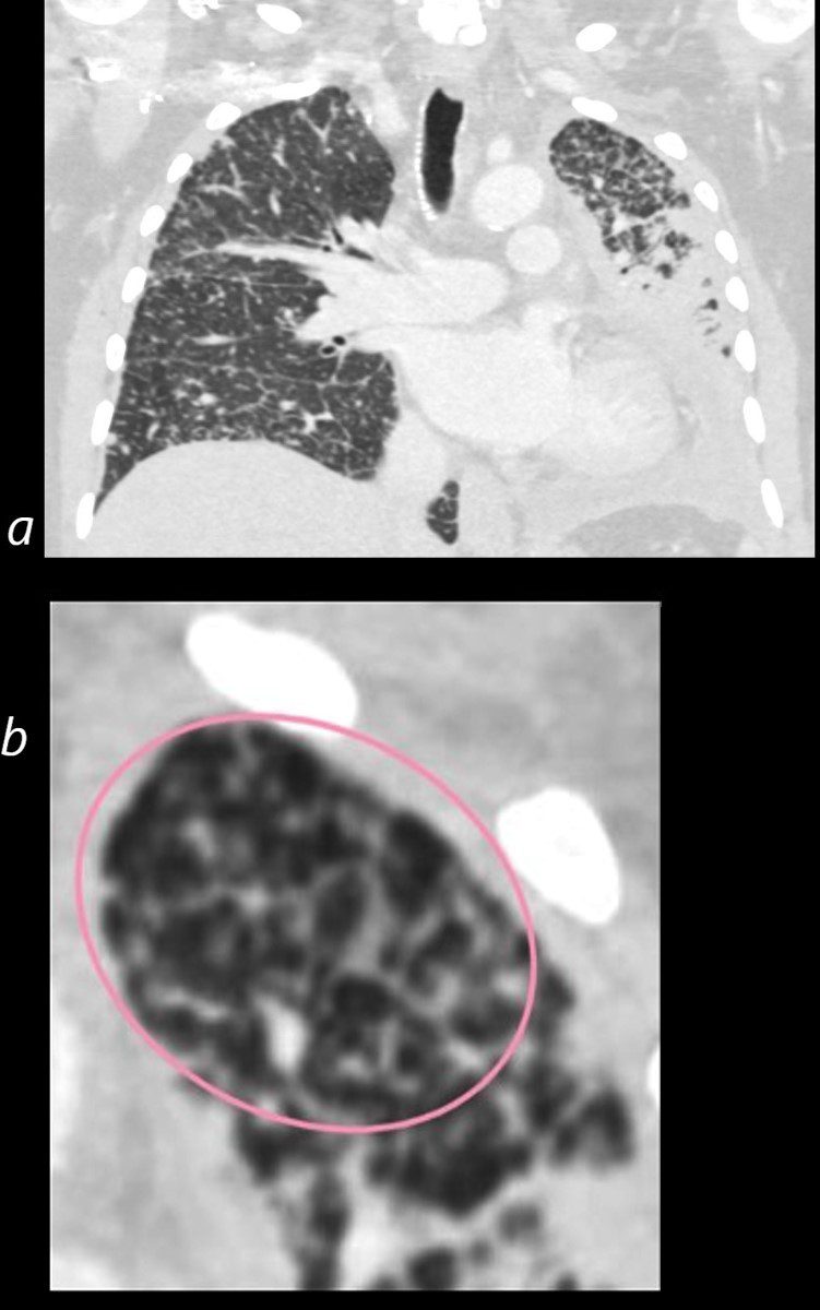
50 year old female with primary adenocarcinoma with the primary lesion presenting as pneumonic consolidation of the left lower lobe, and diffuse reticulonodular changes bilaterally
Image b is a magnified view of the left upper lobe and shows nodular thickening of the interlobular septa representing lymphatic spread along the lymphovascular bundles (pink oval)
The right lung shows interlobular septal thickening centrilobular nodules, and nodular thickening of the minor fissure
These findings are consistent with the diagnosis of lymphangitis carcinomatosa
Ashley Davidoff MD TheCommonVein.net 158Lu 131023c01L
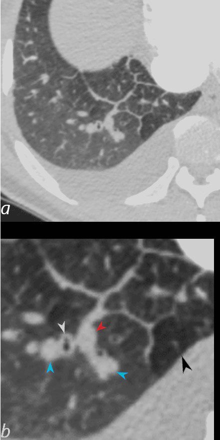
50-year-old female with diabetes, chronic renal failure and congestive heart failure. CT in the axial plane through the right posterior recess, shows thickened interlobular septa at the right base, congested arterioles (light blue arrowheads, b), alongside the bronchioles, peribronchial cuffing (white arrowheads, b), a congested pulmonary venule in the interlobular septum (red arrowhead arrowheads, b), ground glass changes and a secondary lobule demonstrating mosaic attenuation (black arrowhead arrowheads, b). The IVC is dilated and a small complex effusion is present.
Ashley Davidoff MD TheCommonvein.net 135783cL 193Lu
74-year-old man presents with dyspnea and orthopnea. CT shows thickening of the interlobular septa (Kerley B lines), peribronchial cuffing, and enlargement of the lobular arteriole in the right lower lobe. There is a suggestion of vasoconstriction of the arteriole as it enters the secondary lobule. ground glass changes in the some of the secondary lobules on the left and perhaps mosaic attenuation vs normal secondary lobule at the right base are noted. Additionally, there are small bilateral effusions right greater than left. The mild irregular shape of the effusions suggests that they are partially loculated. These findings indicate moderate congestive heart failure with interstitial edema.
Ashley Davidoff MD TheCommonVein.net 135775c01
Links and References
Fleischner Society
beaded septum sign
CT scans.—This sign consists of irregular and nodular thickening of interlobular septa reminiscent of a row of beads (,Fig 10). It is frequently seen in lymphangitic spread of cancer and less often in sarcoidosis (,24).