
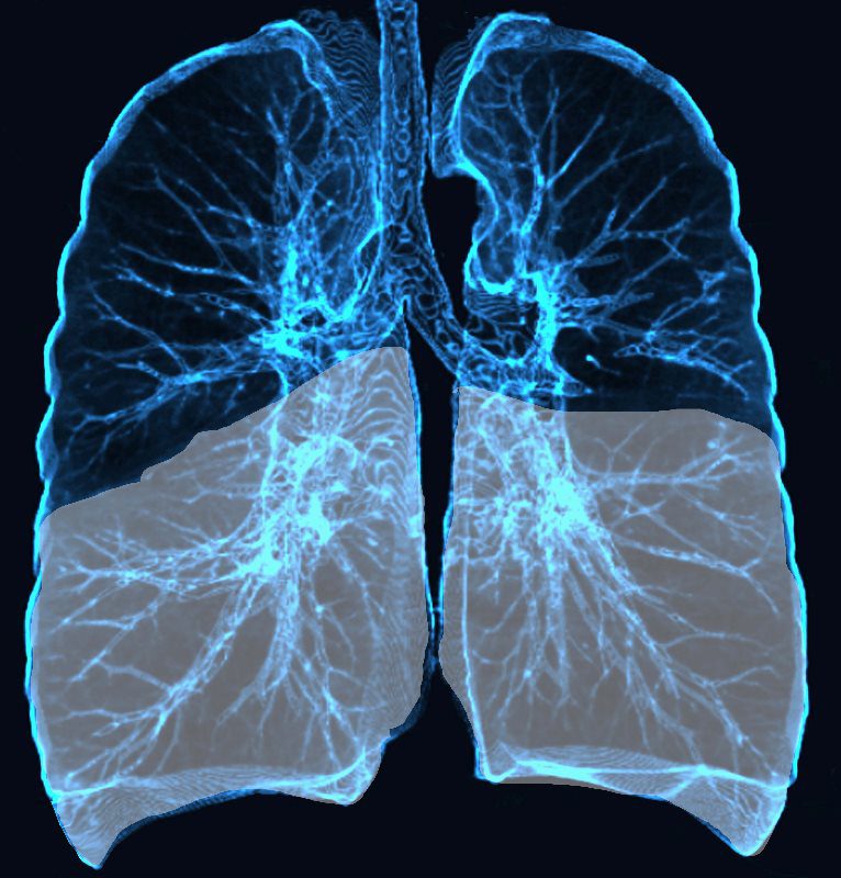
Lower Lobe distribution
Ashley Davidoff MD TheCommonvein.net lungs-0771
Infection
Inflammation
Aspiration
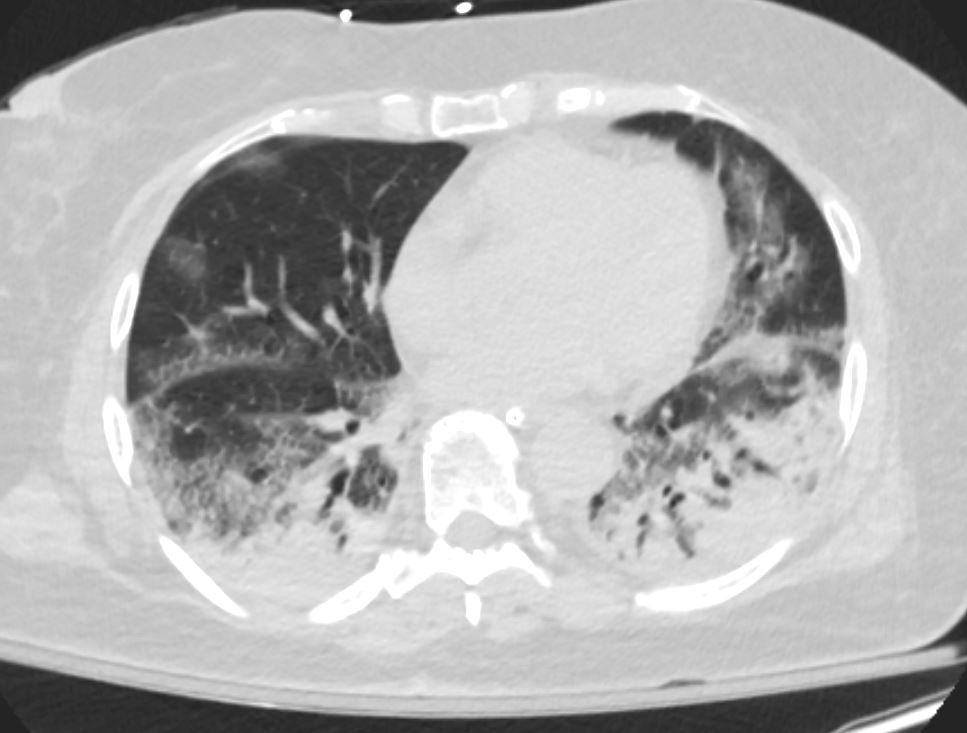
Ashley Davidoff MD TheCommonVein.net crazy paving ICU 003
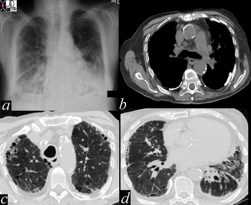
DIP
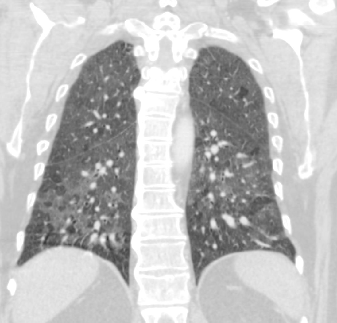
60-year-old male smoker with a history of progressive dyspnea. Coronal CT through the posterior lung fields at the level of the vertebral column shows extensive patchy ground glass changes and mosaic attenuation. The lower lobes show more prominent parenchymal heterogeneity.
Pathology confirmed a diagnosis of DIP
Ashley Davidoff MD TheCommonVein.net 253Lu 136012
UIP
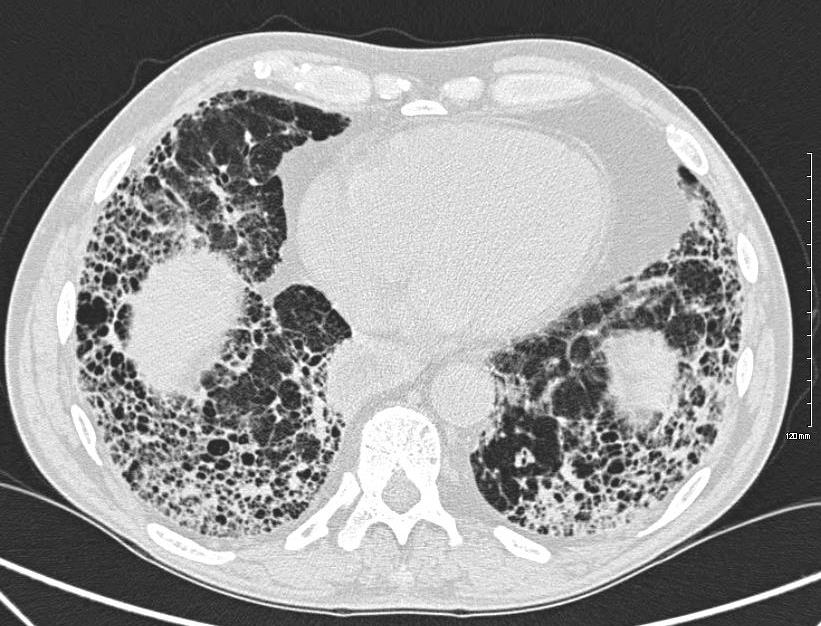
Ashley Davidoff MD thecommonvein.net 134902-lungs UIP
NSIP

Coronal CT shows symmetrical bibasilar reticular change, bronchovascular thickening , ground glass changes and subpleural sparing, all features characteristic of NSIP Also note the volume loss of the lower lobes
Ashley Davidoff MD TheCommonVein.net
Inflammation Inhalation
Aspiration
Ashley Davidoff MD TheCommonVein.net crazy paving ICU 003
Asbestosis
Ashley Davidoff MD thecommonvein.net 47060c01
keywords chest lung fx shaggy heart border reticular changes interstitial lung disease interstitium honeycombing pleural calcification fibrosis dx asbestosis
Malignancy Metastases
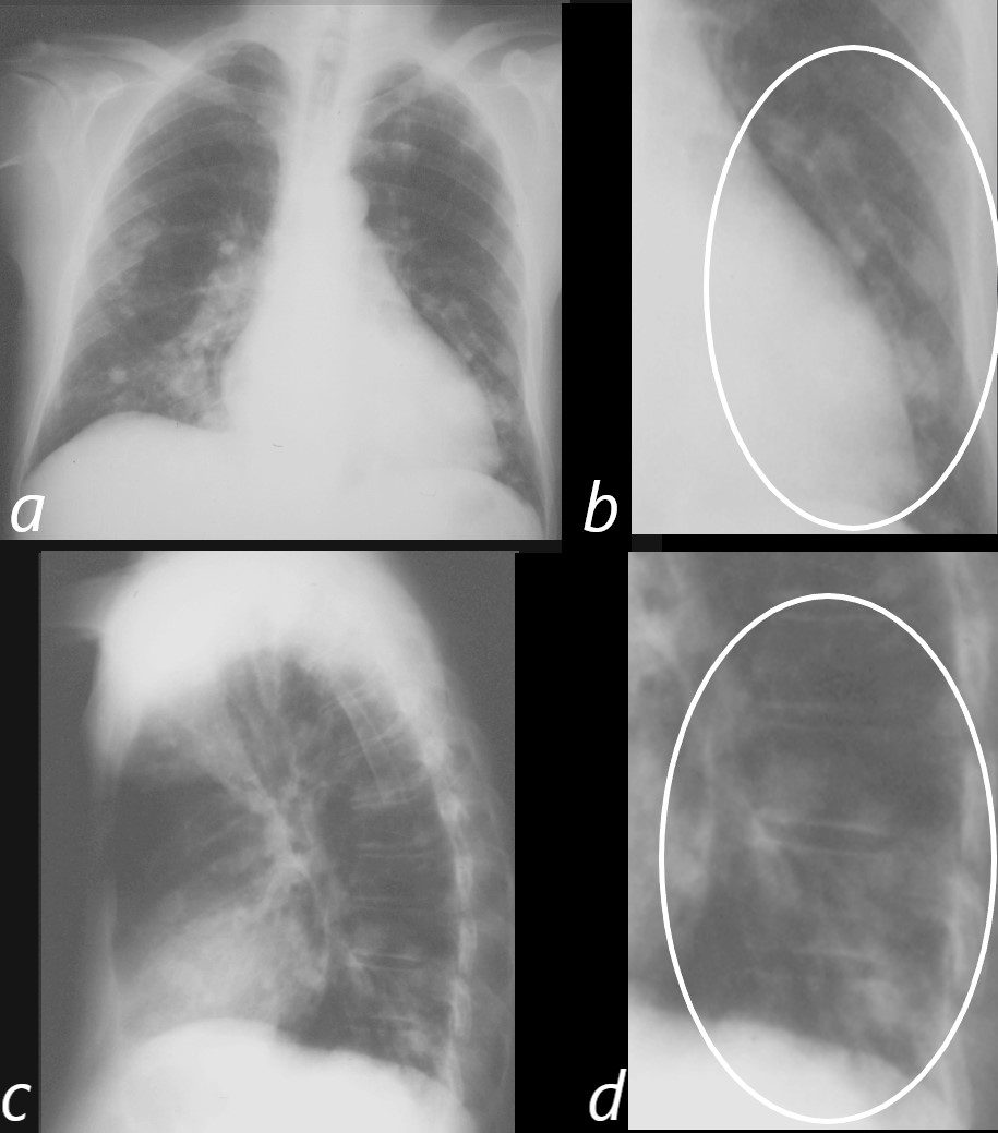
54 year old male with history of right sided renal cell carcinoma.
Frontal and lateral CXR show multiple metastases to the lungs with predominant lower lung distribution. The primary lesion was an RCC. Magnified view of the left lower lobe from the frontal projection (b ringed in white) shows multiple nodular metastases with similar changes noted in the lower lobes on the magnified lateral view (d white ring)
Ashley Davidoff MD TheCommonVein.net 05769cL
Mechanical/Atelectasis
Trauma Metabolic
Circulatory- Pulmonary Embolus (PE)
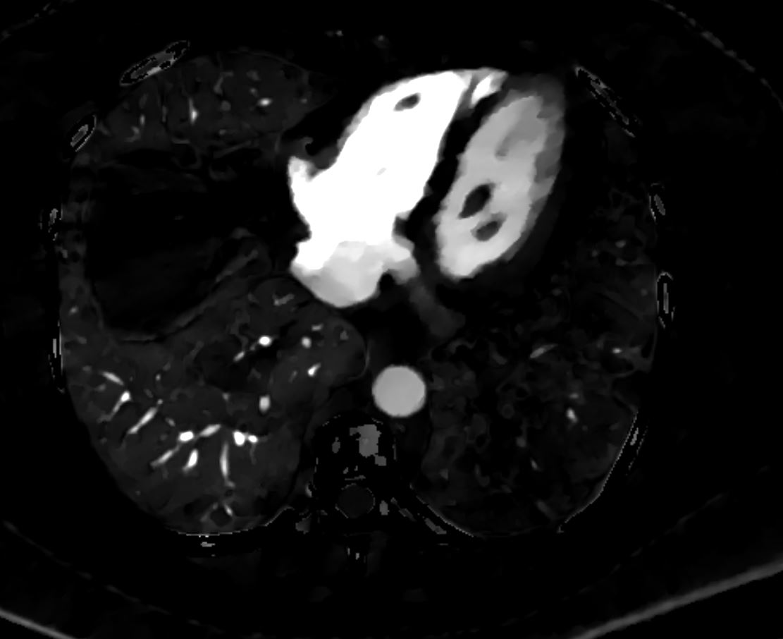
Pulmonary Embolus Left Lower Lobe
56 -year-old female with a history of amyloidosis presenting with tachycardia and dyspnea. CTPA shows an occlusive embolus (PE) in the left lower lobe pulmonary artery.
Ashley Davidoff MD TheCommonVein.net 135738c
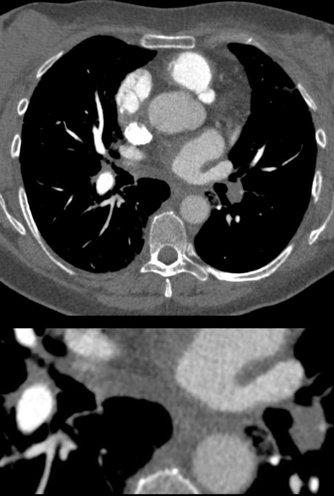
PE and No Enhancement of the Left Lower Lobe-
Dual Energy Iodine Map
Perfusion Defect of the Left Lower Lobe from Occlusive Pulmonary Embolus
56 -year-old female with a history of amyloidosis presenting with tachycardia and dyspnea. Dual energy CT with an iodine map shows shows an almost lobar perfusion defect of the left lower lobe compared
Ashley Davidoff MD TheCommonVein.net 135740
Hampton’s Hump – Middle Lobe
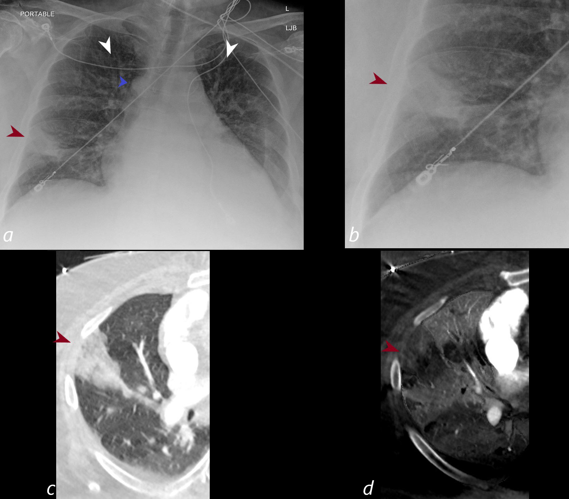
CXR shows a wedge shaped infiltrate in the middle lobe of the lung secondary to a pulmonary embolus (PE) characteristic of a Hampton’s hump (maroon arrowheads a,b) The infarction is confirmed on the CT with contrast (maroon arrowhead c) as well as the region of a perfusion defect (d- maroon arrowhead) In addition there is evidence of CHF on the CXR with cephalization of the vessels (white arrowheads c) cardiomegaly with left atrial enlargement, and enlargement of the azygous vein (blue arrowhead a)
Ashley Davidoff MD TheCommonVein.net)
Hemorrhage
Immune
Infiltrative
Idiopathic
Iatrogenic
Idiopathic