60 year old immunocompromise female 1 year prior to an episode of miliary TB
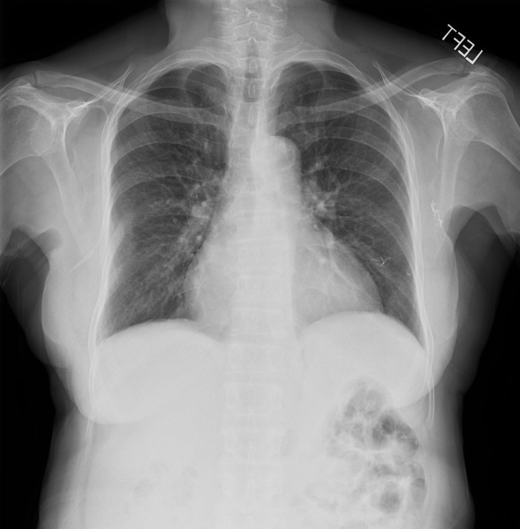
Frontal CXR of a 60 year old immunocompromise female 1 year prior to an episode of miliary TB shows a normal CXR
Ashley Davidoff MD TheCommonVein.net 265Lu 136195
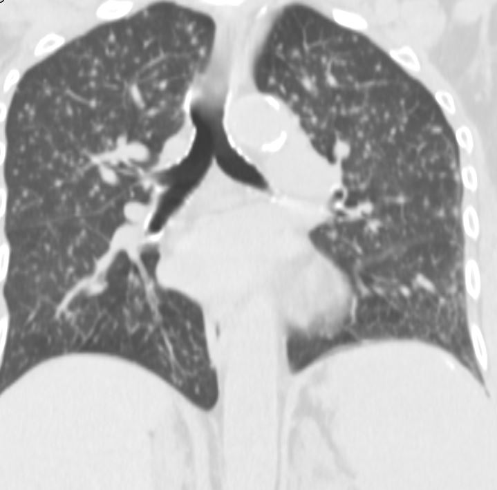
Axial CT of a 60-year-old immunocompromised female 1 year prior to an episode of miliary TB shows a normal examination
Ashley Davidoff MD TheCommonVein.net 265Lu 136196
7 Months Later Presents with Cough and Weight Loss
CXR Miliary Pattern
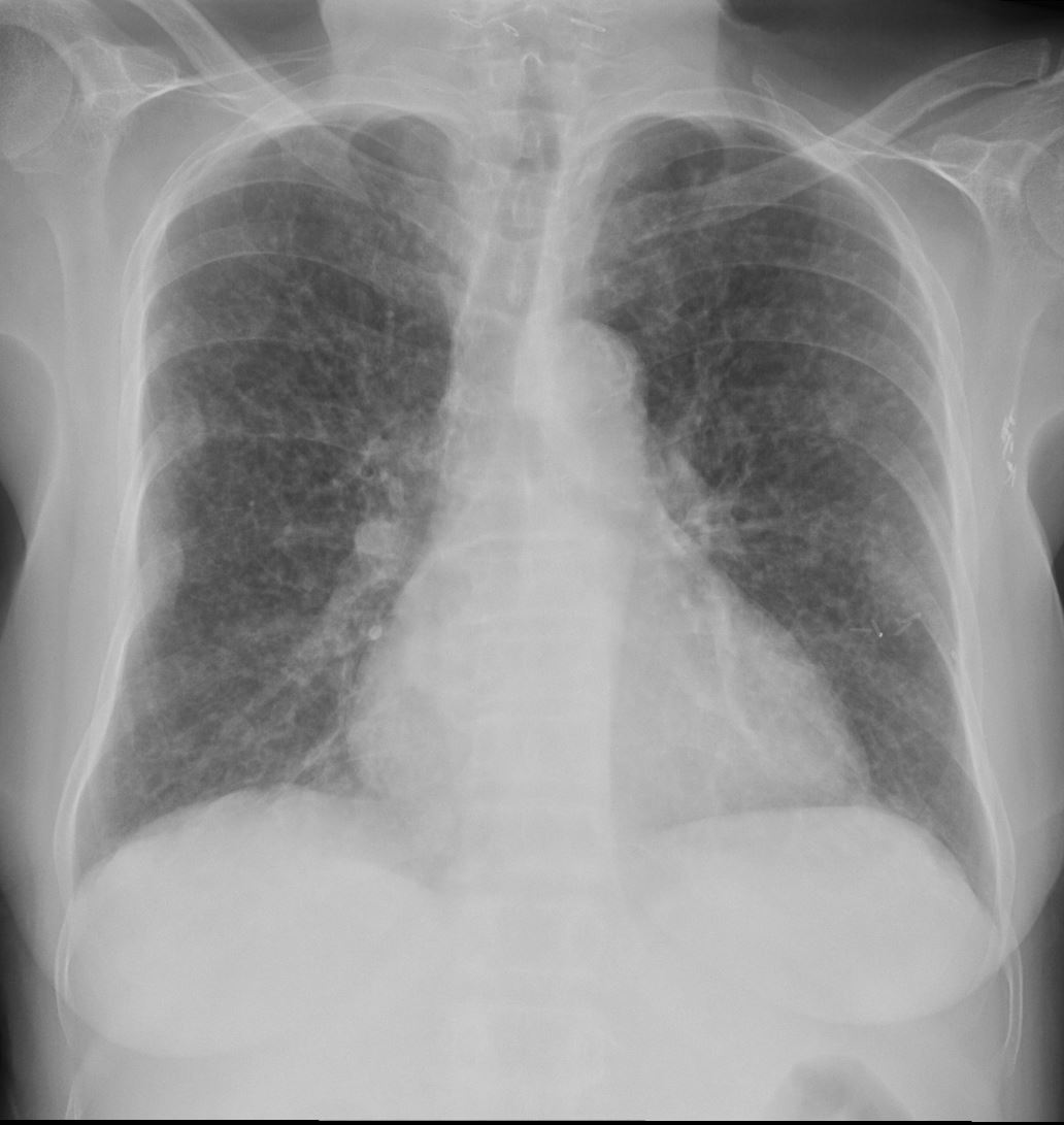
60-year-old immunocompromise female presents with a cough and weight loss CXR shows a diffuse miliary pattern. Final diagnosis was mycobacterium tuberculosis. Associated findings include healed right sided rib fractures and surgical clips in the left axilla
Ashley Davidoff MD TheCommonVein.net 265Lu 136197
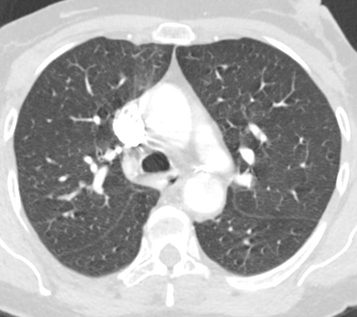
CT Miliary Tuberculosis
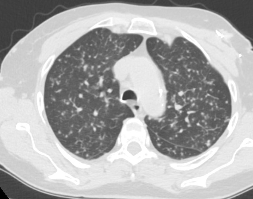
60-year-old immunocompromised female presents with a cough and weight loss. Axial CT shows miliary nodules throughout both lung fields. She responded well to treatment and final diagnosis was mycobacterium tuberculosis.
Ashley Davidoff MD TheCommonVein.net 265Lu 136198
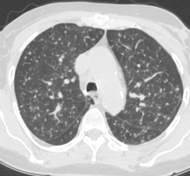
60-year-old immunocompromised female presents with a cough and weight loss. Axial CT shows miliary nodules throughout both lung fields. She responded well to treatment and final diagnosis was mycobacterium tuberculosis.
Ashley Davidoff MD TheCommonVein.net 265Lu 136199
CT Miliary Tuberculosis Centrilobular and Fissural Nodules
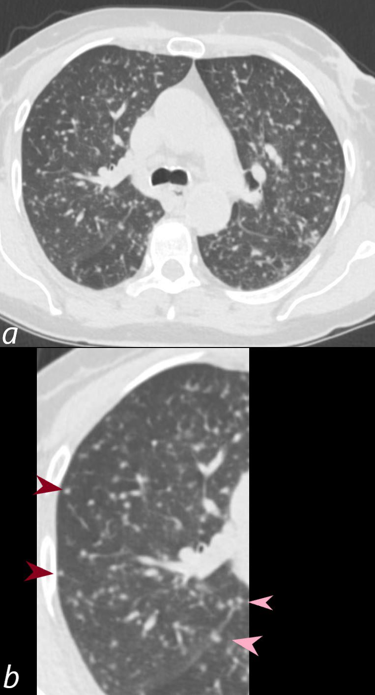
60-year-old female presents with a cough and weight loss. Axial CT shows miliary nodules throughout both lung fields. Some of these nodules are are centrilobular or distributed along the bronchovascular bundles (b, maroon arrowheads) and others are fissural based (b, pink arrowheads). She responded well to treatment and final diagnosis was mycobacterium tuberculosis.
Ashley Davidoff MD TheCommonVein.net 265Lu 136201cL
CT Miliary Tuberculosis Centrilobular Fissural Nodules and Pleural Nodules
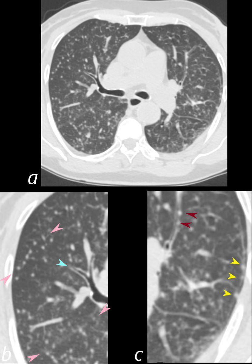
60-year-old immunocompromised female presents with a cough and weight loss. Axial CT shows miliary nodules throughout both lung fields. Some of these nodules are centrilobular or distributed along the bronchovascular bundles (c, maroon arrowheads) and others are fissural based (b, pink arrowheads) and along the pleura (yellow arrowheads suggesting at least a lymphatic distribution. There is bronchial wall thickening (b teal arrowhead). She responded well to treatment and final diagnosis was mycobacterium tuberculosis.
Ashley Davidoff MD TheCommonVein.net 265Lu 136202cL
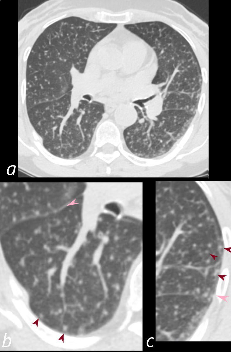
60-year-old immunocompromised female presents with a cough and weight loss. Axial CT shows miliary nodules throughout both lung fields. Some of these nodules are centrilobular or distributed along the bronchovascular bundles (b, c, maroon arrowheads) and others are fissural based (b and c pink arrowheads) , suggesting both bronchovascular and lymphatic distribution. She responded well to treatment and final diagnosis was mycobacterium tuberculosis. There is a healing right sided posterolateral rib fracture.
Ashley Davidoff MD TheCommonVein.net 265Lu 136202cL
CT Miliary Tuberculosis
Centrilobular Nodules Suggesting
Arterial Small Airway and or Lymphatic Involvement
Also Fissural Nodules and Pleural Nodules
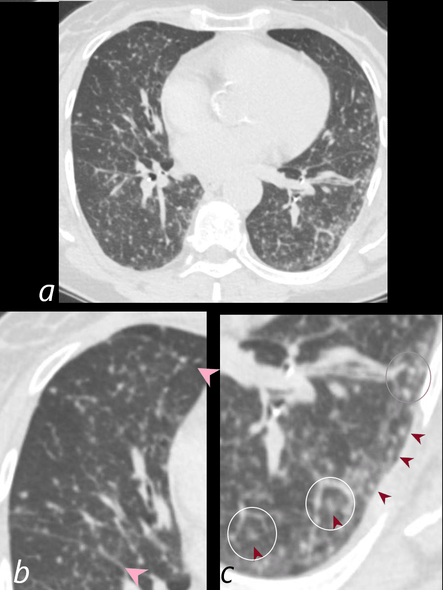
60-year-old immunocompromised female presents with a cough and weight loss. Axial CT shows miliary nodules throughout both lung fields. Some of these nodules are centrilobular (c, maroon arrowheads) and others are fissural based (b, pink arrowheads). In some of the secondary lobules there are 2 centrilobular nodules indicating involvement of the airway and arteriole and or the lymphatics (c white rings). One lobule shows centrilobular and interlobular nodules (c gray ring anteriorly). She responded well to treatment and final diagnosis was mycobacterium tuberculosis.
Ashley Davidoff MD TheCommonVein.net 265Lu 136204cL