
Malignancy
Right Upper Lobe Collapse
Squamous Cell Causing
Obstruction but Airways Filled with
Tumor or Infection or Mucus
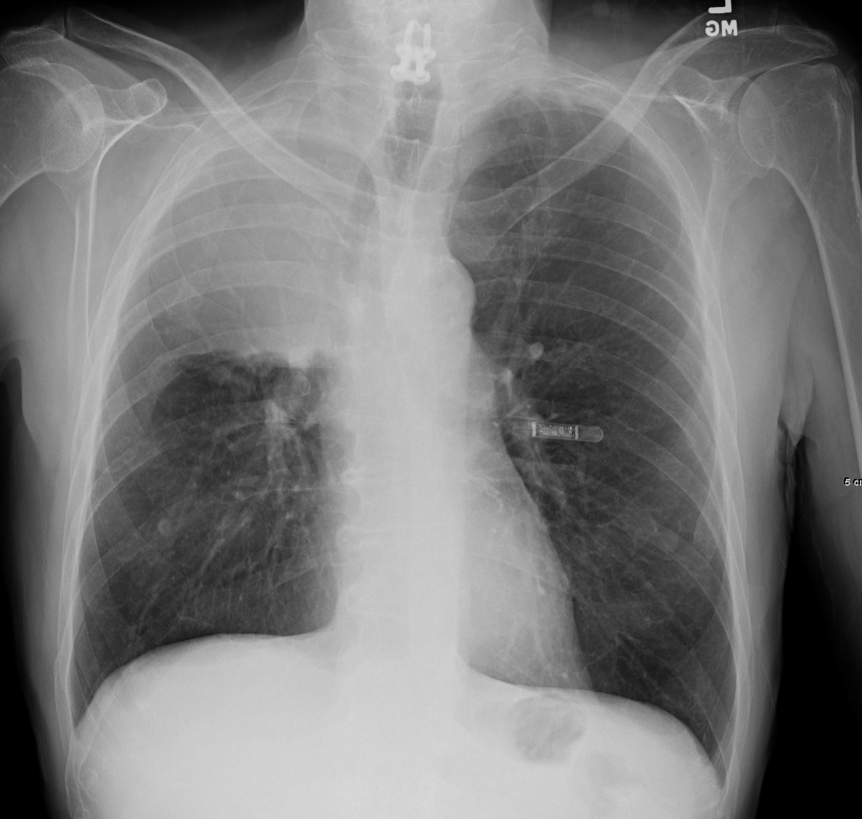
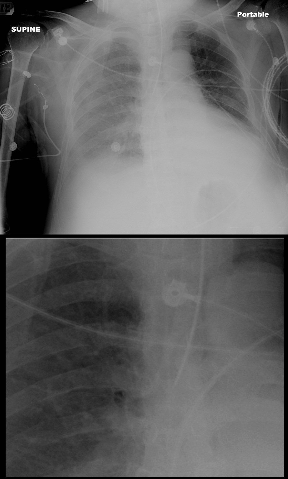
55-year-old male presenting with dyspnea
Frontal CXR shows right upper lobe (RUL) atelectasis characterized by rightward deviation of the trachea elevation of the right hemidiaphragm and opacification of the right upper lobe. Final diagnosis was a central RUL proximal squamous cell carcinoma with extensive filling of the distal bronchi-ectatic segmental and subsegmental airways
Ashley Davidoff TheCommonVein.net 212Lu 136430
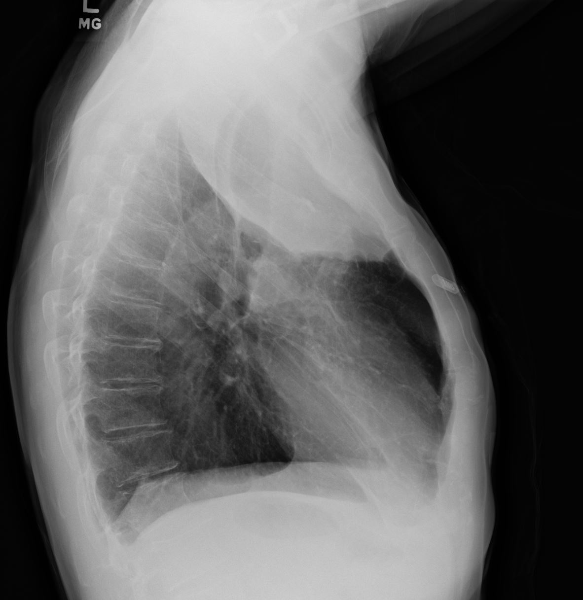
55-year-old male presenting with dyspnea
Lateral CXR confirms atelectasis of the RUL characterized by pie shaped consolidation of the anteriorly position right upper lobe, hyperinflation of the right lower lobe mild elevation of the right hemidiaphragm. Final diagnosis was a central RUL proximal squamous cell carcinoma with extensive filling of the distal bronchi-ectatic segmental and subsegmental airways
Ashley Davidoff TheCommonVein.net 212Lu 136430
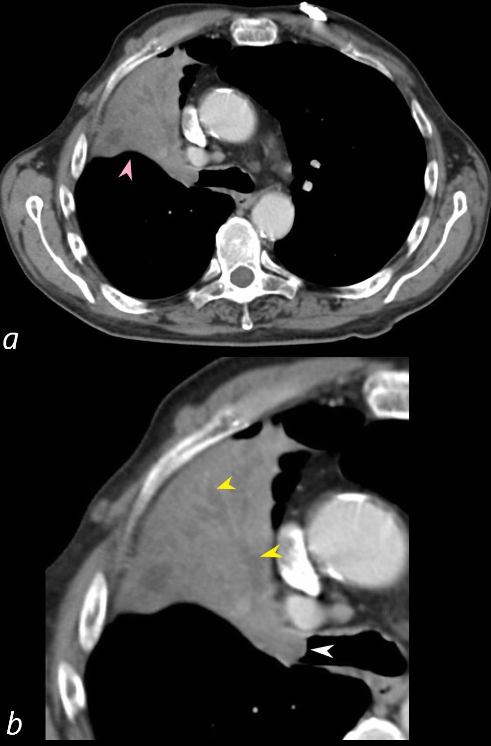
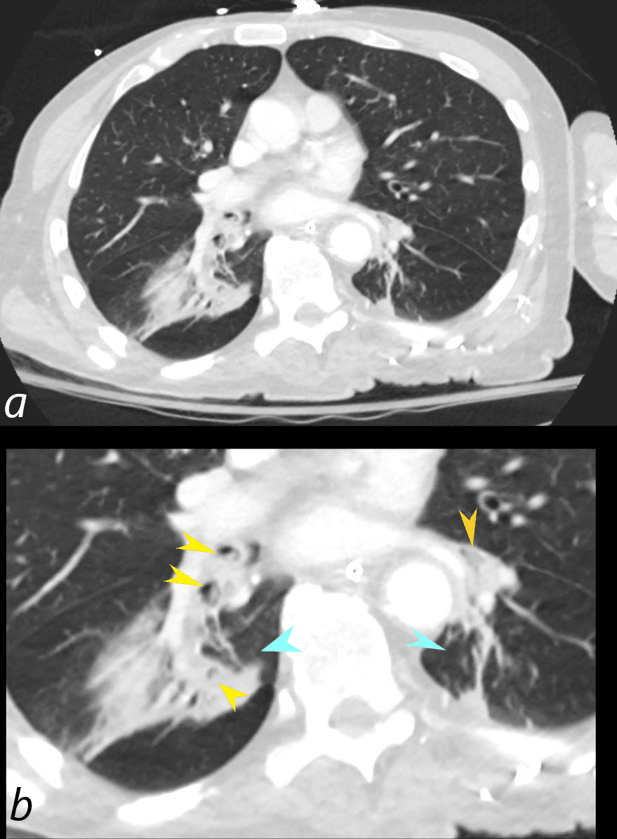
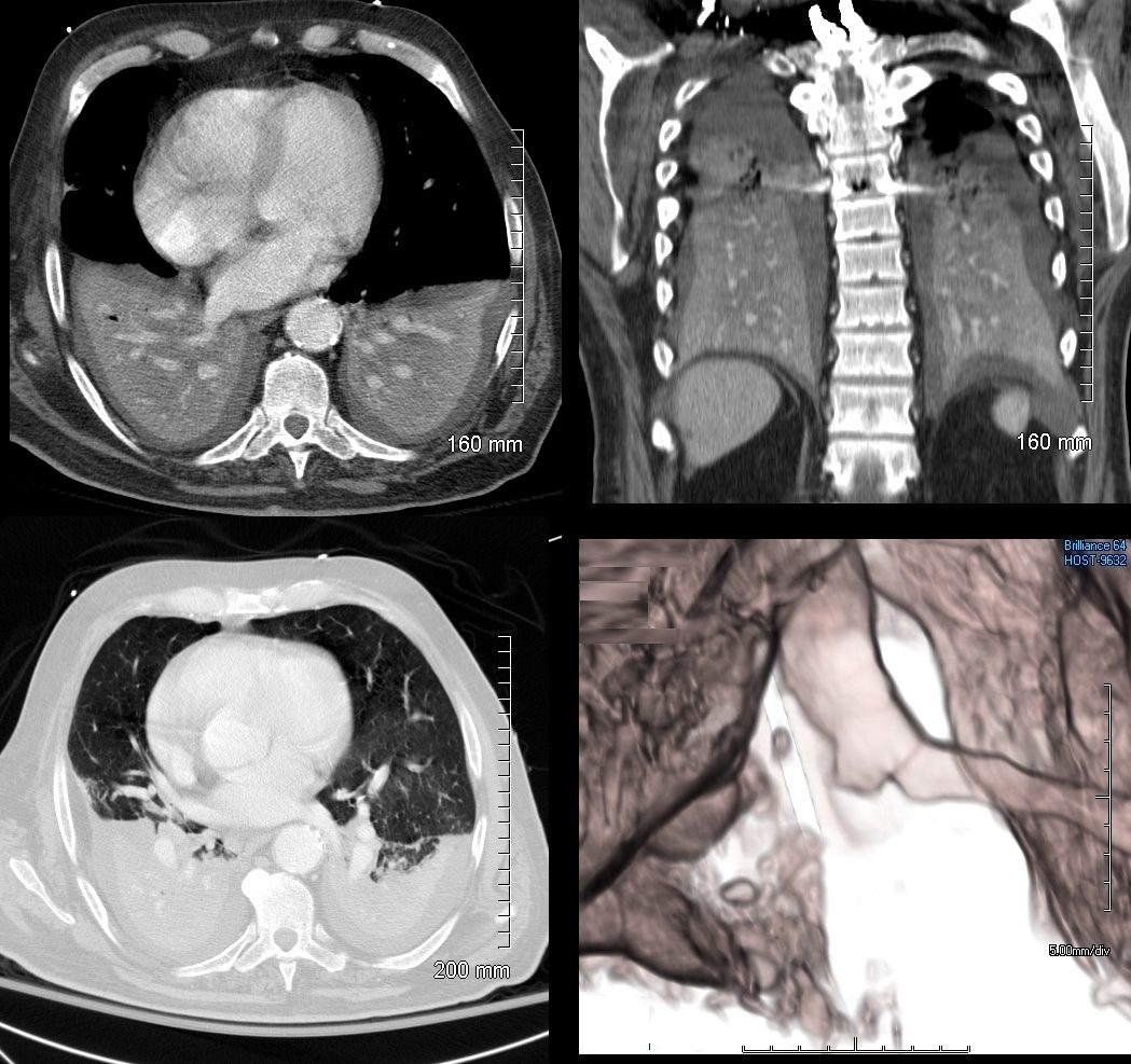
55-year-old male presenting with dyspnea
Axial CT at the level of the carina shows atelectasis of the RUL caused by a central obstructing lesion in the right upper lobe bronchus (b, white arrowhead) resulting in atelectasis of the RUL characterized by a wedge-shaped consolidation of the anteriorly positioned right upper lobe. The major fissure is displaced anteriorly (a, pink arrowhead). There is extensive filling of the distal bronchiectatic segmental and subsegmental airways of the RUL (b, yellow arrowheads). Final diagnosis was a central RUL proximal squamous cell carcinoma.
Ashley Davidoff TheCommonVein.net 212Lu 136432cL
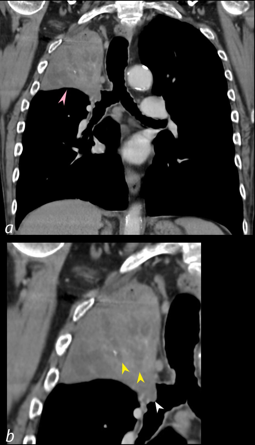
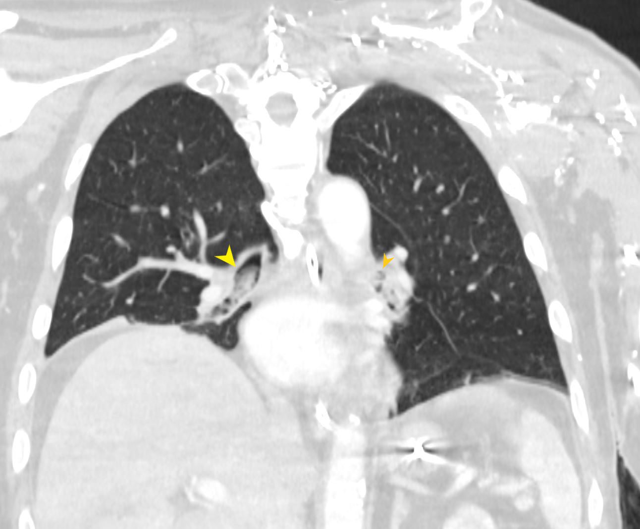
55-year-old male presenting with dyspnea
Coronal CT at the level of the trachea and mainstem bronchi, shows atelectasis of the RUL caused by a central obstructing lesion in the right upper lobe bronchus (b, white arrowhead) resulting in atelectasis of the RUL characterized by a wedge-shaped consolidation of the right upper lobe with superiorly displaced major fissure (a, pink arrowhead). There is extensive filling of the distal bronchiectatic segmental and subsegmental airways of the RUL (b, yellow arrowheads). Final diagnosis was a central RUL proximal squamous cell carcinoma.
Ashley Davidoff TheCommonVein.net 212Lu 136433cL
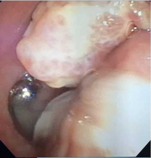
Endoscopic image of a central squamous cell carcinoma (SCC) with extensive
Ashley Davidoff TheCommonVein.net 212Lu 136434
-

Reversed S sign of Golden
The scout film performed prior to a CT scan from a 76-year-old man with chest pain and shortness of breath. The appearance suggests atelectasis of the right upper lobe with the normal position of the minor fissure (yellow) altered so that the upper portion (light green above the yellow line) is shifted upward caused by volume loss of an atelectatic right upper lobe (pink). The lower portion of the fissure (light green below the yellow line) is bulging rightward and outward caused by an implied mass (dark green). The “reversed S sign of Golden” is demonstrated in this case and infers a central mass causing obstruction and resulting in the shape described by the light green line of the minor fissure.
Courtesy: Ashley Davidoff, M.D.

Mechanical
Mucus or Aspirated Material
Ashley Davidoff MD TheCommonVein.net
70231cL
Aspiration
Aspiration of Solid Food Right and Left lower Lobe Bronchi
71-year-old male presents with acute respiratory difficulty. CT in the coronal plain shows solid food particles in right mainstem bronchus (yellow arrowhead), extending into the right lower lobe bronchus, as well as segmental airways to the left lower lobe (orange arrowheads). The right hemidiaphragm is elevated secondary to the atelectasis
Ashley Davidoff MD TheCommonVein.net 271Lu 136241
71-year-old male presents with acute respiratory difficulty. CT in the axial plain shows solid food particles in the apical segment of the right lower lobe of the lung (b, yellow arrowheads) and in the left lower lobe (b, orange arrowheads) associated with bilateral basilar subsegmental atelectasis right greater than left (b, teal arrowheads).
Ashley Davidoff MD TheCommonVein.net 271Lu 136237cL
Iatrogenic
Post
Obstructive Atelectasis of the left Lower Lobe
Ashley Davidoff MD TheCommonVein.net
42077
Bilateral Lower Lobar Atelectasis with
Occlusion of the Right Main Stem Bronchus
74 year old male alcoholic with bilateral basilar lobar atelectasis caused by bilateral aspiration
CT scan shows airless lower lobes with small bilateral effusions. 3D reconstruction shows total obstruction of the right mainstem bronchus, and patent proximal mainstem bronchus
Ashley Davidoff MD TheCommonVein.net