
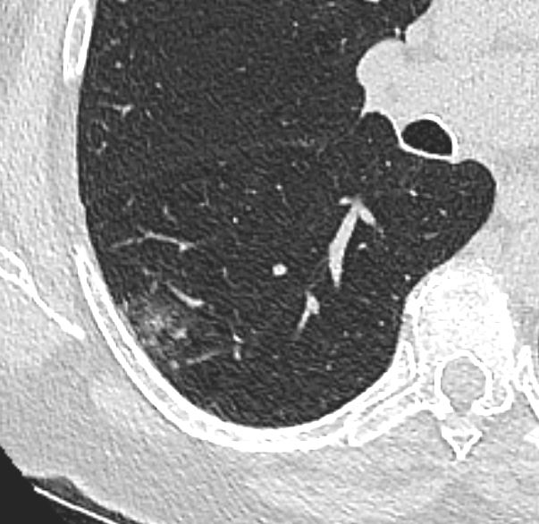
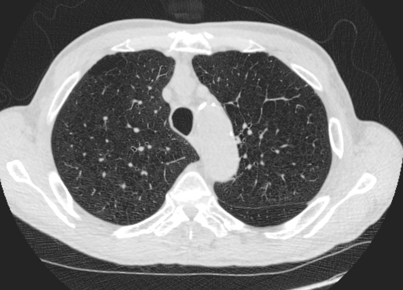
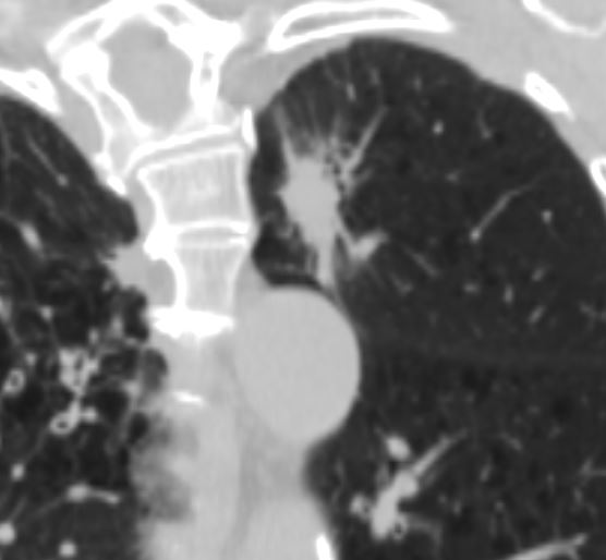
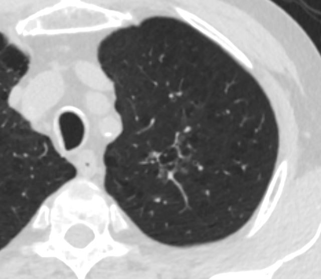
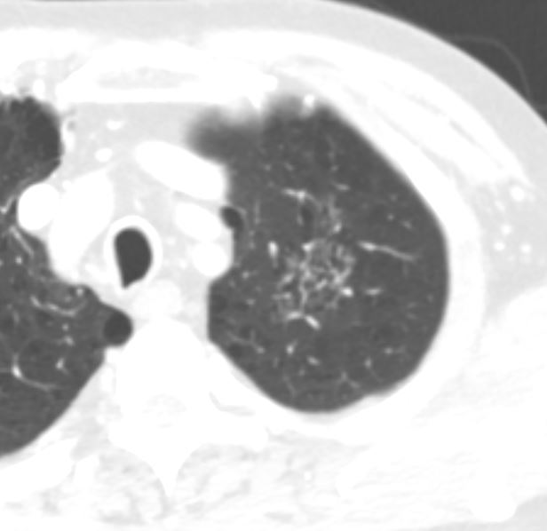
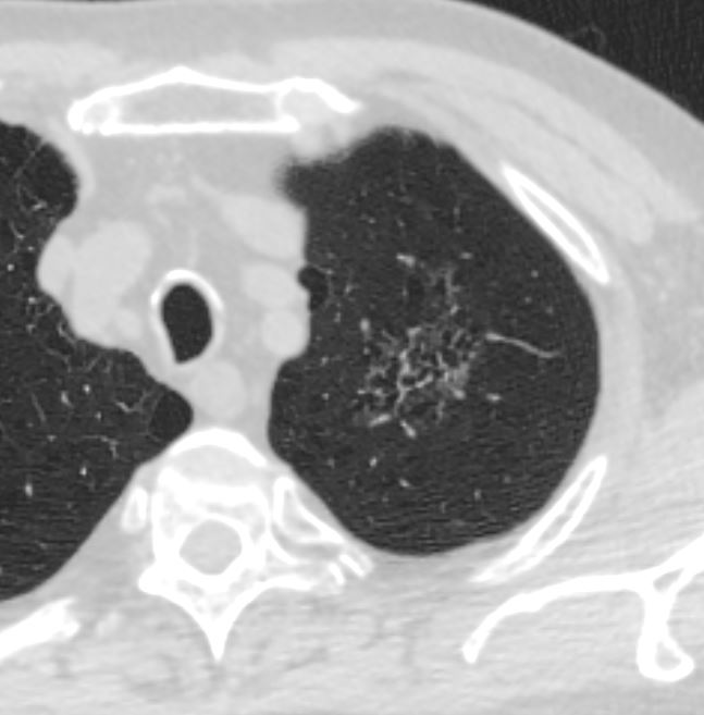
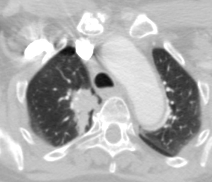
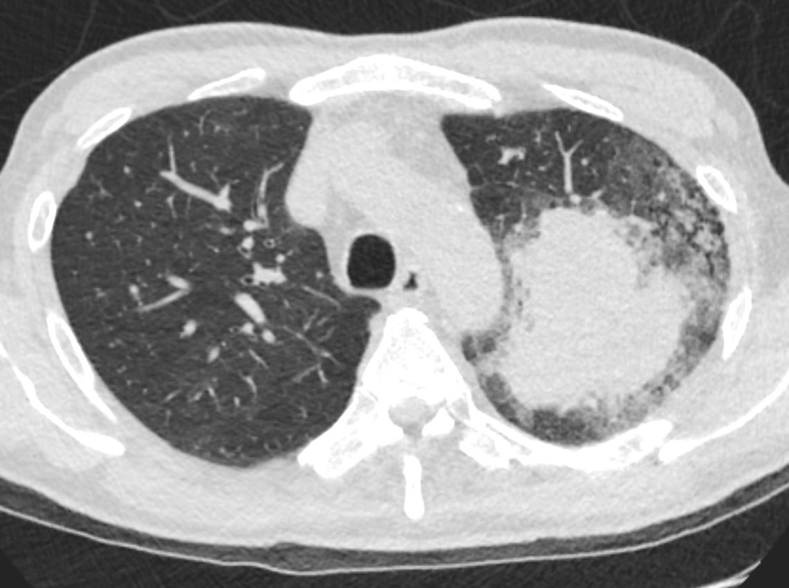
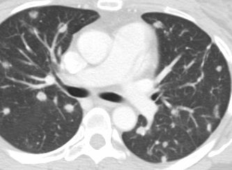
Cigarettes and Adenocarcinoma
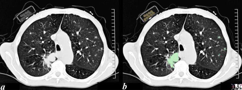
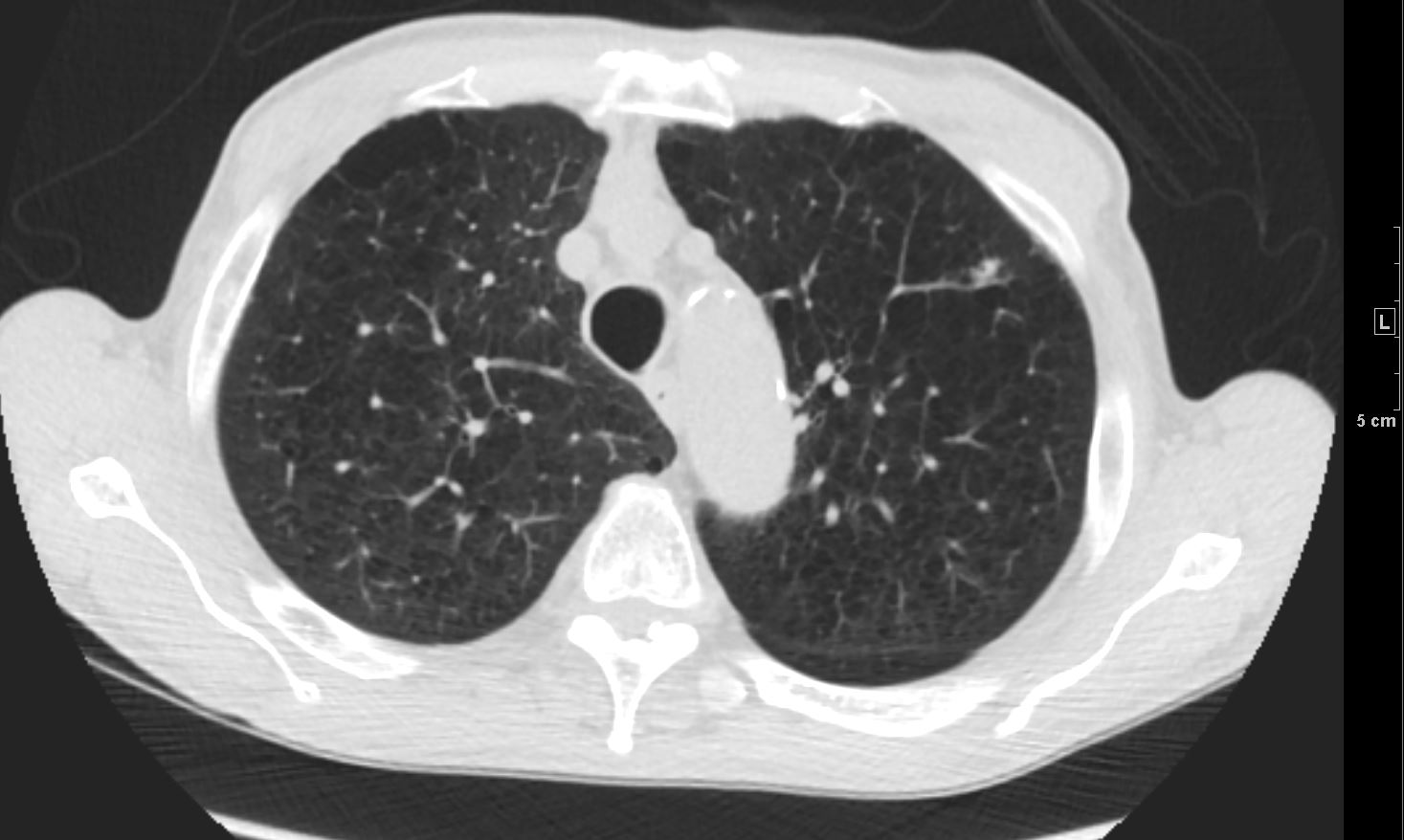
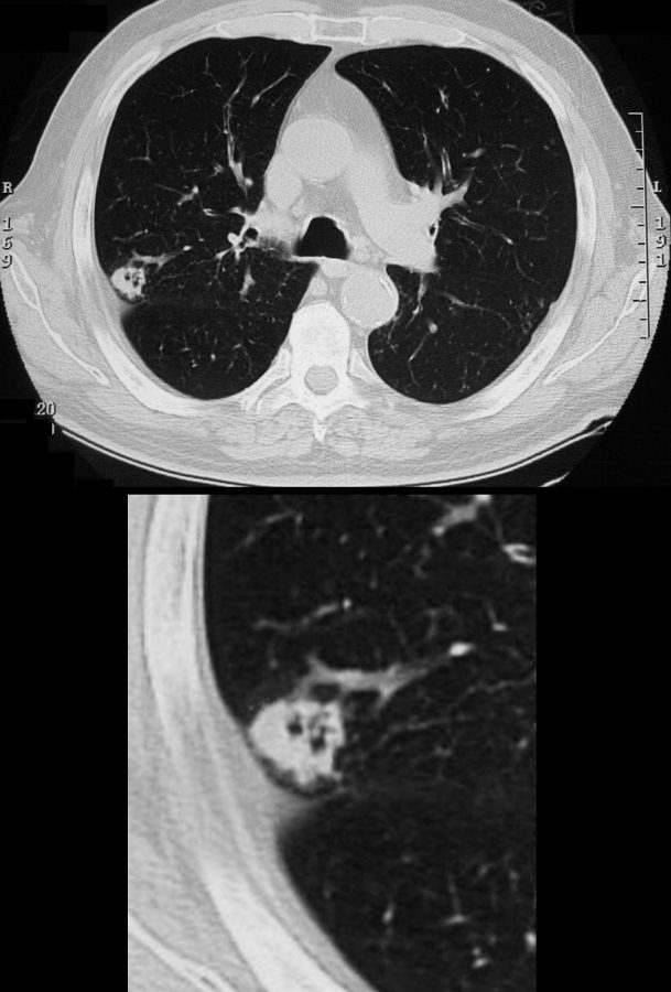
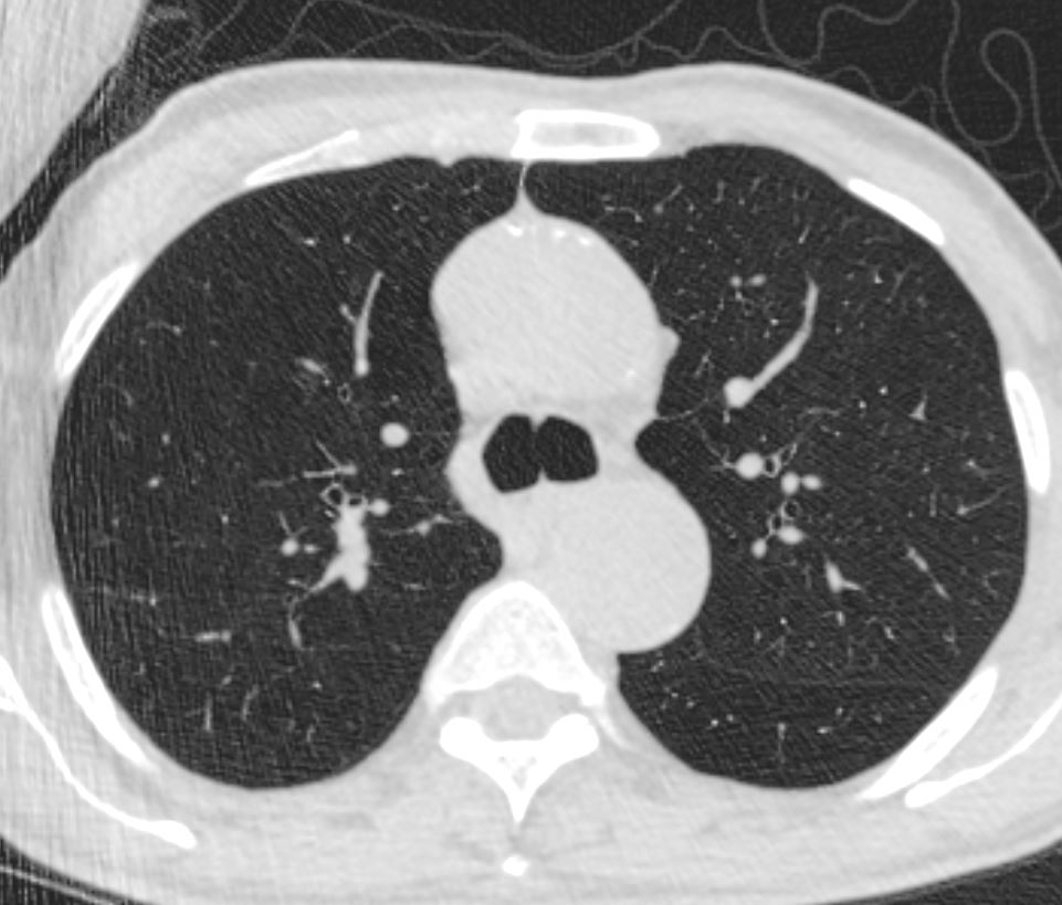
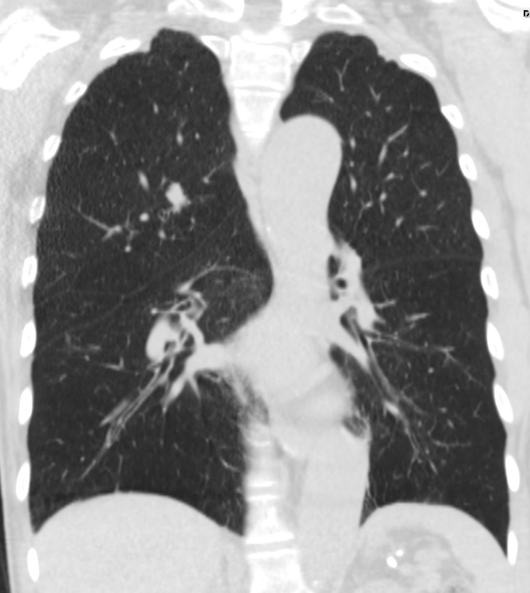
The CT scan through the chest of an 80 year old male shows a large lung mass in the posterior aspect of the right upper lobe (overlaid in green) and3 small nodules in the left upper lobe (overlaid in green). The patient is obviously a smoker and the incriminating pack of cigarettes is identified in his right shirt pocket containing 9 cigarettes. The lung cancer was shown to be an adenocarcinoma The pathology of the nodules may either represent metastatic disease or multicentric foci of bronchioloalveolar cell carcinoma. Associated finding of a thinned anterior junction line suggests hyperinflation and emphysema, and the thickened bronchial walls noted in the right lung suggest chronic bronchitis. Saber shaped trachea is also reminiscent of emphysema. The patient is emaciated, a finding that relates both to his chronic lung disease and his cancer.
Ashley Davidoff MD 87831c01b.8s
TheCommonVein.net
Parts
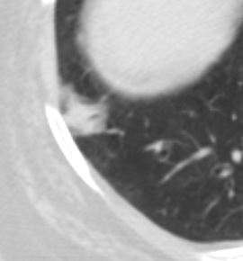
Size
Small
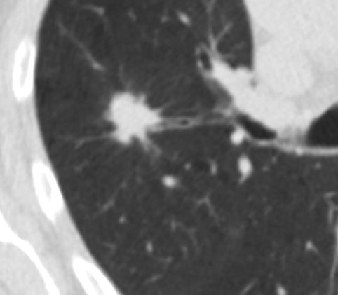
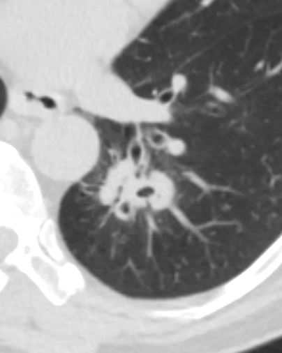
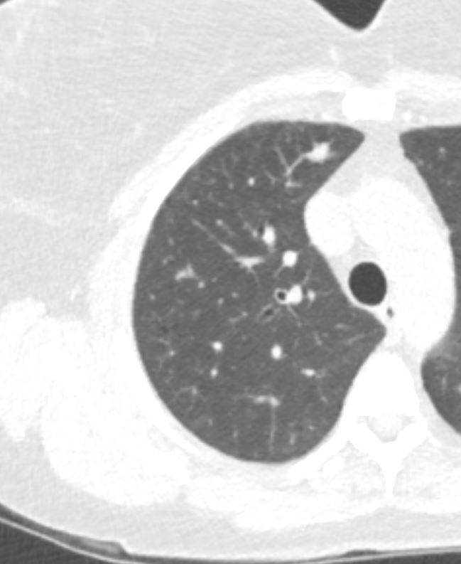
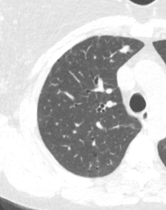
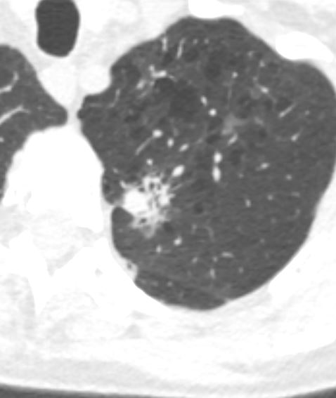
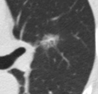
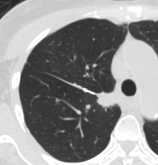
Adenocarcinoma Near the Fissure
(See Below in “Time” Section for Growth)
8mm Irregular Nodule LUL
(See Below in “Time” Section for Growth)

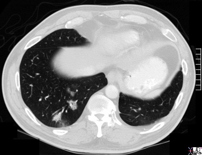
Shape
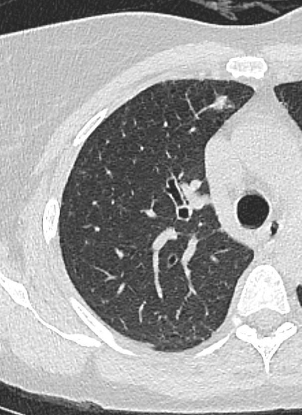
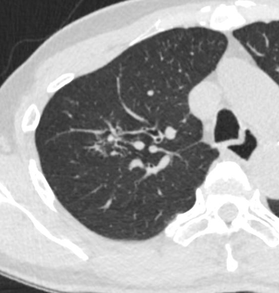
Classical Lobulated Peripheral Nodule or Mass
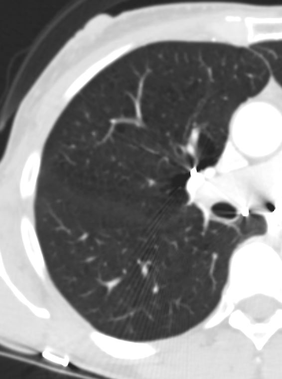
Axial images of the case shown above through the lung mass in a 51-year-old female who presents with nausea vomiting and abdominal distension and who was shown to have a small bowel obstruction. The mass is situated in the left lower lobe and was an incidental asymptomatic finding. It measures in the 3cm range, is lobulated and peripherally located. Pathology showed an adenocarcinoma.
Courtesy: Ashley Davidoff, M.D.TheCommonVein.net
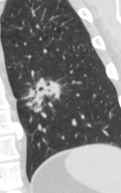
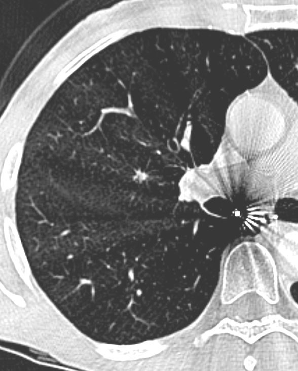
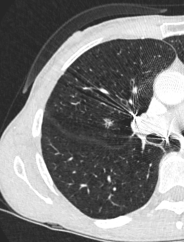
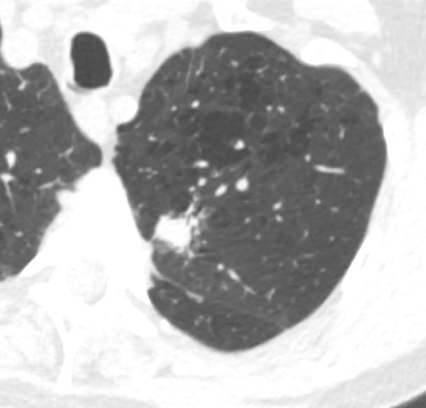
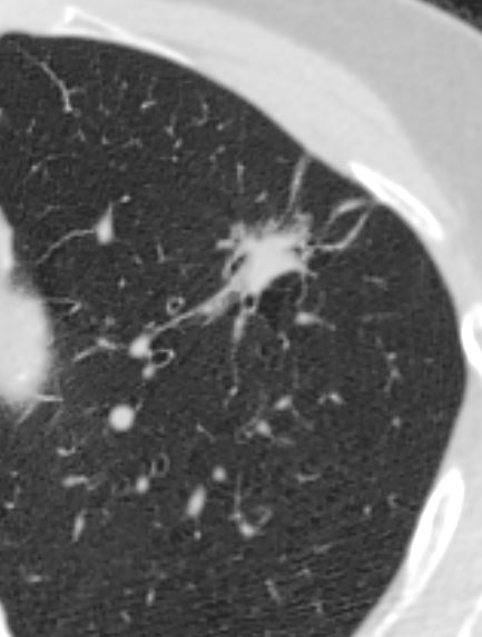
Spiculated Nodule
Ashley Davidoff MD TheCommonVein.net
Adenocarcinoma 001 lung
Position
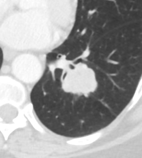
Classical Lobulated Peripheral Nodule or Mass
Axial images of the case shown above through the lung mass in a 51-year-old female who presents with nausea vomiting and abdominal distension and who was shown to have a small bowel obstruction. The mass is situated in the left lower lobe and was an incidental asymptomatic finding. It measures in the 3cm range, is lobulated and peripherally located. Pathology showed an adenocarcinoma.
Courtesy: Ashley Davidoff, M.D.TheCommonVein.net
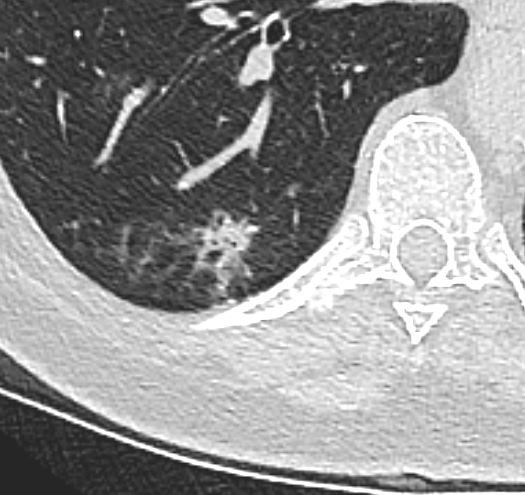
Can Be More Central
Ashley Davidoff MD TheCommonVein.net
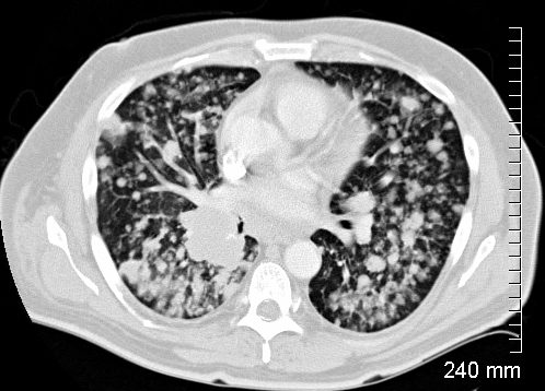
MULTICENTRIC
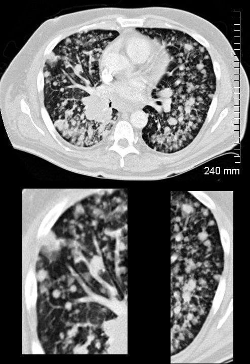
Adenocarcinoma as an Consolidation Focal and Segmental
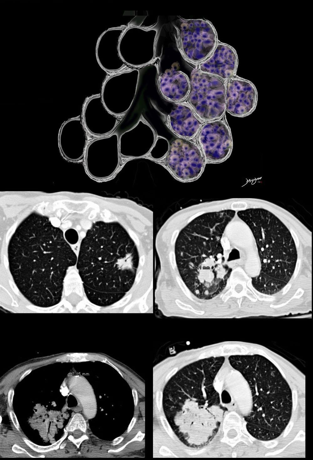
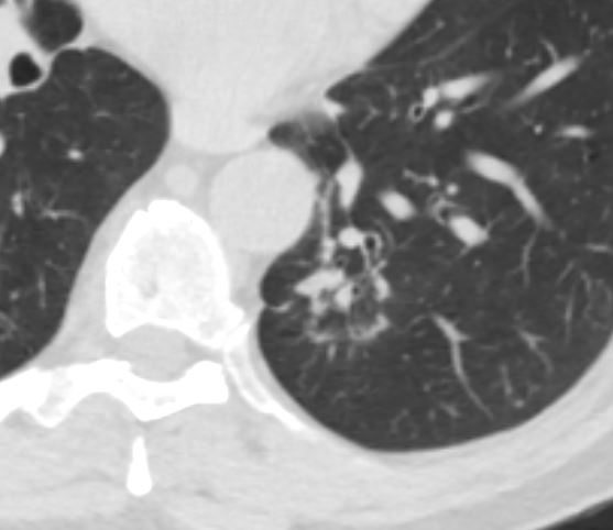
The focal nodules (middle row) and segmental consolidation in the right upper lobe (lower row) in this case is caused by total filling of the alveoli with malignant cells. This results in opacification of the alveoli and the “white” density in contrast to the “black” airways, enable the airways to be visualised as air bronchograms
Ashley Davidoff MD TheCommonVein.net 87770c01
Character
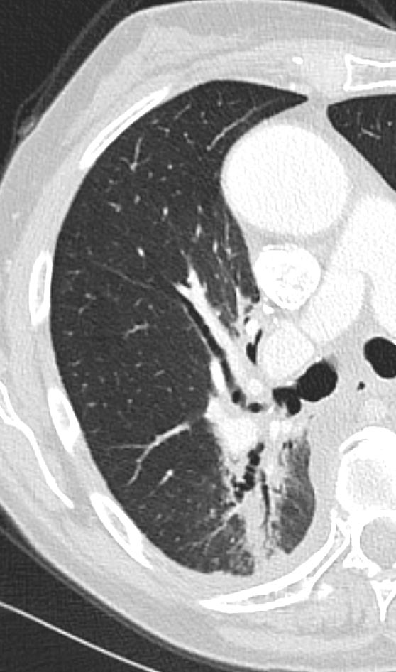
GGO
Ground Glass Nodule
Adenocarcinoma with Lepidic Growth
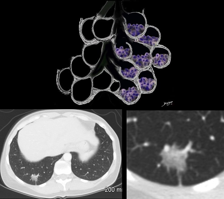
The Ground Glass Opacity (GGO) in this case is caused by partial filling of the alveolus with malignant cells Ground glass opacification may be caused by partial filling of the alveolus with cellular material resulting in partial replacement of air with solid material. The net density is gray rather than white in the situation where the alveolus is fully replaced with cells or fluid. There is blending of the black of the subtending airways and the white of the vessels with the gray density of the cellular infiltrate and hence the normal vessels are not visualized in ground glass opacities.
Ashley Davidoff MD TheCommonVein.net 134375b01
Papillary Adenocarcinoma with Lymphatic Invasion
Ground Glass Nodule with Small Solid Components
Ashley Davidoff MD TheCommonVein.net 02 2nd site
Consolidation
Adenocarcinoma as an Consolidation Focal and Segmental
The focal nodules (middle row) and segmental consolidation in the right upper lobe (lower row) in this case is caused by total filling of the alveoli with malignant cells. This results in opacification of the alveoli and the “white” density in contrast to the “black” airways, enable the airways to be visualised as air bronchograms
Ashley Davidoff MD TheCommonVein.net 87770c01
AIR
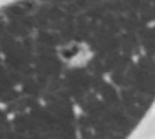
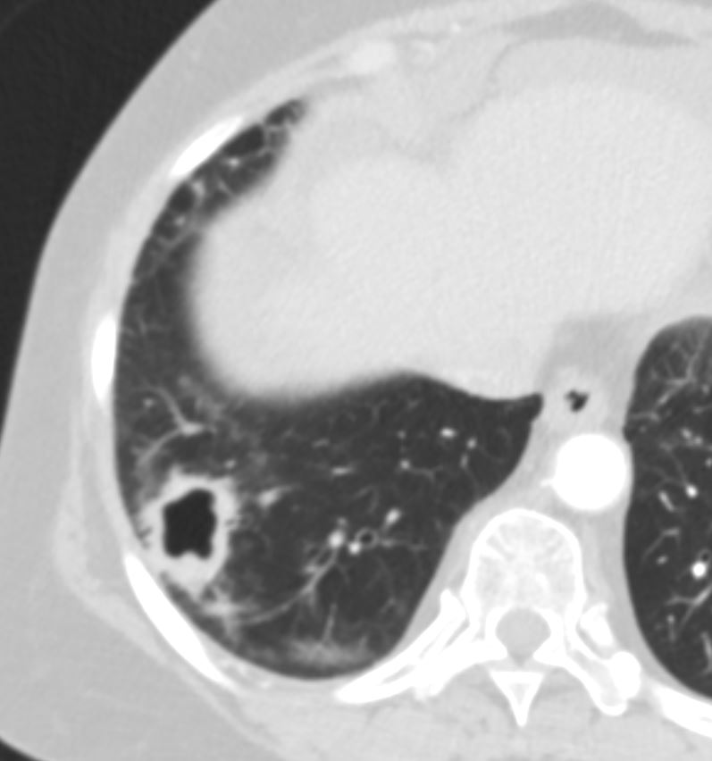
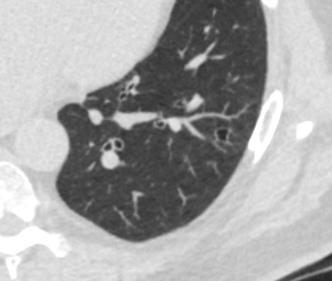
“Pseudocavitation”
Caused by Encasement of Several Bronchioles
Ashley Davidoff TheCommonVein.net adenocarcinoma-pseudocavitation-000a
Ashley Davidoff TheCommonVein.net adenocarcinoma-pseudocavitation-000b
Ashley Davidoff TheCommonVein.net adenocarcinoma-pseudocavitation-001
Ashley Davidoff TheCommonVein.net adenocarcinoma-pseudocavitation-001
Ashley Davidoff TheCommonVein.net adenocarcinoma-pseudocavitation-002
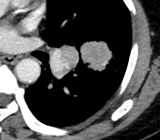
CALCIFICATION
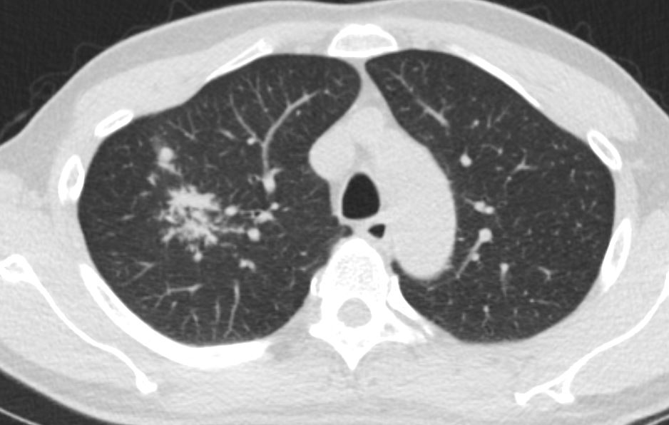
Nodule or Mass with Eccentric Calcification
Spiculated mass with eccentric calcification and nodal involvement
Ashley Davidoff MD TheCommonVein.net adenocarcinoma 001
Spiculated mass with eccentric calcification and nodal involvement
Ashley Davidoff MD TheCommonVein.net adenocarcinoma 001b
Time
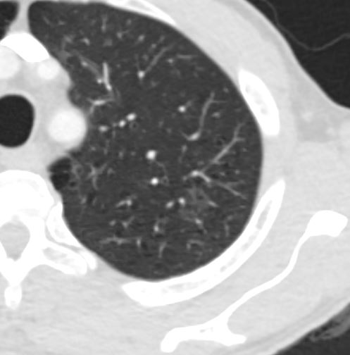
Time 1 Year
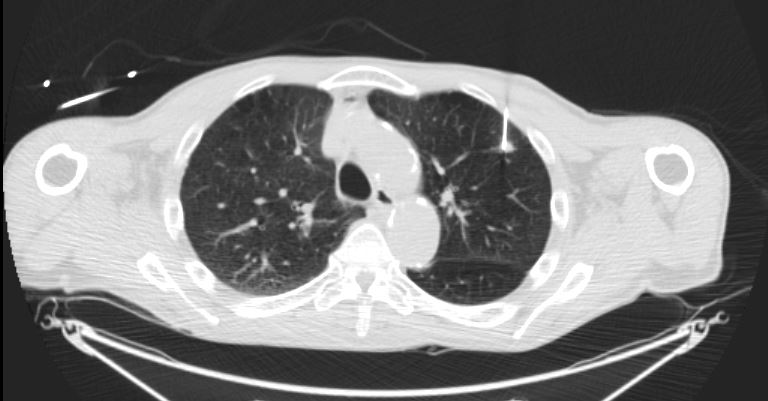
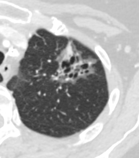
1 Year Prior Negative
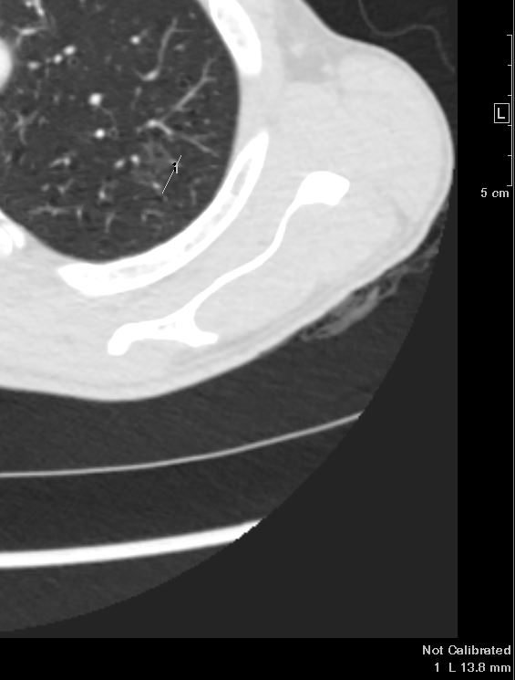
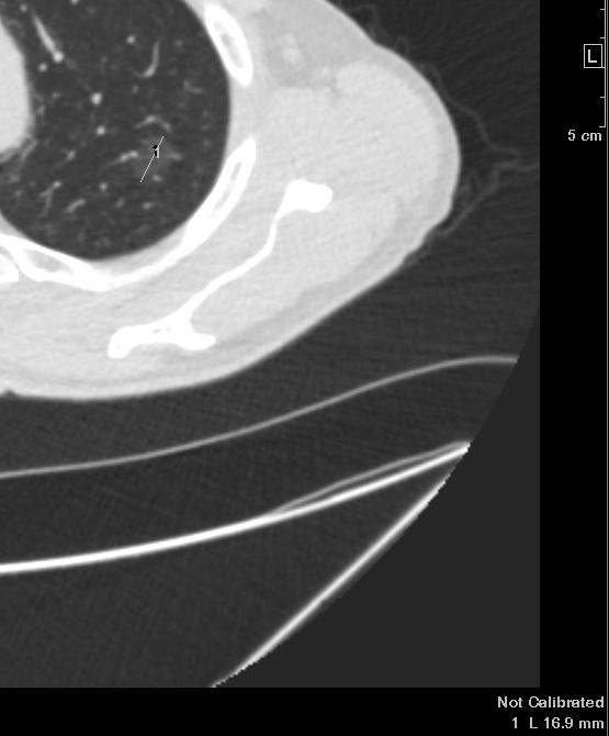
1 Year Later 8mm Irregular Nodule
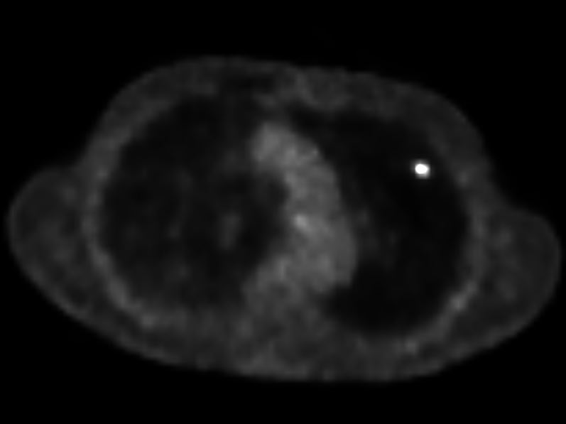
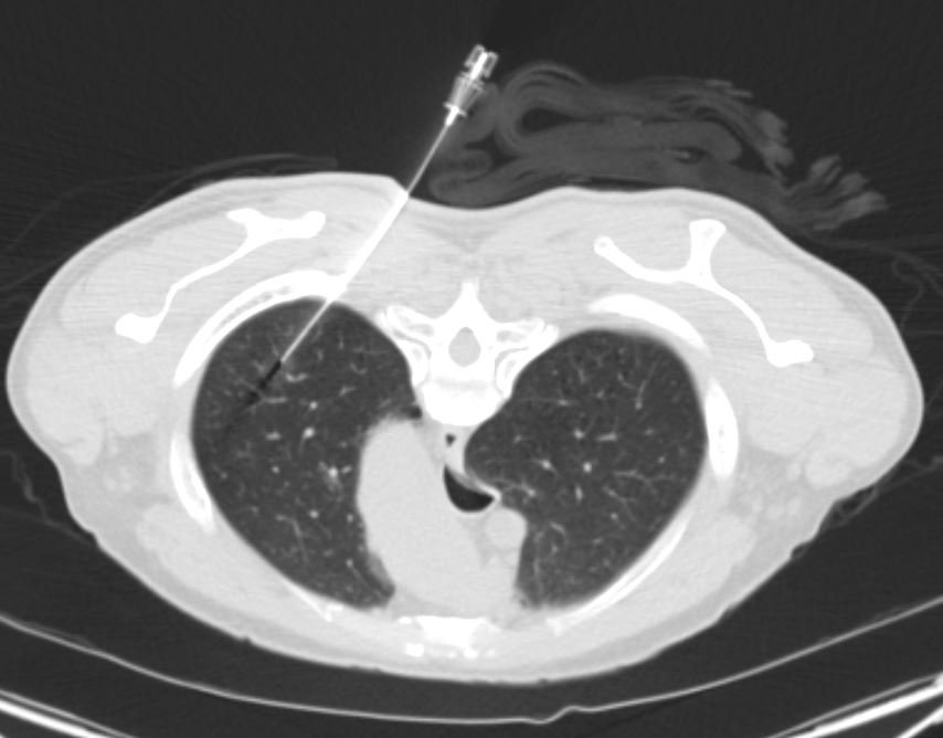
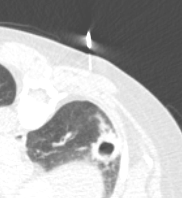
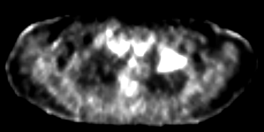
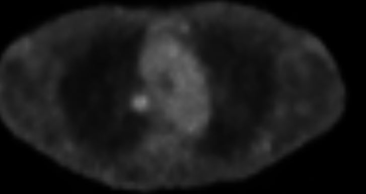
PET scan
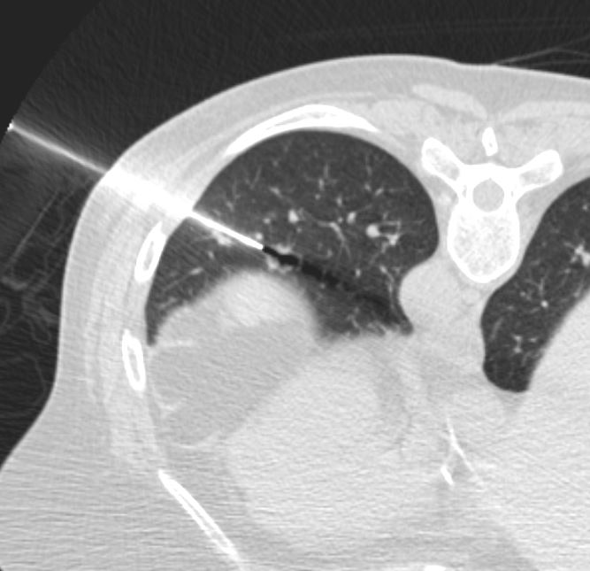
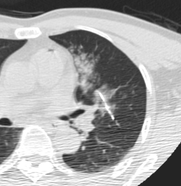
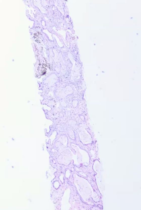
Biopsy and Placement of Fiduciary Markers
Spiculated Spiculated Nodule Another Case Over Time
Small Smooth Peripheral Nodule with 1 year Growth
Associated Findings
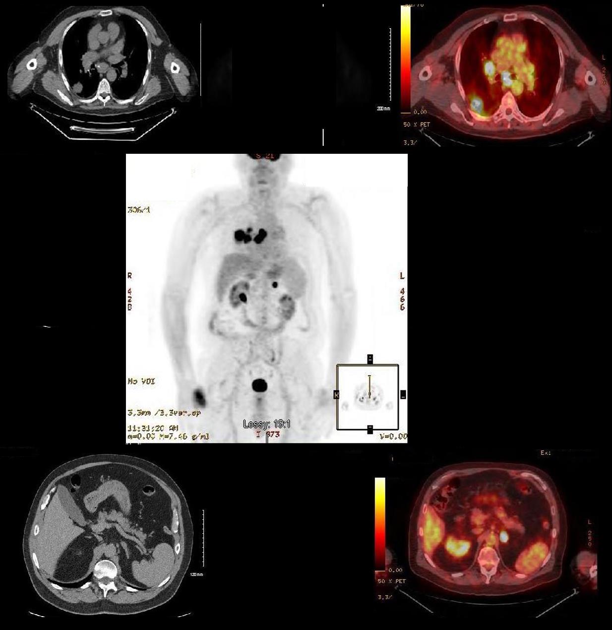
Satellite Nodule
Spiculated Consolidation and a Satellite Nodule
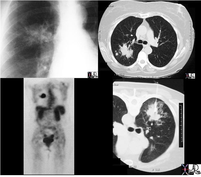
The plain film (top left) and CT (top right, bottom right) features of this mass in a 66-year-female who is a smoker are concerning, with size being greater than 3cm, and the shape having spiculated borders and satellite nodules seen in the top right image. The PET scan (bottom left) was positive and biopsy confirmed the diagnosis. The (bottom right) was performed during a CT biopsy and the patient is lying prone.
Ashley Davidoff, M.D. TheCommonVein.net Lung cancer P 036
Spiculated mass with eccentric calcification and nodal involvement
Ashley Davidoff MD TheCommonVein.net adenocarcinoma 001c
Adenocarcinoma with Lepidic Growth
Adenocarcinoma as an Consolidation Focal and Segmental
The focal nodules (middle row) and segmental consolidation in the right upper lobe (lower row) in this case is caused by total filling of the alveoli with malignant cells. This results in opacification of the alveoli and the “white” density in contrast to the “black” airways, enable the airways to be visualised as air bronchograms
Ashley Davidoff MD TheCommonVein.net 87770c01
Ashley Davidoff TheCommonVein.net
Minimal Changes in a GGO Adenocarcinoma with Lepidic Growth
1 year prior
Ashley Davidoff MD
TheCommonVein.net
1 year prior
Ashley Davidoff MD
TheCommonVein.net
Current
Ashley Davidoff MD
TheCommonVein.net
Current

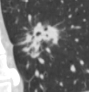
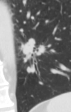
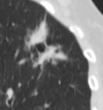
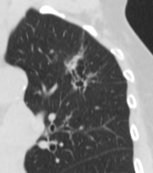
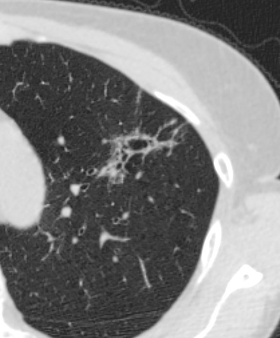
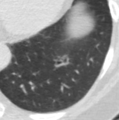
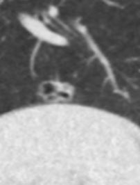
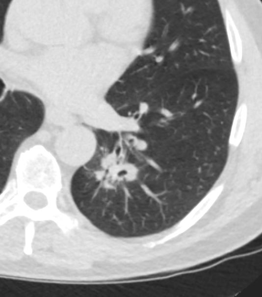
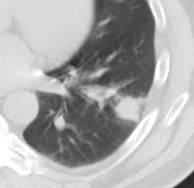
Bronchocentric or Pseudocavitation
Adenocarcinoma Acinar Predominant
The mass seems to have incorporated and constricted several small airways
Ashley Davidoff MD TheCommonVein.net 78f 001
The mass seems to have incorporated and constricted several small airways
Ashley Davidoff MD TheCommonVein.net 78f 002
The mass seems to have incorporated and constricted several small airways
Ashley Davidoff MD TheCommonVein.net 78f 003
Ashley Davidoff MD TheCommonVein.net
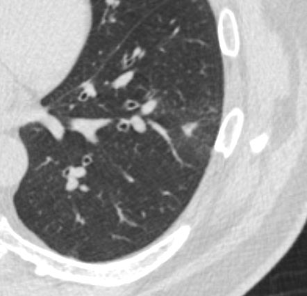
Moderately differentiated Adenocarcinoma with Bronchiectasis
Ashley Davidoff MD The CommonVein.net adenocarcinoma 01 CT
Ashley Davidoff MD The CommonVein.net adenocarcinoma 02 CT
Ashley Davidoff MD The CommonVein.net adenocarcinoma 03 CT
Ashley Davidoff MD The CommonVein.net adenocarcinoma 04 CT
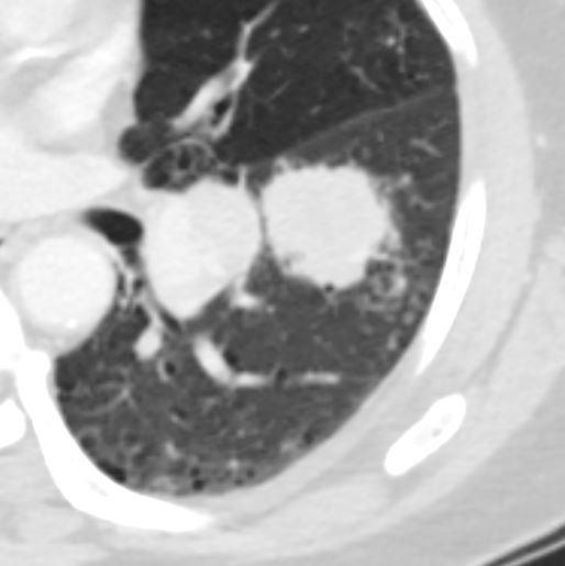
Bubble Lucencies – Pseudocavitation
Cavitating Squamous Cell Carcinoma
65 year male with peripheral lung nodule characterized by cavitation that was not present 2 years earlier . Pathology revealed squamous cell carcinoma
Ashley Davidoff
TheCommonVein.net
Ashley Davidoff MD TheCommonVein.net
Ashley Davidoff MD TheCommonVein.net
Ashley Davidoff MD TheCommonVein.net
Ashley Davidoff MD TheCommonVein.net
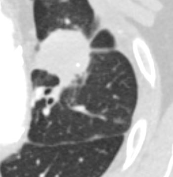
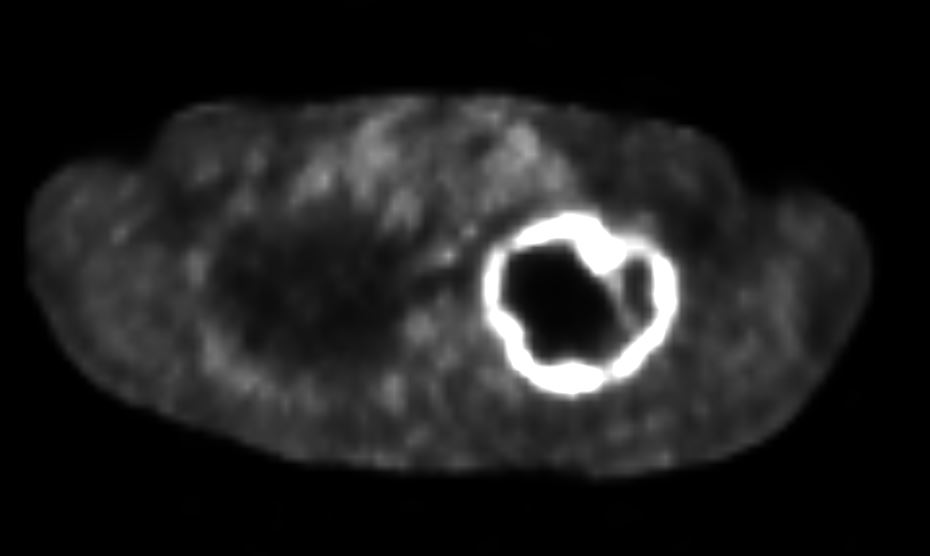
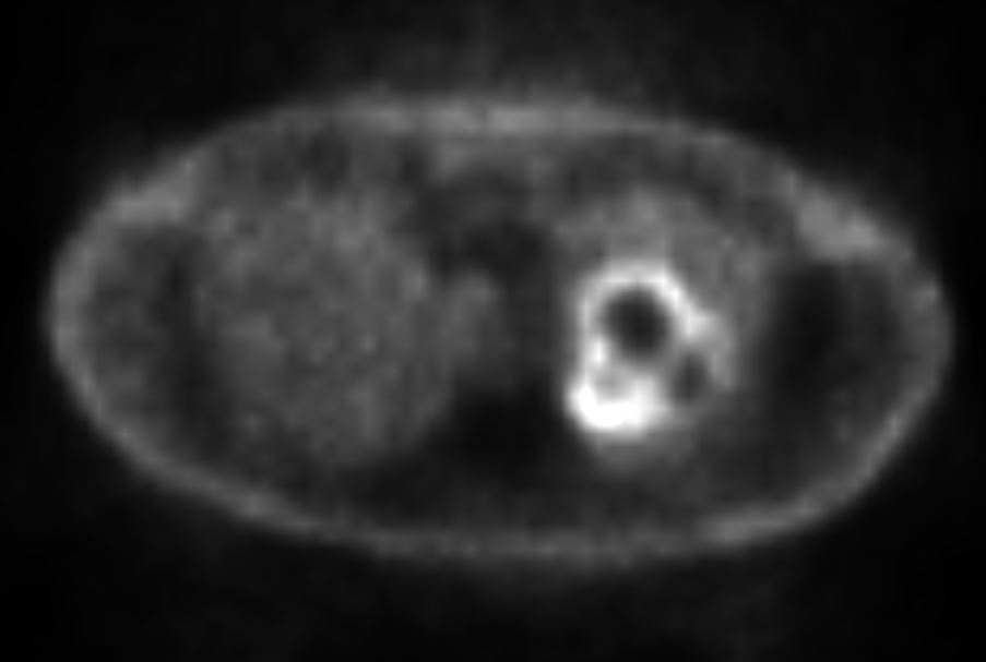
Adenocarcinoma with “Pseudocavitation” Minimal PET Activity
Ashley Davidoff TheCommonVein.net pseudocavitation-001-CT
Ashley Davidoff TheCommonVein.net pseudocavitation-002-CT
Bronchovascular Infiltrate
Adenocarcinoma
Atypical presentation along the bronchovascular bundle
Ashley Davidoff TheCommonVein.net
Extreme Form of Pseudocavitation
Current Invasive-Adenoca
Adenocarcinoma Presenting as an Infiltrate
Ashley Davidoff MD TheCommonVein.net
adenocarcinoma 79M
Hard to See Lesion Next to Vessels
Ashley Davidoff MD TheCommonVein.net
Ashley Davidoff MD TheCommonVein.net
Positive PET Scan
Necrotic Mass
Diagnosis – adenocarcinoma of the lung with extensive necrosis of the tumor
Ashley Davidoff MD The CommonVein.net
Diagnosis – adenocarcinoma of the lung with extensive necrosis of the tumor
Ashley Davidoff MD The CommonVein.net
Diagnosis – adenocarcinoma of the lung with extensive necrosis of the tumor
Ashley Davidoff MD The CommonVein.net
Diagnosis – adenocarcinoma of the lung with extensive necrosis of the tumor
Ashley Davidoff MD The CommonVein.net
Cavitating Well Differentiated Mucinous Adenocarcinoma
The mass was not present on this image taken 2 years prior. Patient had a history of TB Lesion was biopsied and showed well differentiated mucinous adenocarcinoma
Ashley Davidoff MD TheCommonVein.net adencacinoma-005
Patient had a history of TB Lesion was biopsied and showed well differentiated mucinous adenocarcinoma
Ashley Davidoff MD TheCommonVein.net adencacinoma-0046 biopsy
Patient had a history of TB Lesion was biopsied and showed well differentiated mucinous adenocarcinoma
Ashley Davidoff MD TheCommonVein.net adenocarcinoma biopsy
Cystic Adenocarcinoma
Presenting as Multiple Nodules
Adenocarcinoma as Multiple
Well Defined Solid Multicentric Nodules
Ashley Davidoff TheCommonVein.net 134362
Adenocarcinoma as Multiple
Well Defined and Poorly Defined Solid Multicentric Nodules
Ashley Davidoff MD TheCommonVein.net 134336
Some of the Nodules are Centrilobular
The lower panels demonstrate centrilobular distribution of someof the nodules
Ashley Davidoff MD
TheCommonVein.net
134336c
Following Treatment
Post Radiation
Radiation Pneumonitis with bronchiectasis 3 months post treatment. Note the straight lateral border inkeeping with the path of the radiation beam
Ashley Davidoff MD TheCommonVein.net post XRT 001
Recurrence Along Surgical Margins
Alongside a scar
Ashley Davidoff MD TheCommonvein.ne
Cancer Look Alikes
5 months prior
Ashley Davidoff MD TheCommonVein.net
3 months prior
Ashley Davidoff MD TheCommonVein.net
Current
Ashley Davidoff MD TheCommonVein.net
TB
Ashley Davidoff TheCommonVein.net
After TB Treatment
Ashley Davidoff TheCommonVein.net
Metastatic Disease to Adrenal and Brain
Ashley Davidoff TheCommonVein.net 292Lu 121892
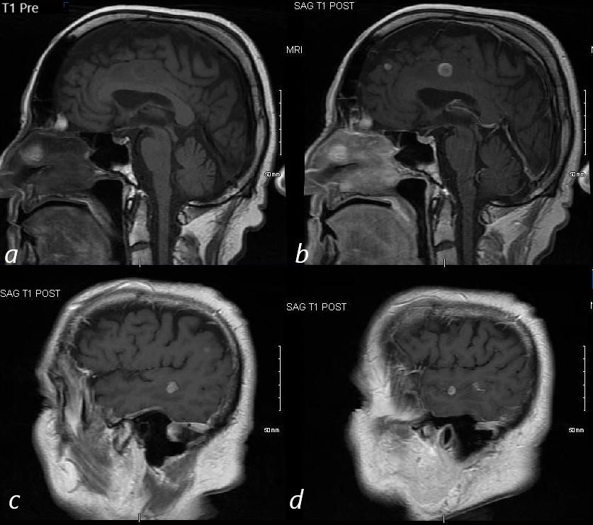
MRI of an 80 year old male with primary adenocarcinoma of the lung show metastatic disease to the brain. I mage a is a non contrast T1 weighted image prior to contrast administration. Images b, c ad are sagittal images of the brain following gadolinium administration and shows multiple enhancing lesions in the frontal parietal and temporal lobes consistent with metastatic disease
Ashley Davidoff MD TheCommonVein.net 292Lu 121893cL