
Parts
Size
Small
Ashley Davidoff MD TheCommonVein.net Squamous Cell carcinoma 001
Ashley Davidoff MD TheCommonVein.net Squamous Cell carcinoma 002
Large
Ashley Davidoff MD TheCommonVein.net
Shape
Wedge Peripheral
Peripheral Squamous Cell Carcinoma that
Looked Like and Infiltrate
Triangular Shaped
Final diagnosis Squamous Cell Carcinoma
Ashley Davidoff MD TheCommonVein.net
Wedge Post Obstructive
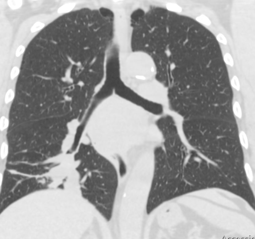
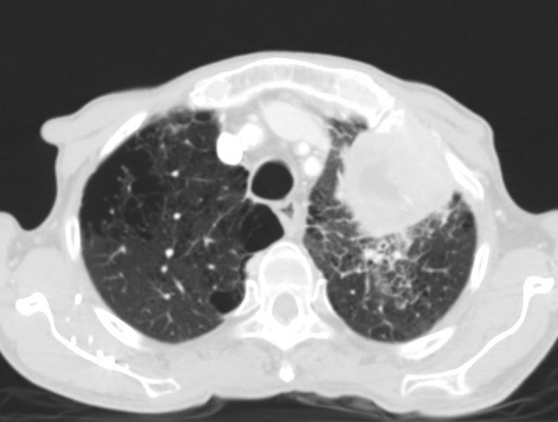
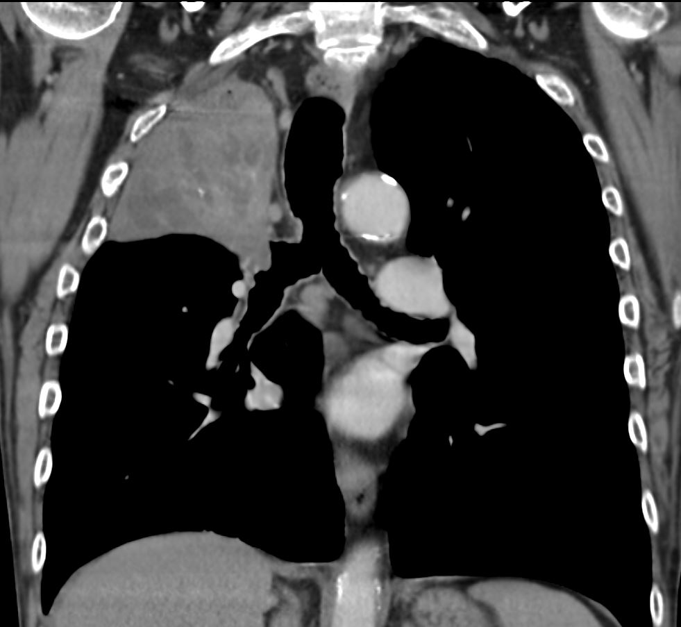
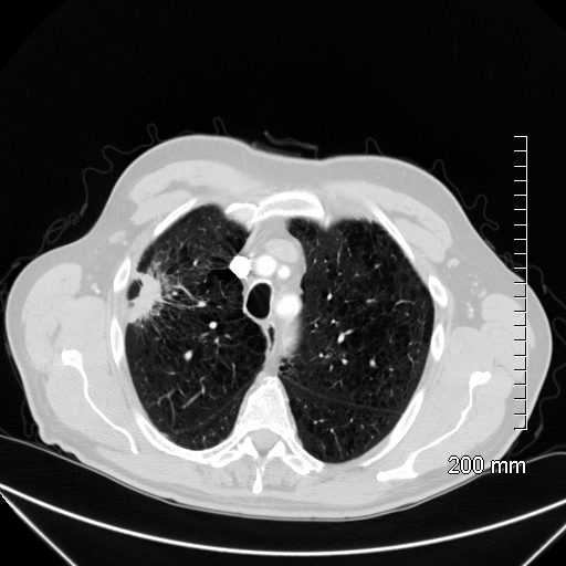
55-year-old male presenting with dyspnea
Axial CT at the level of the carina shows atelectasis of the RUL caused by a central obstructing lesion in the right upper lobe bronchus resulting in atelectasis of the RUL characterized by a wedge-shaped consolidation of the anteriorly positioned right upper lobe. The major fissure is displaced anteriorly. There is extensive filling of the distal bronchiectatic segmental and subsegmental airways of the RUL. Final diagnosis was a central RUL proximal squamous cell carcinoma.
Ashley Davidoff TheCommonVein.net 212Lu 136432
Position
Central

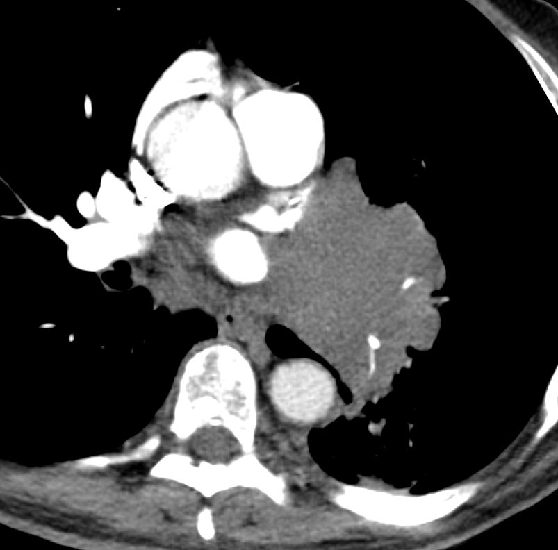
PET scan shows endobronchial mass within the
right mainstem bronchus with intense FDG uptake, corresponding to
biopsy-proven squamous cell carcinoma.
Ashley Davidoff MD TheCommonVein.net
Character
Cavitating
Pseudocavitation
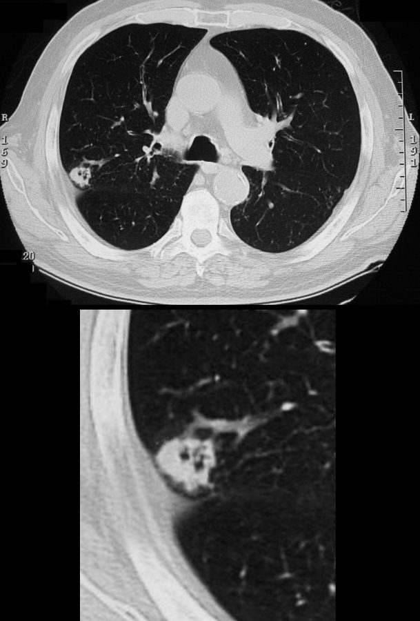
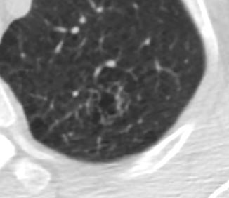
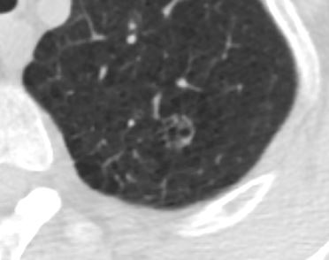
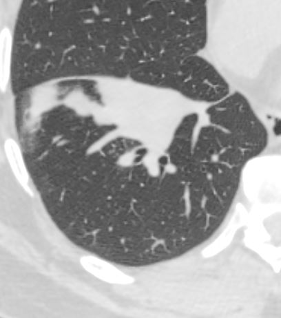
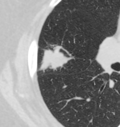
65 year male with peripheral lung nodule characterized by cavitation that was not present 2 years earlier . Pathology revealed squamous cell carcinoma
Ashley Davidoff
TheCommonVein.net
Final diagnosis Squamous Cell Carcinoma
Ashley Davidoff MD TheCommonVein.net
Heterogeneous Liquefaction
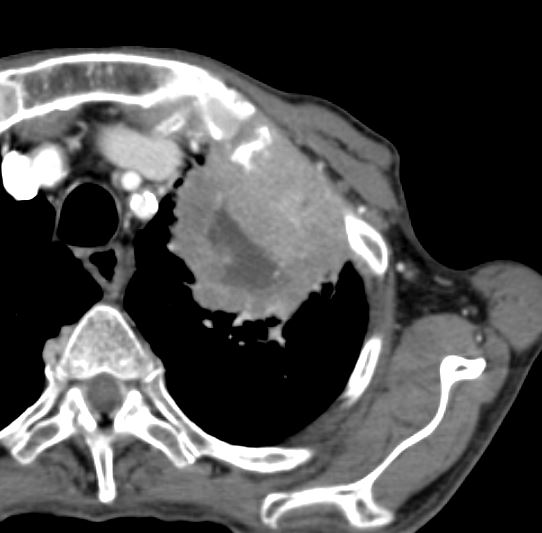
Ashley Davidoff MD TheCommonVein.net
Ashley Davidoff MD TheCommonVein.net
Necrotic Squamous Cell Carcinoma
52 year old male with known squamous cell carcinoma of the lung
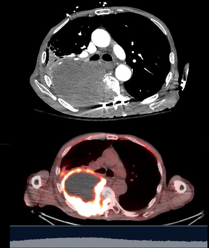
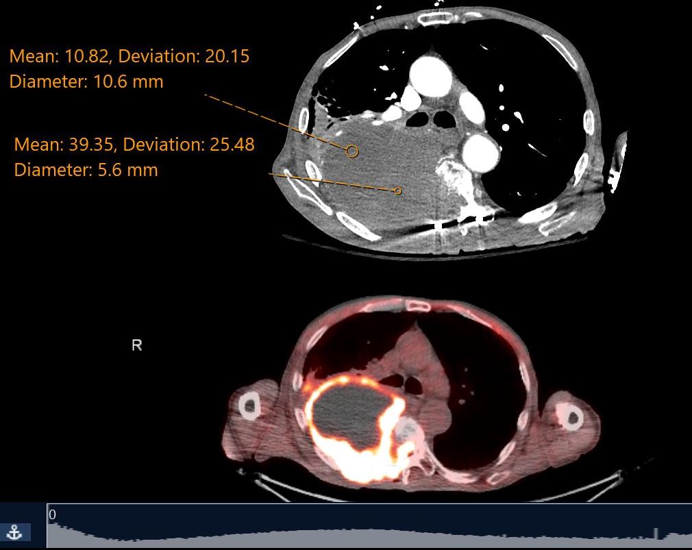
CT scan in the axial projection shows a large low density mass in the right upper lobe with invasion into the neural foramen of the abutting vertebral body. Antero-laterally, the mass is low density and postero-medially it is slightly higher density. PET CT shows a rind of intense activity surround the necrotic center and invading the vertebra. Findings are consistent with a squamous cell carcinoma
Ashley Davidoff MD TheCommonVein.net 136490
52 year old male with known squamous cell carcinoma of the lung
CT scan in the axial projection shows a large low density mass in the right upper lobe with invasion into the neural forman of the abutting vertebral body. Antero-laterally, the mass is low density (11 HU) and postero-medially it is slightly higher density ( 39HU). PET CT shows a rind of intense activity surround ythe necrotic center and invading the vertebra. Eindings are consistent with a aquamous cell carcinoma
Ashley Davidoff MD TheCommonVein.net 136489
Time
Starting
6 Months Prior left Upper Lobe Complex Cystic Lesion Pseudocavitation
Final diagnosis Squamous Cell Carcinoma
Ashley Davidoff MD TheCommonVein.net
Growth of Cystic Lesion Over 6months
Final diagnosis Squamous Cell Carcinoma
Ashley Davidoff MD TheCommonVein.net
PET CT shows Hyperintense Lesion
Despite Lack of Soft Tissue Component
Final diagnosis Squamous Cell Carcinoma
Ashley Davidoff MD TheCommonVein.net
Significant Progression of Soft Tissue Growth
2 Months Later
Prior to Biopsy
Final diagnosis Squamous Cell Carcinoma
Ashley Davidoff MD TheCommonVein.net
Associated Findings
Bronchiectasis and Bronchial Invasion
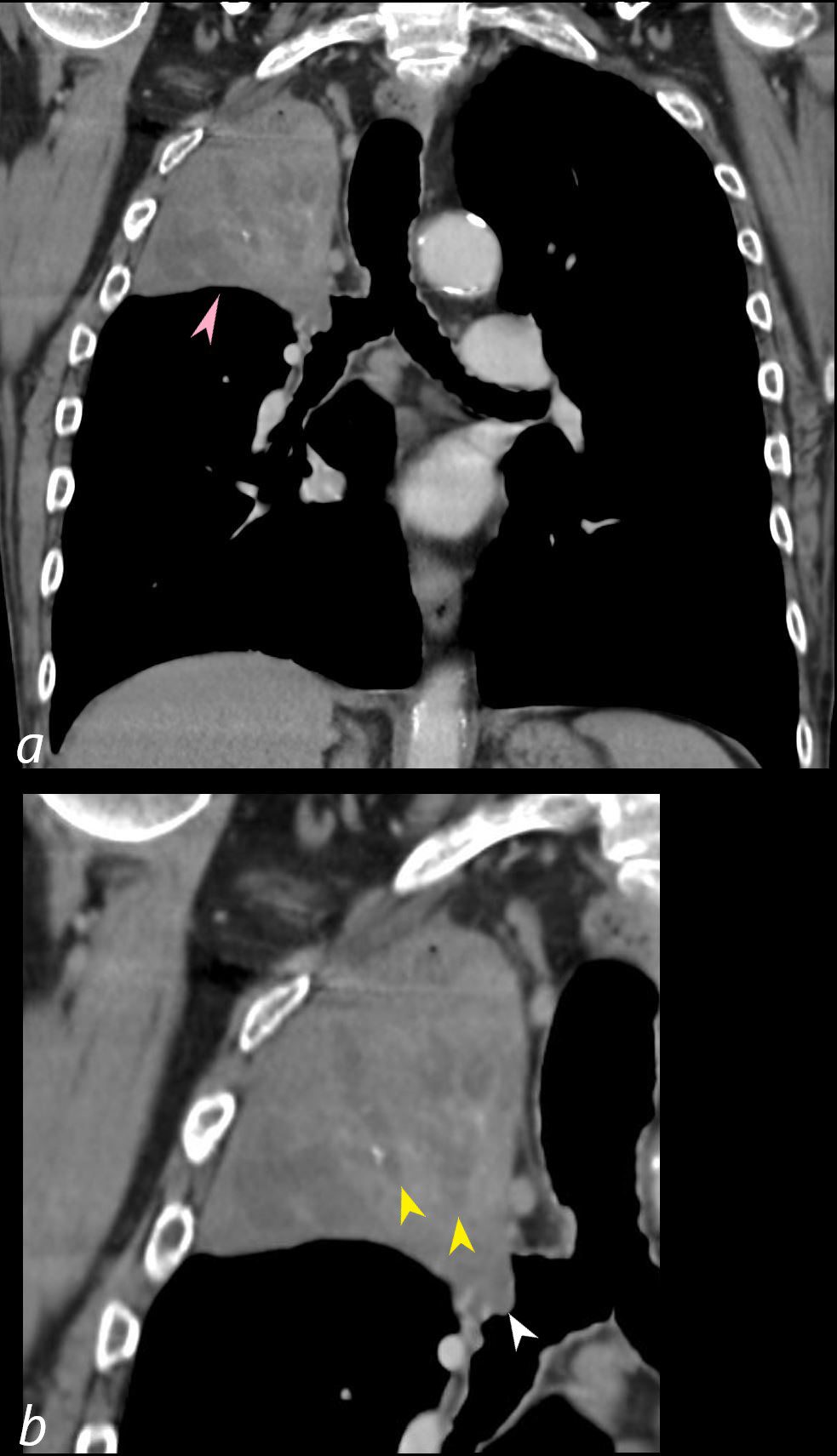
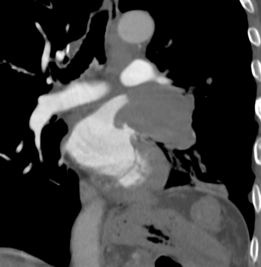
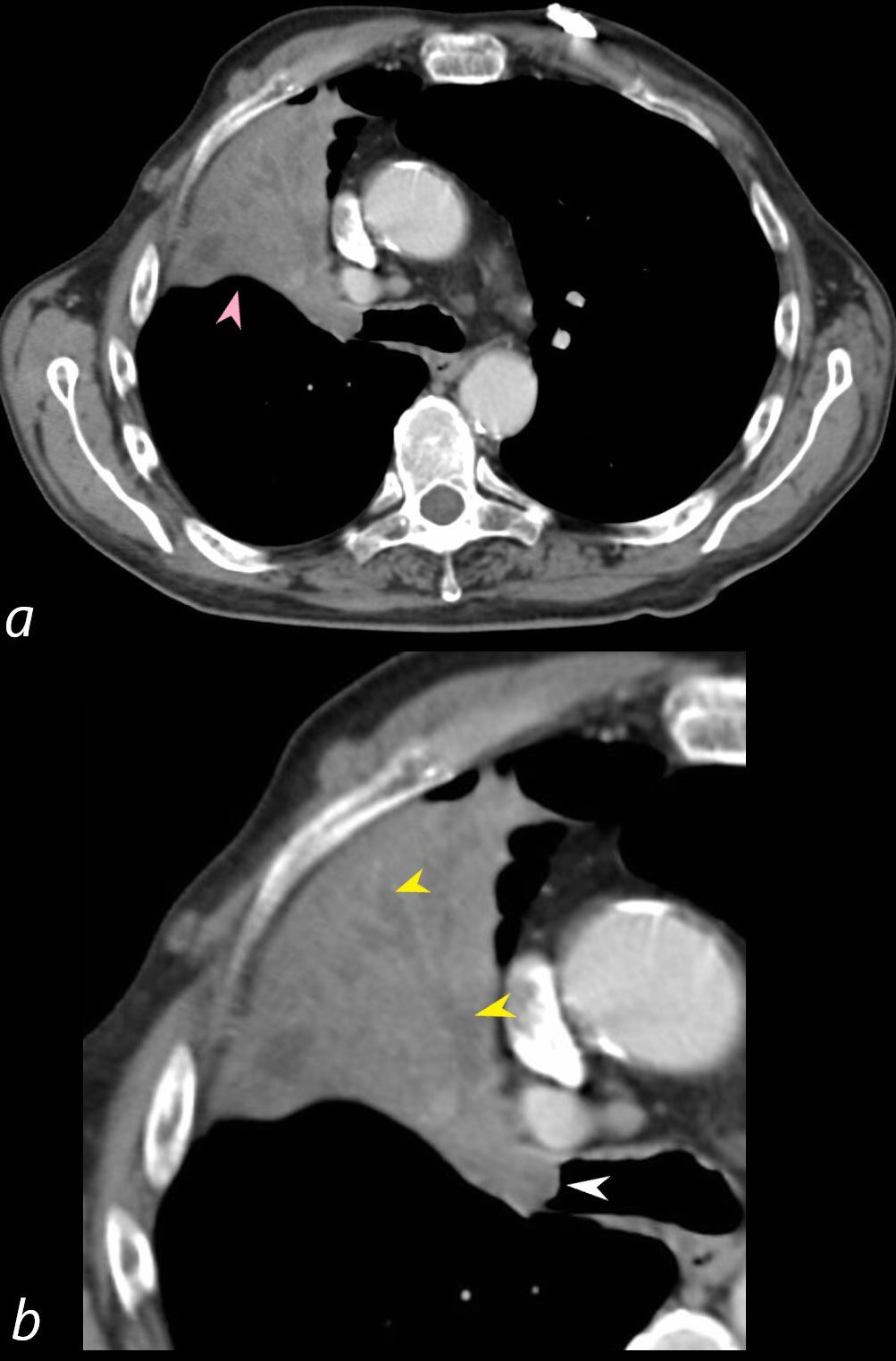
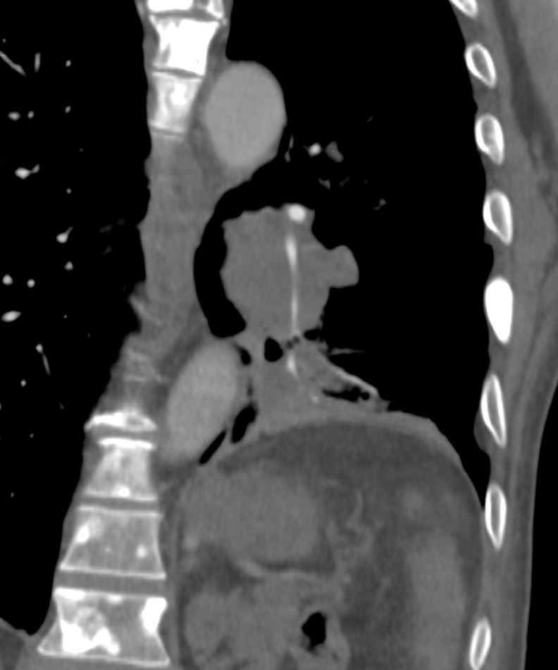
55-year-old male presenting with dyspnea
Coronal CT at the level of the trachea and mainstem bronchi, shows atelectasis of the RUL caused by a central obstructing lesion in the right upper lobe bronchus (b, white arrowhead) resulting in atelectasis of the RUL characterized by a wedge-shaped consolidation of the right upper lobe with superiorly displaced major fissure (a, pink arrowhead). There is extensive filling of the distal bronchiectatic segmental and subsegmental airways of the RUL (b, yellow arrowheads). Final diagnosis was a central RUL proximal squamous cell carcinoma.
Ashley Davidoff TheCommonVein.net 212Lu 136433cL
Squamous Cell Carcinoma Masquerading as ABPA
56-year-old male presents with chronic cough dyspnea and weight loss. CT scan in coronal projection shows an appearance reminiscent of finger in glove in the right lower lobe. There s segmental and subsegmental thickening of the airways in the upper lobes, and paraseptal emphysema. Micronodules in the upper lobes suggest smoker’s bronchiolitis. The subcarinal esophageal mass was diagnosed as a leiomyoma, Pathology of the right lower process was a squamous cell carcinoma
Ashley Davidoff MD TheCommonVein.net 267Lu 136219
56-year-old male presents with chronic cough dyspnea and weight loss. CT scan in axial projection shows an appearance reminiscent of finger in glove in the right lower lobe. There s a para-fissural soft tissue mass that seems “soft” since it does not displace nor deform the fissure.. Pathology of the right lower process was a squamous cell carcinoma
Ashley Davidoff MD TheCommonVein.net 267Lu 136221
Lymphangitis Carcinomatosa
Ashley Davidoff MD TheCommonVein.net
Occluded Veins
Ashley Davidoff MD TheCommonVein.net occluded-pulm-vein-001
Squamous Cell Carcinoma
Central
PET scan shows endobronchial mass within the
right mainstem bronchus with intense FDG uptake, corresponding to
biopsy-proven squamous cell carcinoma.
Ashley Davidoff MD TheCommonVein.net
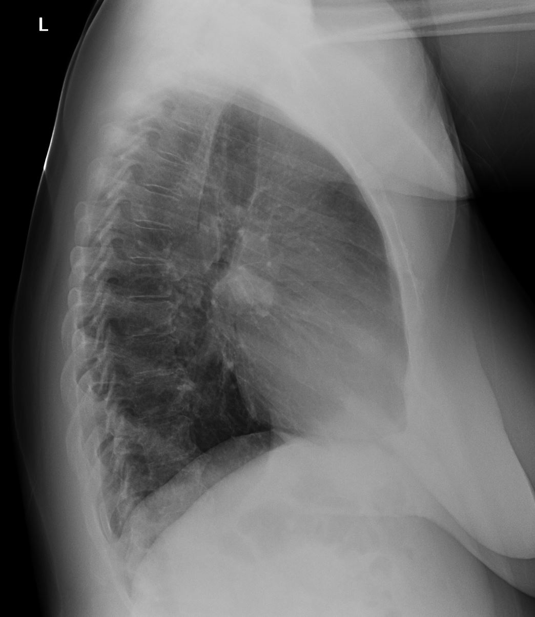
CXR
Ashley Davidoff MD TheCommonVein.net
Ashley Davidoff MD TheCommonVein.net
Ashley Davidoff MD TheCommonVein.net
Ashley Davidoff MD TheCommonVein.net
Ashley Davidoff MD TheCommonVein.net
Cavitating
Lower Lobe
Ashley Davidoff MD TheCommonVein.net squamous cell carcinoma cavitating 002
Poorly Differentiated
Ashley Davidoff MD TheCommonVein.net
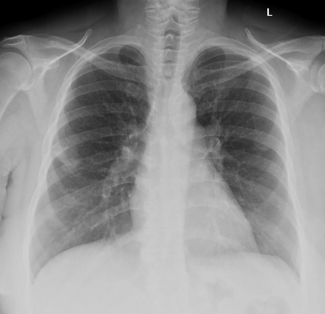
Central with Post Obstructive Atelectasis
55-year-old male presenting with dyspnea
Axial CT at the level of the carina shows atelectasis of the RUL caused by a central obstructing lesion in the right upper lobe bronchus (b, white arrowhead) resulting in atelectasis of the RUL characterized by a wedge-shaped consolidation of the anteriorly positioned right upper lobe. The major fissure is displaced anteriorly (a, pink arrowhead). There is extensive filling of the distal bronchiectatic segmental and subsegmental airways of the RUL (b, yellow arrowheads). Final diagnosis was a central RUL proximal squamous cell carcinoma.
Ashley Davidoff TheCommonVein.net 212Lu 136432cL
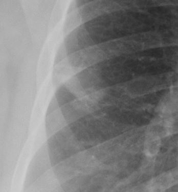
55-year-old male presenting with dyspnea
Coronal CT at the level of the trachea and mainstem bronchi, shows atelectasis of the RUL caused by a central obstructing lesion in the right upper lobe bronchus resulting in atelectasis of the RUL characterized by a wedge-shaped consolidation of the right upper lobe with superiorly displaced major fissure. There is extensive filling of the distal bronchiectatic segmental and subsegmental airways of the RUL. Final diagnosis was a central RUL proximal squamous cell carcinoma.
Ashley Davidoff TheCommonVein.net 212Lu 136433
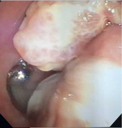
Endoscopic image of a central squamous cell carcinoma (SCC) with extensive
Ashley Davidoff TheCommonVein.net 212Lu 136434
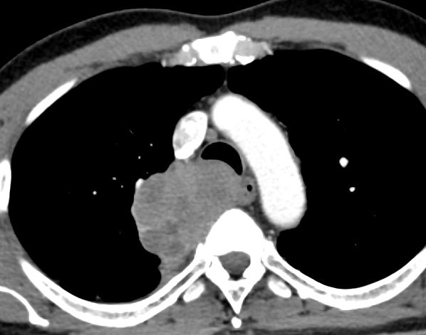
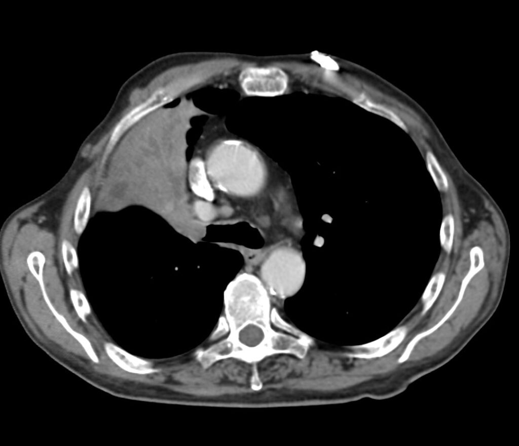
Large Central Mass with Obstruction of the Pulmonary Vein and Encasement of the Arteries – Squamous Cell Carcinoma
Ashley Davidoff MD TheCommonVein.net occluded-pulm-vein-005
Occluded Pulmonary Vein
Ashley Davidoff MD TheCommonVein.net occluded-pulm-vein-001
Encased Pulmonary Artery
Ashley Davidoff MD TheCommonVein.net occluded-pulm-vein-003
Encased Pulmonary Artery

Ashley Davidoff MD TheCommonVein.net occluded-pulm-vein-001
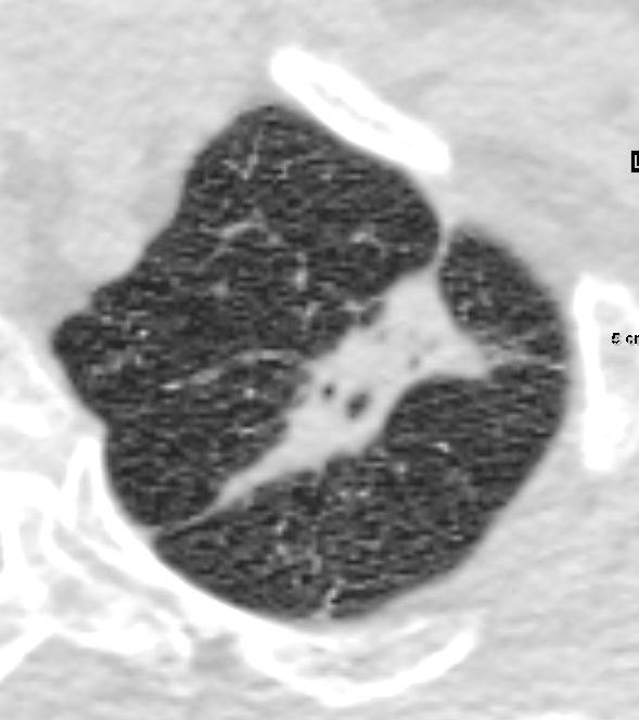
Peripheral with Cavitation
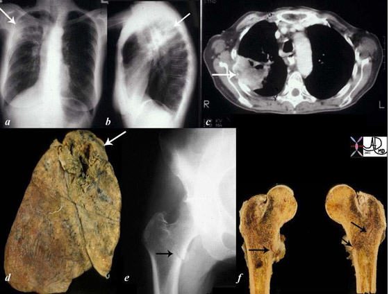
The collage of images reflects a patient with stage IV, cavitating, primary, squamous carcinoma of the right upper lobe (RUL) (a, b, c, d – white arrows) with COPD. A metastatic lesion to the right femur was complicated by a pathological fracture. (e, f black arrows).
Courtesy Ashley Davidoff, M.D. TheCommonVein.net Lung cancer P 018
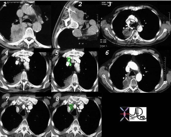
This is a case of poorly differentiated non small cell carcinoma presenting as a large necrotic mass, with a percutaneous biopsy (2) treated with radiation therapy (3), with associated small nodes (4) overlaid in green, (5) with a response as seen by shrinkage of the tumor 4 months later (6) as well shrinkage of the nodes (7, 8).
Ashley Davidoff, M.D. TheCommonVein.net Lung cancer P 037b
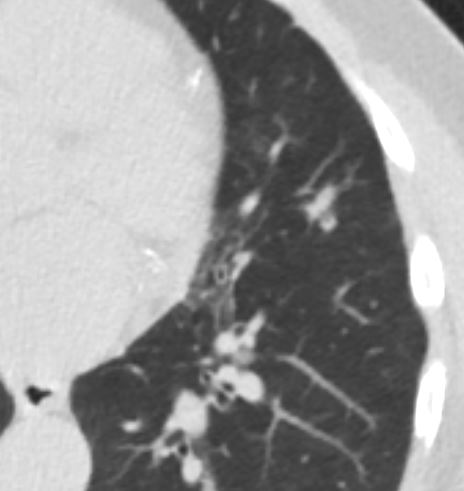
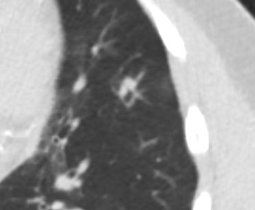
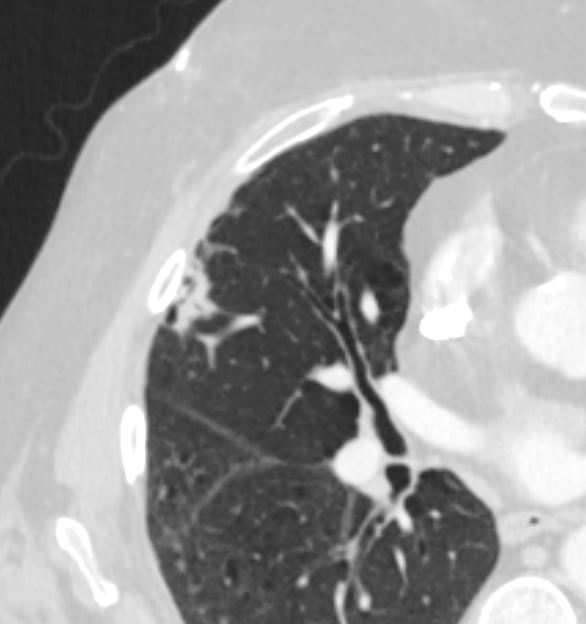
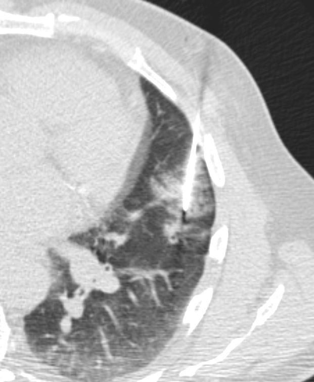
Peripheral Squamous Cell Carcinoma
Ashley Davidoff TheCommonVein.net
Small Peripheral Growth Over 5 Months
5 Months Prior
Ashley Davidoff MD TheCommonVein.net Squamous Cell carcinoma 001
Ashley Davidoff MD TheCommonVein.net Squamous Cell carcinoma 002
Ashley Davidoff MD TheCommonVein.net Squamous Cell carcinoma 004
Ashley Davidoff MD TheCommonVein.net Squamous Cell carcinoma 005
Ashley Davidoff MD TheCommonVein.net Squamous Cell carcinoma 006
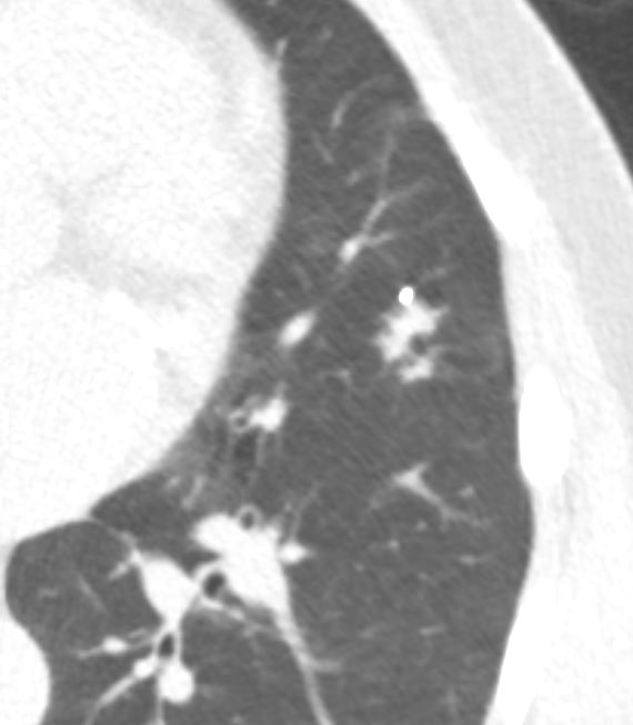
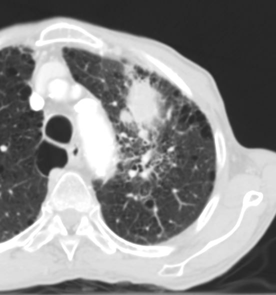
Peripheral Squamous Cell Carcinoma that Looked Like and Infiltrate
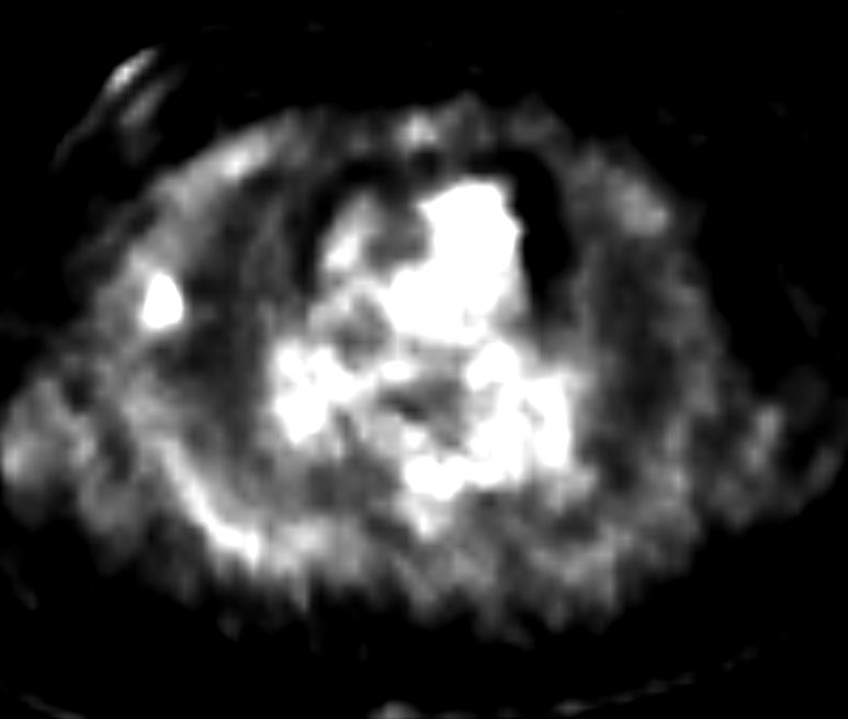
PET Positive Peripheral Parenchymal infiltrate in the lung
Biopsy confirmed the presence of a squamous cell carcinoma.
Ashley Davidoff MD TheCommonVein.net
Ashley Davidoff MD TheCommonVein.net
Ashley Davidoff MD TheCommonVein.net
peripheral Mass with Lymphangitis Carcinomatosa
Ashley Davidoff MD TheCommonVein.net
Ashley Davidoff MD TheCommonVein.net
Cavitating Masses
Ashley Davidoff MD TheCommonVein.net
Necrotic Squamous Cell Carcinoma
52 year old male with known squamous cell carcinoma of the lung
CT scan in the axial projection shows a large low density mass in the right upper lobe with invasion into the neural foramen of the abutting vertebral body. Antero-laterally, the mass is low density and postero-medially it is slightly higher density. PET CT shows a rind of intense activity surround the necrotic center and invading the vertebra. Findings are consistent with a squamous cell carcinoma
Ashley Davidoff MD TheCommonVein.net 136490
52 year old male with known squamous cell carcinoma of the lung
CT scan in the axial projection shows a large low density mass in the right upper lobe with invasion into the neural forman of the abutting vertebral body. Antero-laterally, the mass is low density (11 HU) and postero-medially it is slightly higher density ( 39HU). PET CT shows a rind of intense activity surround ythe necrotic center and invading the vertebra. Eindings are consistent with a aquamous cell carcinoma
Ashley Davidoff MD TheCommonVein.net 136489
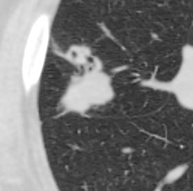
Cavitating and Spiculated
Spiculated and Cavitating Nodule
Ashley Davidoff
TheCommonVein.net
65 year male with peripheral lung nodule characterized by cavitation that was not present 2 years earlier . Pathology revealed squamous cell carcinoma
Ashley Davidoff
TheCommonVein.net
Pseudocavitation
6 Months Prior left Upper Lobe Complex Cystic Lesion
Final diagnosis Squamous Cell Carcinoma
Ashley Davidoff MD TheCommonVein.net
Growth of Cystic Lesion Over 6months
Final diagnosis Squamous Cell Carcinoma
Ashley Davidoff MD TheCommonVein.net
PET CT shows Hyperintense Lesion
Despite Lack of Soft Tissue Component
Final diagnosis Squamous Cell Carcinoma
Ashley Davidoff MD TheCommonVein.net
Significant Progression of Soft Tissue Growth
2 Months Later
Prior to Biopsy
Final diagnosis Squamous Cell Carcinoma
Ashley Davidoff MD TheCommonVein.net