
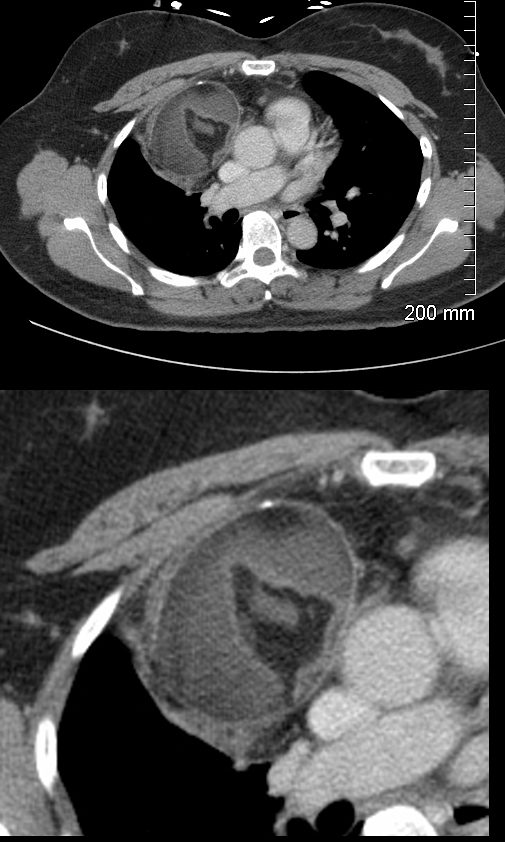
47-year-old female presents with an abnormal CXR
CT scan in the axial projection shows a 6.3X 5.9cms mass in the anterior mediastinum that shows central fatty elements, surrounded by soft tissue elements and a anterior peripheral calcification. These findings are consistent with a teratoma
Ashley Davidoff MD TheCommonVein.net 275Lu 136333c
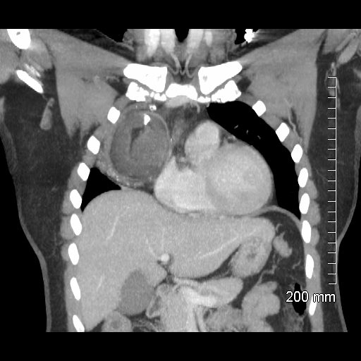
47-year-old female presents with an abnormal CXR
CT scan in the coronal projection shows a 6.3X 5.9 x 6.8cms mass in the anterior mediastinum that shows central fatty elements, surrounded by intermediate density of homogeneous soft tissue elements and a prominent calcification, and a more heterogeneous infero-lateral rim of soft tissue density with punctate calcifications.. These findings are consistent with a teratoma
Ashley Davidoff MD TheCommonVein.net 275Lu 136340
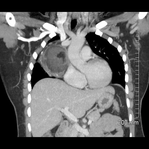
Ashley Davidoff MD TheCommonVein.net 275Lu 136341
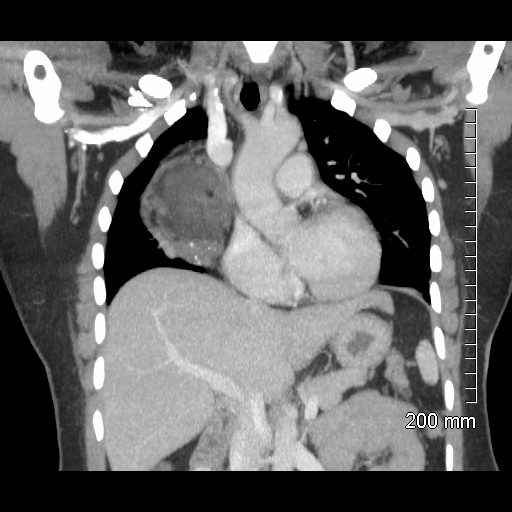
CT scan in the coronal projection more shows a complex mass in the anterior mediastinum that shows punctate fatty element, surrounded by intermediate density of homogeneous soft tissue elements, and a more heterogeneous infero-lateral rim of soft tissue density with punctate calcifications. The SVC is mildly dilated with persistence of contrast in the right subclavian vein, without collateral vessels, suggesting mild obstruction of the SVC
These findings are consistent with a teratoma
Ashley Davidoff MD TheCommonVein.net 275Lu 136342
47 year Old female presents with an abnormal CXR