
Parts
Size
Shape
Position
Character
Time Associated Findings
Infection
Inflammation
Malignancy
Small Cell Carcinoma
62-year-old female presents with acute dyspnea and chest pain
Frontal CXR shows a “white out” of the left hemithorax. The left hemidiaphragm is elevated and there is leftward mediastinal shift indicating volume loss
She was subsequently diagnosed with a small cell lung carcinoma that was obstructing the left main stem bronchus
Ashley Davidoff MD TheCommonVein.net 298Lu 136700
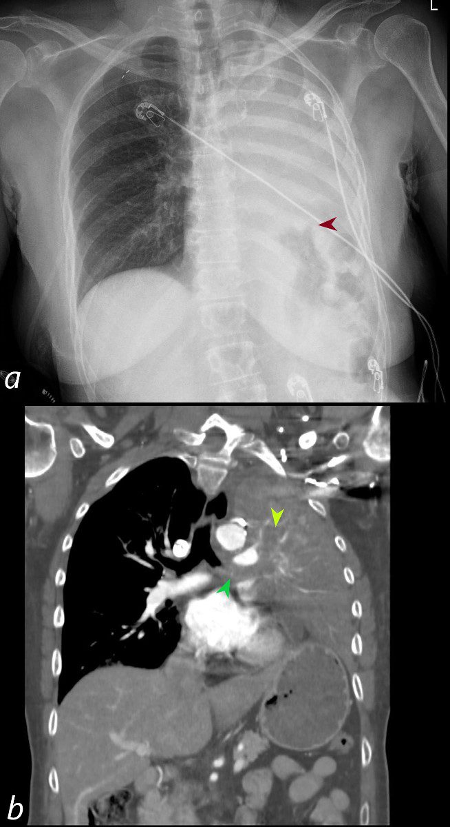
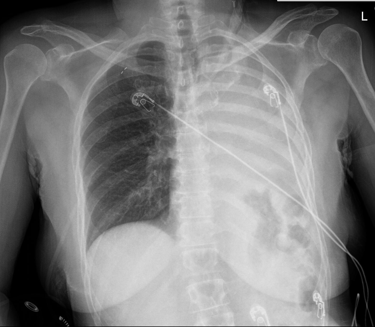
62-year-old female presents with acute dyspnea and chest pain
Frontal CXR shows a “white out” of the left hemithorax. The left hemidiaphragm is elevated (maroon arrowhead) and there is leftward mediastinal shift indicating volume loss.
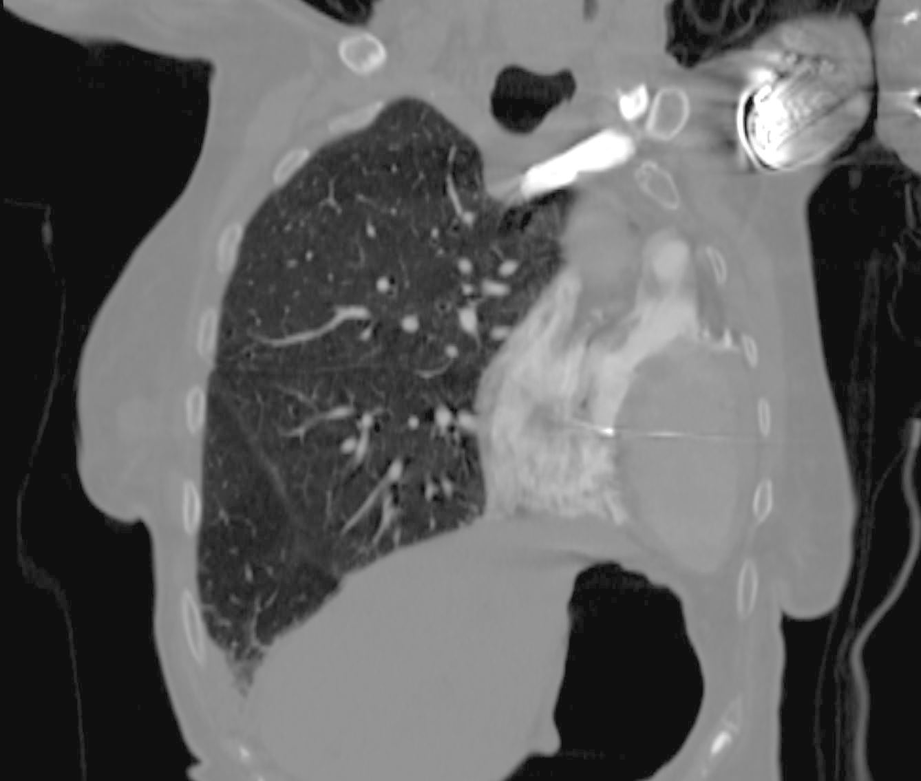
Coronal CT confirms the presence of an obstructing lesion in the left mainstem bronchus,(b, dark green arrowhead), with extension of the soft tissue into an upper lobe bronchus (b light green arrowhead). There is total collapse of the left lung and an elevated left hemidiaphragm
Subsequent pathological diagnosis of small cell lung carcinoma was established
Ashley Davidoff MD TheCommonVein.net 298Lu 136702
Mechanical
Atelectasis
Mucus or Aspirated Material
Ashley Davidoff MD TheCommonVein.net
70231cL
Trauma
Metabolic
Circulatory-
Hemorrhage
Immune Infiltrative Idiopathic
Iatrogenic
White Out S/P Left Pneumonectomy
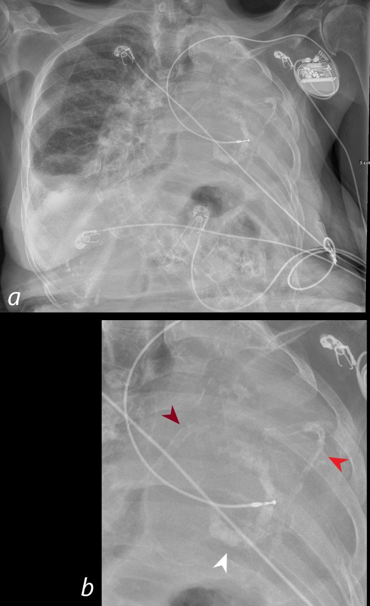
Frontal CXR of a 98-year-old woman showing a left sided white out secondary to a pneumonectomy. The soft tissue structures of the mediastinum have all shifted into the left hemithorax accounting for the white out. The calcified mitral annulus (b, white arrowhead), calcified right coronary artery – RCA (b, maroon arrowhead) and left anterior descending (LAD) – (b, bright red arrowhead) and right ventricle (RV pacemaker lead) confirm the diagnosis of acquired dextrocardia. There is hyperinflation of the right lung which crosses the midline associated with a small right effusion. A significant dextro-thoracic scoliosis with a compensatory levoscoliosis of the lumbar spine is present
Ashley Davidoff MD TheCommonVein.net 269Lu136234cL
Coronal CT of a 98-year-old woman showing a left sided white out on CXR secondary to a pneumonectomy shows a leftward shift of cardiac structures including the contrast filled right ventricle (RV) and the oval shaped left ventricle (LV) which occupy the left hemithorax. The tip of the pacing lead is noted in the RV septum. There is hyperinflation of the right lung which crosses the midline associated with a small right effusion.
Ashley Davidoff MD TheCommonVein.net 269Lu 136235b