
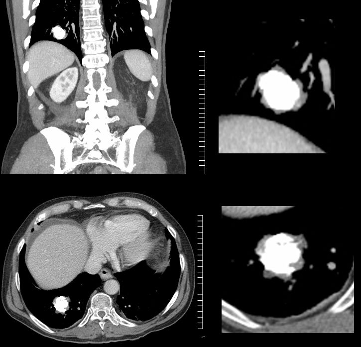
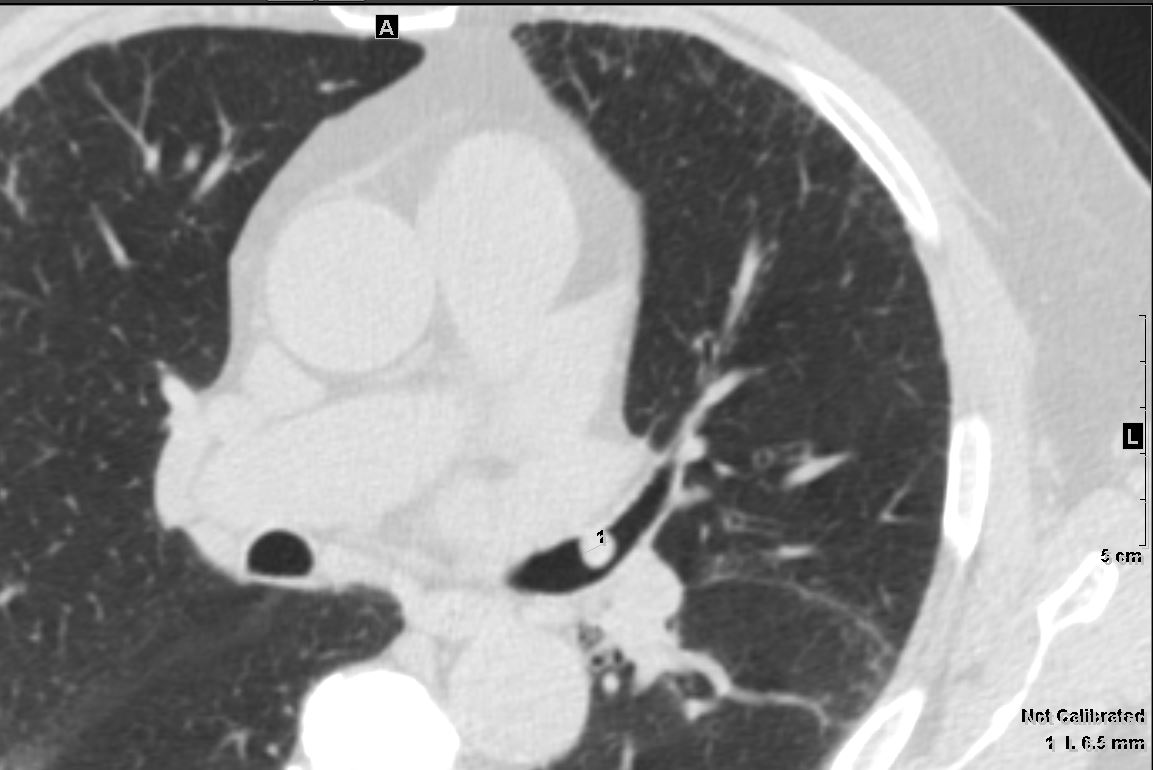
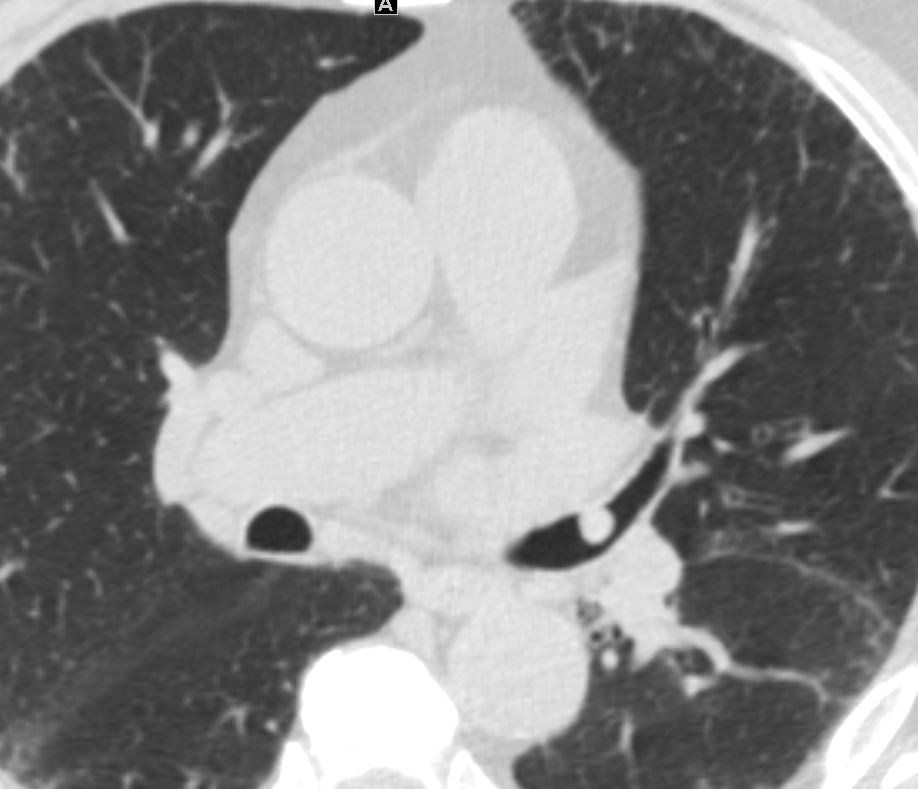
Fat Containing Heterogeneous Nodule in the Lung – Hamartoma
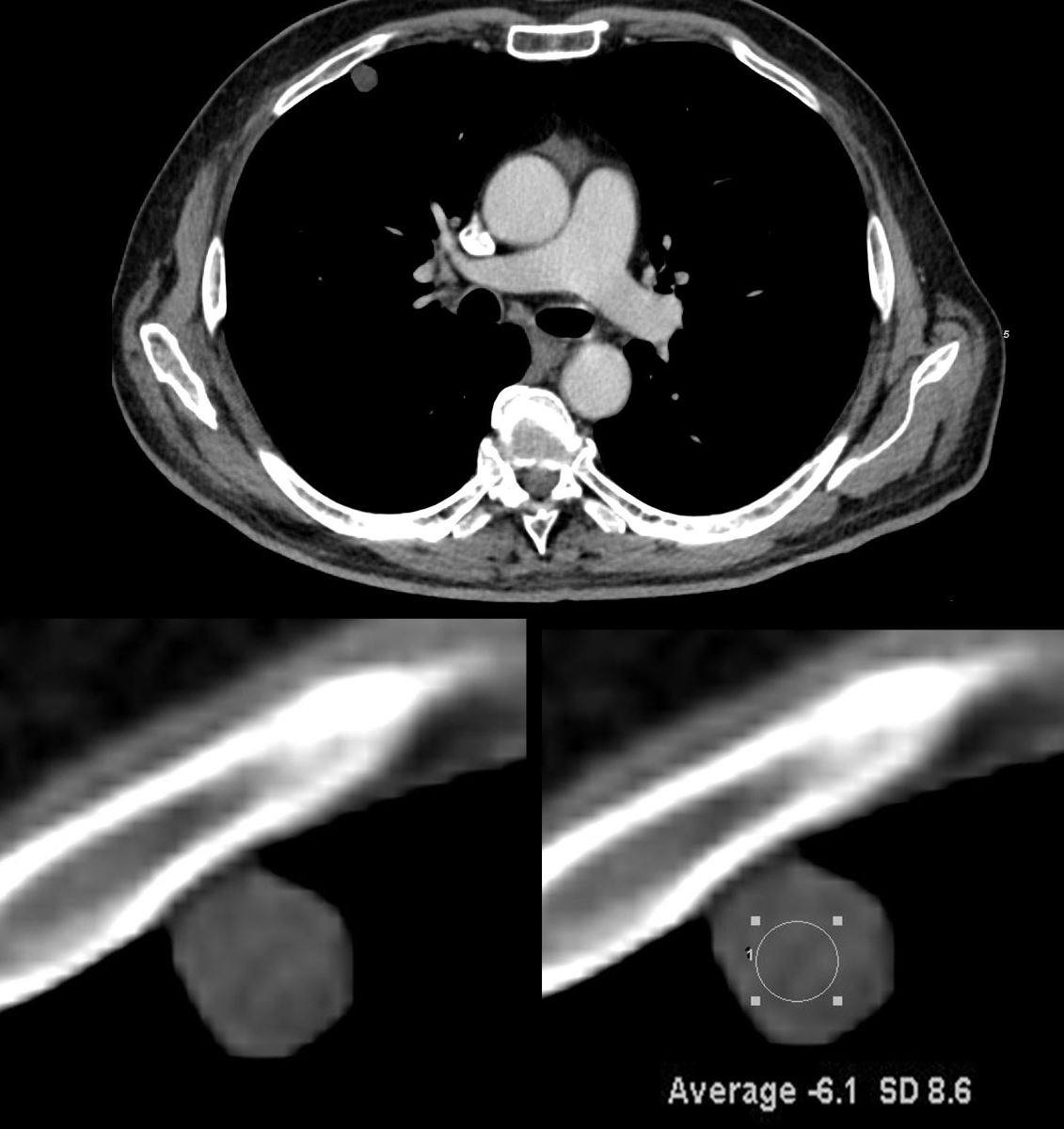
CT in the axial plane of a 42-year-old male shows a 9mm nodule in the anterior segment of right upper lobe that is heterogeneous (lower right panel) and fat containing (-6.1HU measurement in lower left panel). Findings are consistent with a pulmonary hamartoma
Ashley Davidoff MD TheCommonVein.net 136031c
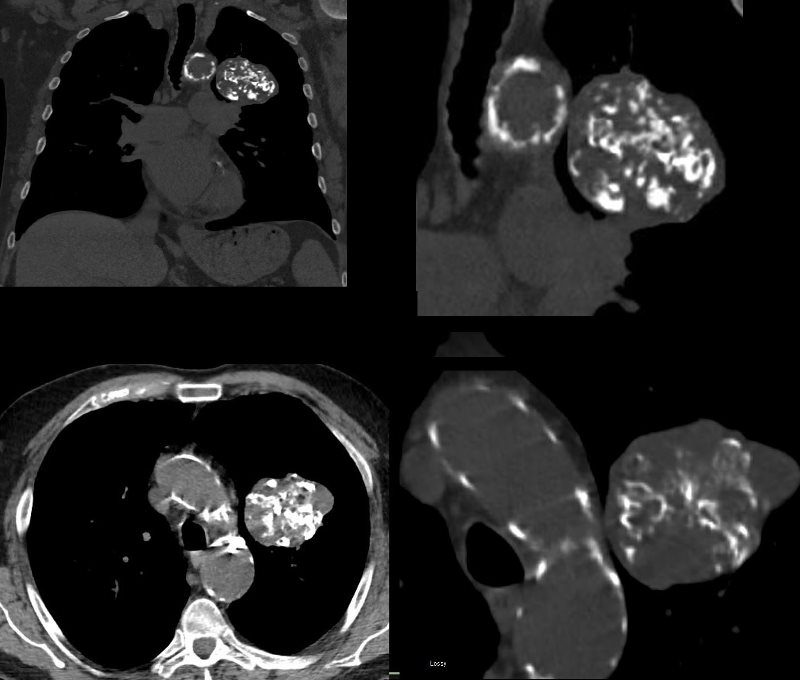
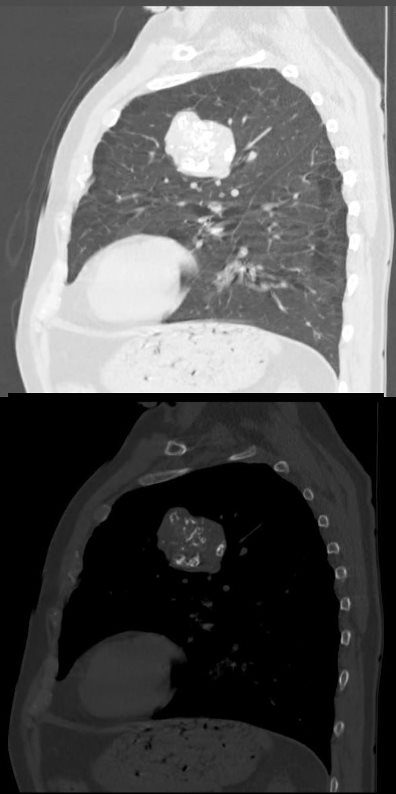
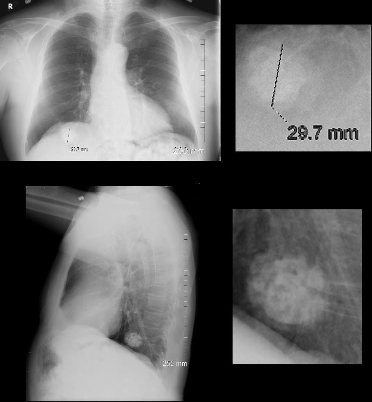
Classical Case with Popcorn Calcification
Ashley Davidoff MD TheCommonVein.net hamartoma 006v.8
Ashley Davidoff MD TheCommonVein.net hamartoma 003c.8
Ashley Davidoff MD TheCommonVein.net hamartoma 004b.8
Diagnosis includes a benign hamartoma
and amyloidoma
Ashley Davidoff TheCommonvein.net
hamartoma calcifications 002c stable
and amyloidoma
Ashley Davidoff TheCommonvein.net
hamartoma calcifications 004c stable
Ashley Davidoff TheCommonvein.net
hamartoma 0001c01 86f
Ashley Davidoff TheCommonvein.net
hamartoma calcifications 002c stable
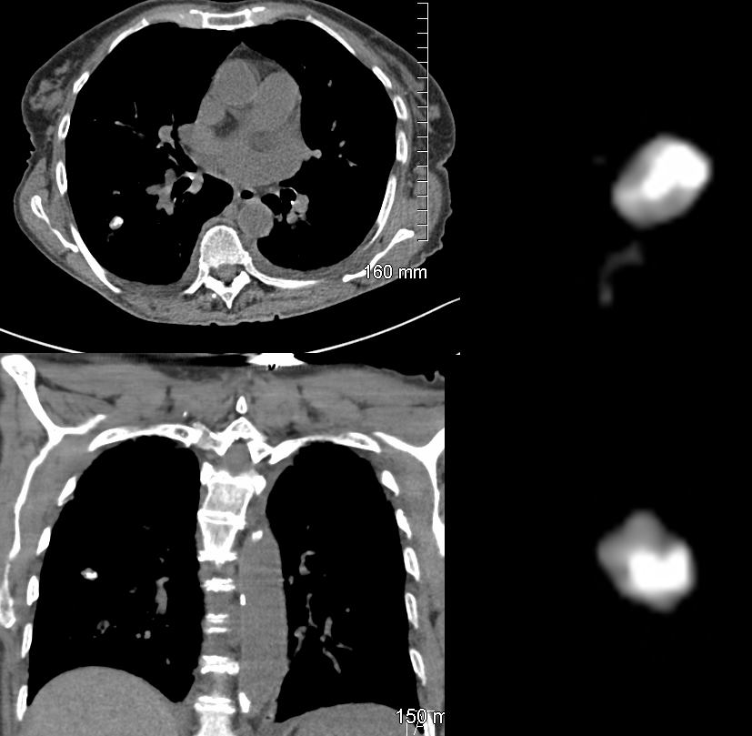
Mildly Heterogeneous Soft Tissue Nodule
Ashley Davidoff MD TheCommonVein.net benign hamartoma 005 33m c01
Ashley Davidoff MD TheCommonVein.net benign hamartoma 004 33m
Ashley Davidoff MD TheCommonvein.net hamartoma
Ashley Davidoff MD TheCommonvein.net hamartoma 005
Ashley Davidoff MD TheCommonvein.net hamartoma 004
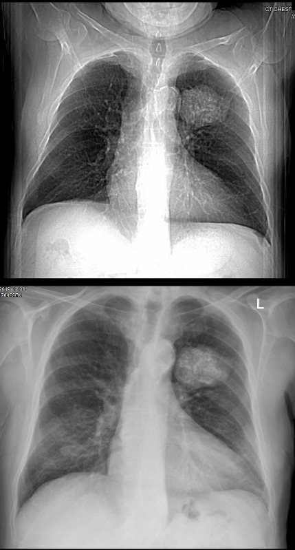
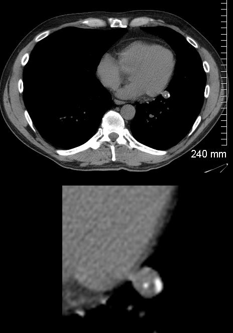
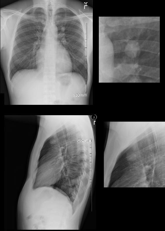
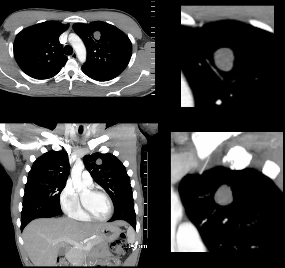
Lingular Atelectasis with Obstructing Nodule
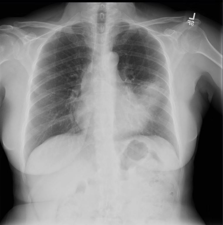
55-year-old female presents with a chronic cough
Frontal CXR shows an infiltrate involving the superior segment of the lingula, with partial silhouetting of the left heart border, and without associated secondary changes of volume loss
Final diagnosis was an obstructing hamartoma of the superior lingula bronchus
Courtesy Ashley Davidoff MD TheCommonVein.net 290 Lu 136563
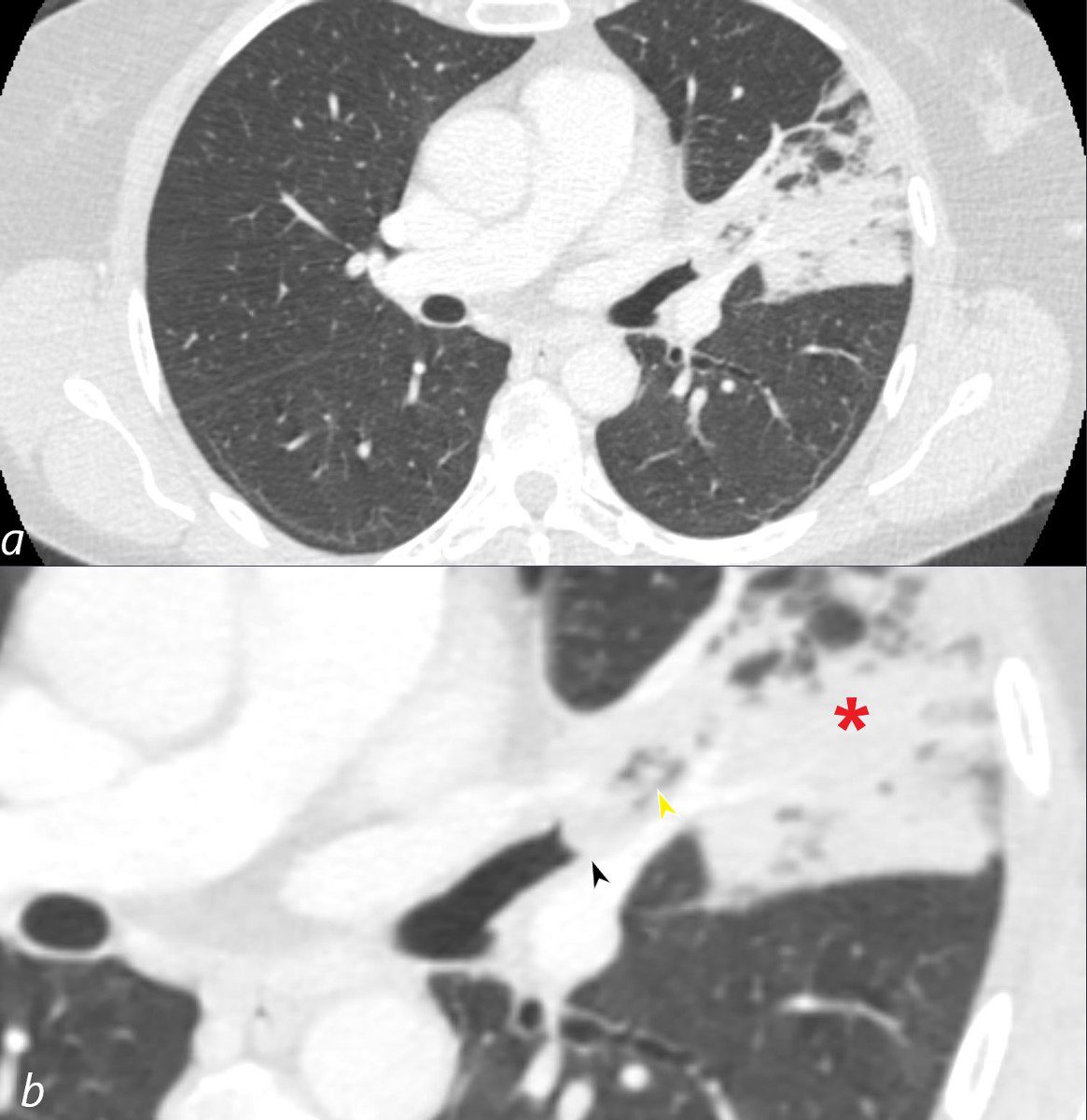
55-year-old female presents with a chronic cough
CT in the axial plane shows an infiltrate involving the superior segment of the lingula, reflecting segmental post obstructive atelectasis (b red asterisk). A rounded soft tissue filling defect is noted in the subtending bronchus (b, black arrowhead) with downstream mucus accumulation (b yellow arrowhead)
Final diagnosis was an obstructing hamartoma of the superior lingula bronchus
Courtesy Ashley Davidoff MD TheCommonVein.net 290 Lu 136566cL