

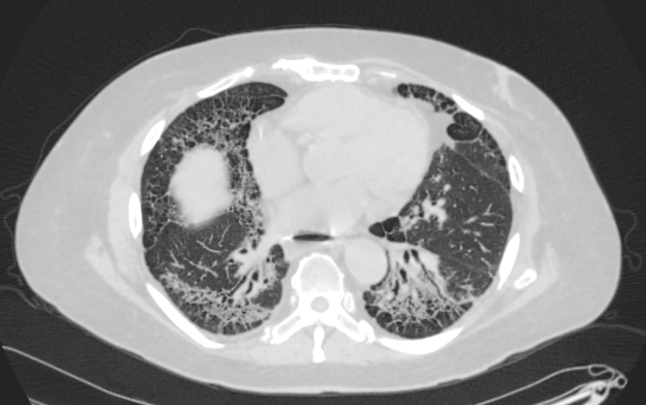
59-year-old male presents with history of scleroderma, Raynaud’s disease, and ILD
CXR shows basilar reticular changes and low lung volumes. There is an air bronchogram in the left lower lobe as a result of traction bronchiectasis and fibrotic change surrounding the bronchovascular bundle
The CT highlights the bronchovascular thickening, and bronchiectasis that results in the air bronchogram. In addition there is volume loss, and subpleural sparing. The fibrotic process has resulted in traction of the secondary lobules in the region of subpleural sparing
Ashley Davidoff MD TheCommonVein.net 110Lu 136592c
Axial CT shows peripheral reticular changes, ground glass, bronchiolectasis at both lung bases, volume loss with crowding of the bronchovascular bundles posteriorly and subpleural sparing posteriorly. Note air-fluid level in the distended esophagus.
Ashley Davidoff MD TheCommonVein.net 110Lu 136598


Ashley Davidoff MD TheCommonVein.net
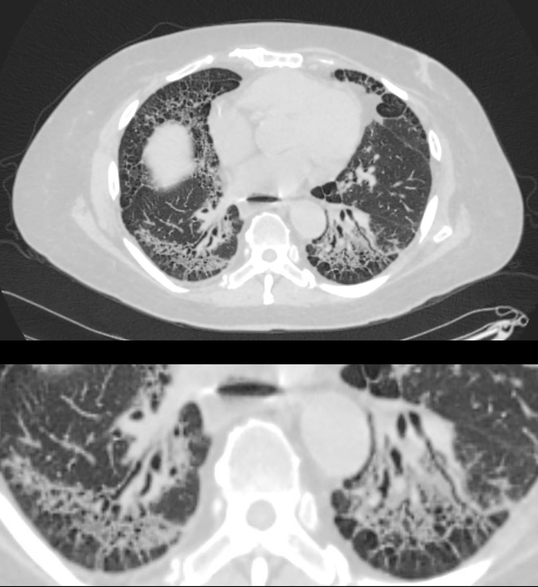
59-year-old male presents with history of scleroderma, Raynaud’s disease, and ILD
Upper Image
Axial CT shows bibasilar ground glass, bronchiectasis, and bronchiolectasis with volume loss and with crowding of the bronchovascular bundles posteriorly. There is subpleural sparing. Note air-fluid level in the distended esophagus.
The lower image focuses on the peripheral sparing. The spared secondary lobules have also undergone enlargement secondary to the fibrotic process
Ashley Davidoff MD TheCommonVein.net 110Lu 136598c01
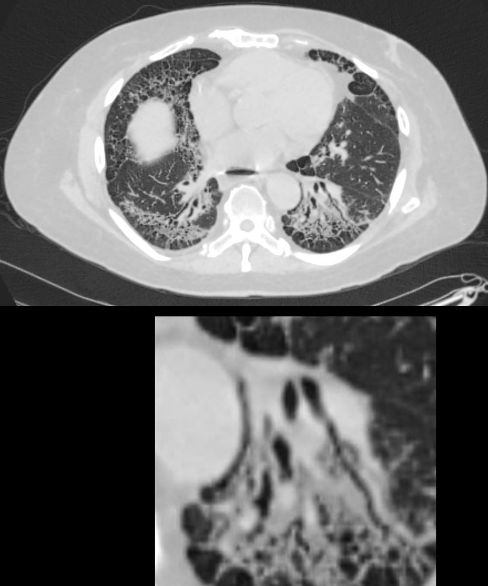
59-year-old male presents with history of scleroderma, Raynaud’s disease, and ILD
Upper Image
Axial CT shows bibasilar peripheral reticular changes, ground glass, bronchiectasis, and bronchiolectasis with volume and with crowding of the bronchovascular bundles posteriorly. There is subpleural sparing posteriorly. Note air-fluid level in the distended esophagus.
Lower Image
The lower image focuses on the traction bronchiectasis caused by the fibrotic process
Ashley Davidoff MD TheCommonVein.net 110Lu 136598c