
All Comers for Word First Image First Sherlock Holmes 1000 Words Where is Waldo
WF
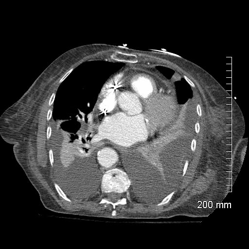
Axial CT through the lung bases with large bilateral effusions with compressive atelectasis. The abnormal shape, particularly of the left lung characterized by pointed tethering and fixation of the left lower parenchyma at 2 points to the pleura indicating a fibrotic adhesive process and loculation
Ashley Davidoff MD TheCommonvein.net RnD
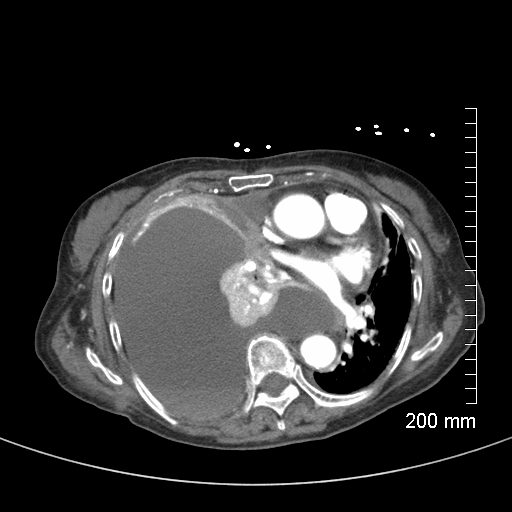
46 year-old female with bilateral pleural effusions, left greater than right. The scout film prior to the CT scan shows a left retrocardiac consolidation and a suggestion of a left sided complex pleural effusion. The liver appears enlarged.
Ashley Davidoff MD TheCommonvein.net 238Lu RnD image
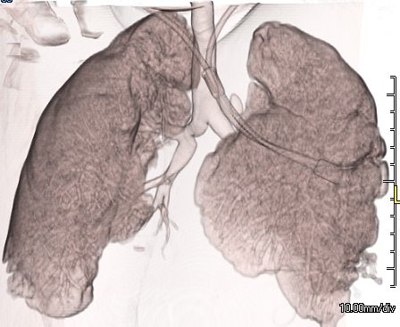
3D CT reconstructions show “naked” segmental airways to the lowers consistent with atelectasis. There is sparing of the superior segmental airways bilaterally.
Ashley Davidoff MD TheCommonVein.net
238Lu RnD image
Ashley Davidoff MD TheCommonVein.net RnD Image 106Lu
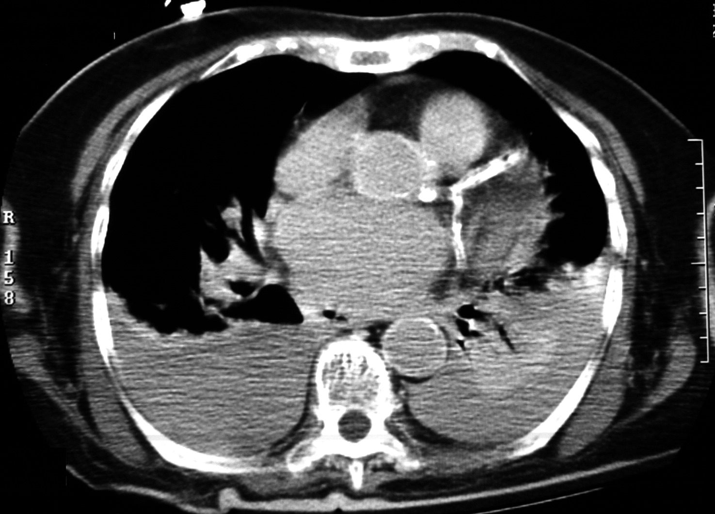
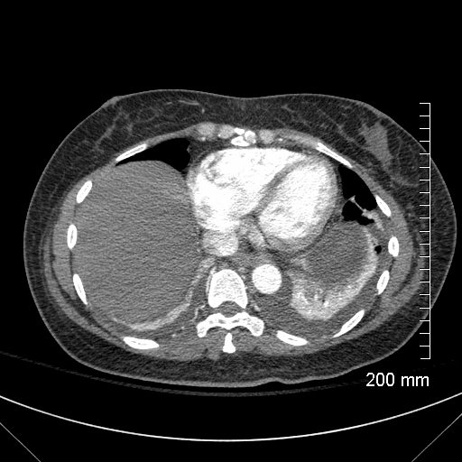
83 year old with bilateral pleural effusions, right larger than left with bibasilar compressive atelectasis. There is significant coronary calcification including left main disease and possible LAD stent. The left atrium is enlarged likely secondary to elevated left ventricular end diastolic pressure. This finding and the pleural effusions suggest CHF. Visualization of the wall of the aorta without the usual coarsened calcification raises the possibility of anemia
Ashley Davidoff MD TheCommonVein.net RnD image
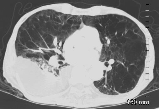
Rounded Atelectasis (aka Folded Lung Syndrome) and Asbestos Related disease
72-year-old male with a history of asbestos exposure presents with a cough. Axial CTscan shows a pleural based nodule with a comet tail and a series of lung markings folded into the nodule. There is subsegmental compensatory hyperinflation of the lateral segment of the right lower lobe Noted bilateral pleural thickening and pleural based calcification which is reminiscent of asbestos related disease. Early evolution of rounded atelectasis is also noted in the left lower lobe
Ashley Davidoff MD TheCommonVein.net RnD 240Lu
WF CHF Kerley B Lines and Thickening of the Interlobular septa
74-year-old female presents in CHF and an echo showing reduced EF (35%)
CT scan from a coronary CTA with a limited field of view above, and magnified below shows thickening of the interlobular septa extending to the periphery and in contact with the pleural surface indicating moderate CHF
Ashley Davidoff MD TheCommonVein.net 003H RnD
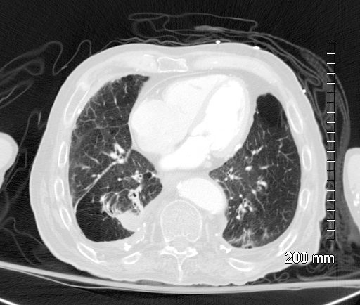
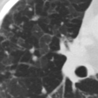
Centrilobular Nodules
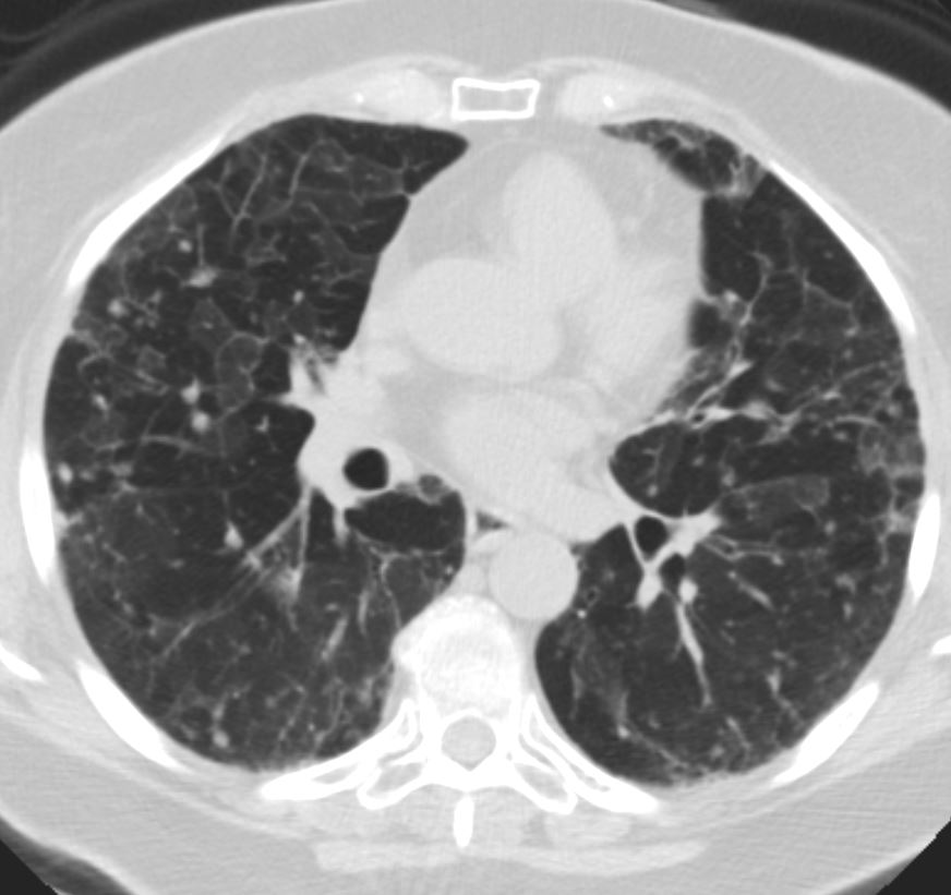
77F with long history of dyspnea and cough showing medium and small airway disease, centrilobular nodules, paraseptal nodules ground glass changes and mosaic attenuation Diagnosis includes Stage 3 sarcoidosis
Ashley Davidoff
TheCommonVein.net RnD
77F with long history of dyspnea and cough showing medium and small airway disease, centrilobular nodules, para-septal nodules ground glass changes and mosaic attenuation Diagnosis includes Stage 3 sarcoidosis
Ashley Davidoff
TheCommonVein.net
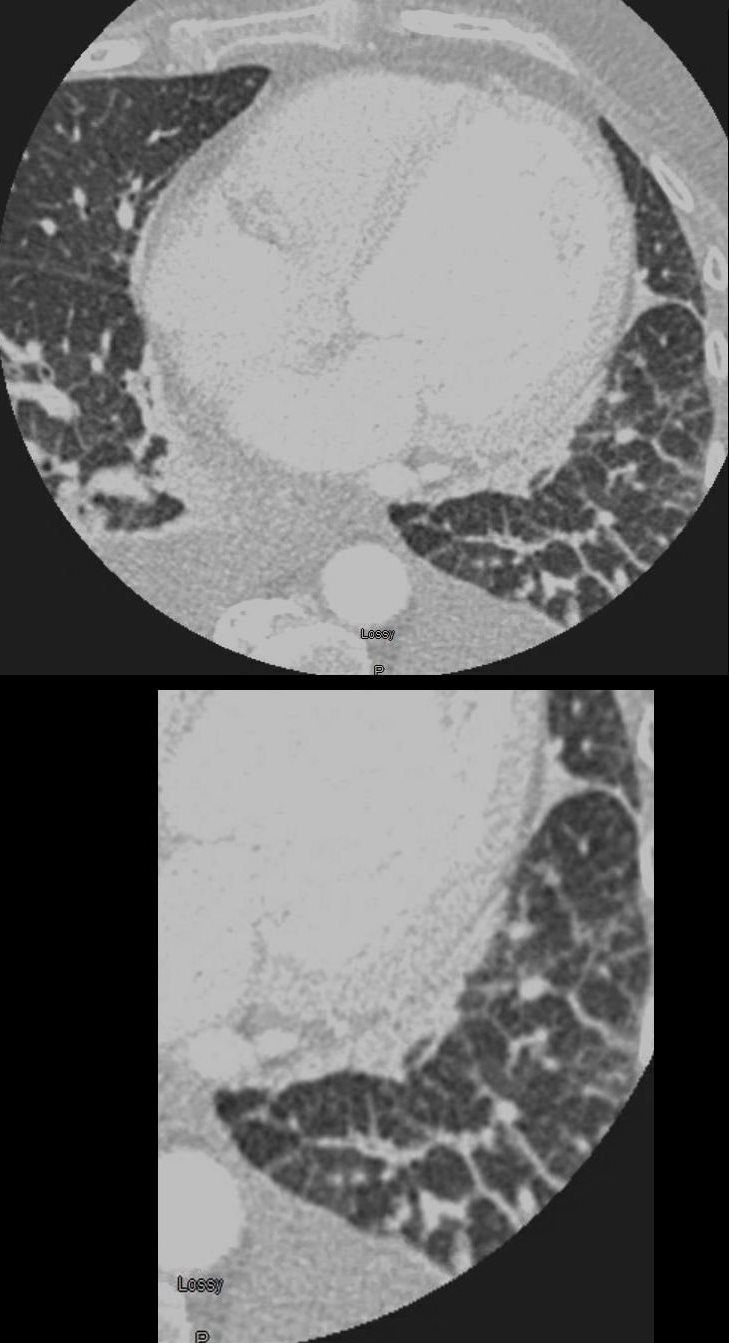
WF Centrilobular Emphysema
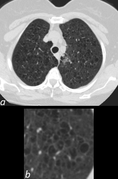
Axial CT (a) with magnified view of the upper lobes of a 66year female with centrilobular emphysema shows an expanded lobule with a centrilobular vessel in the middle characteristic of centrilobular emphysema
Ashley Davidoff MD TheCommonvein.net RnD
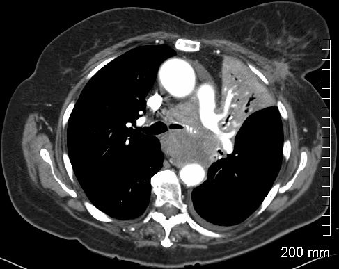
WF Panlobular Emphysema
CT scan of a 78 year old male with history of alpha-1 antitrypsin deficiency with extensive and severe emphysematous change uncharacteristically more prominent in the upper and midlung regions. Associated complex right pleural effusion with compressive atelectasis
Ashley Davidoff TheCommonVein.net RnD
1000 Words
Left Upper Lobe Atelectasis (Collapse) Caused by Central Squamous Cell Carcinoma and Left Breast Lesion
82-year-old female with dyspnea presents with an obstructing and infiltrating central squamous cell carcinoma of the left main stem bronchus with secondary post obstructive atelectasis of the left upper lobe of the lung. In addition, there is encasement of the left pulmonary artery and a small left effusion. A spiculated lesion at the base of the left breast in close association with the left pectoralis muscle The lesion also extends beyond the muscle to abut the rib. There is a small amount of fluid in the pericardial recess, and an small left pleural effusion.
Ashley Davidoff MD TheCommonVein.net RnD case 239Lu
1000 words CXR White Out S/P Left Pneumonectomy
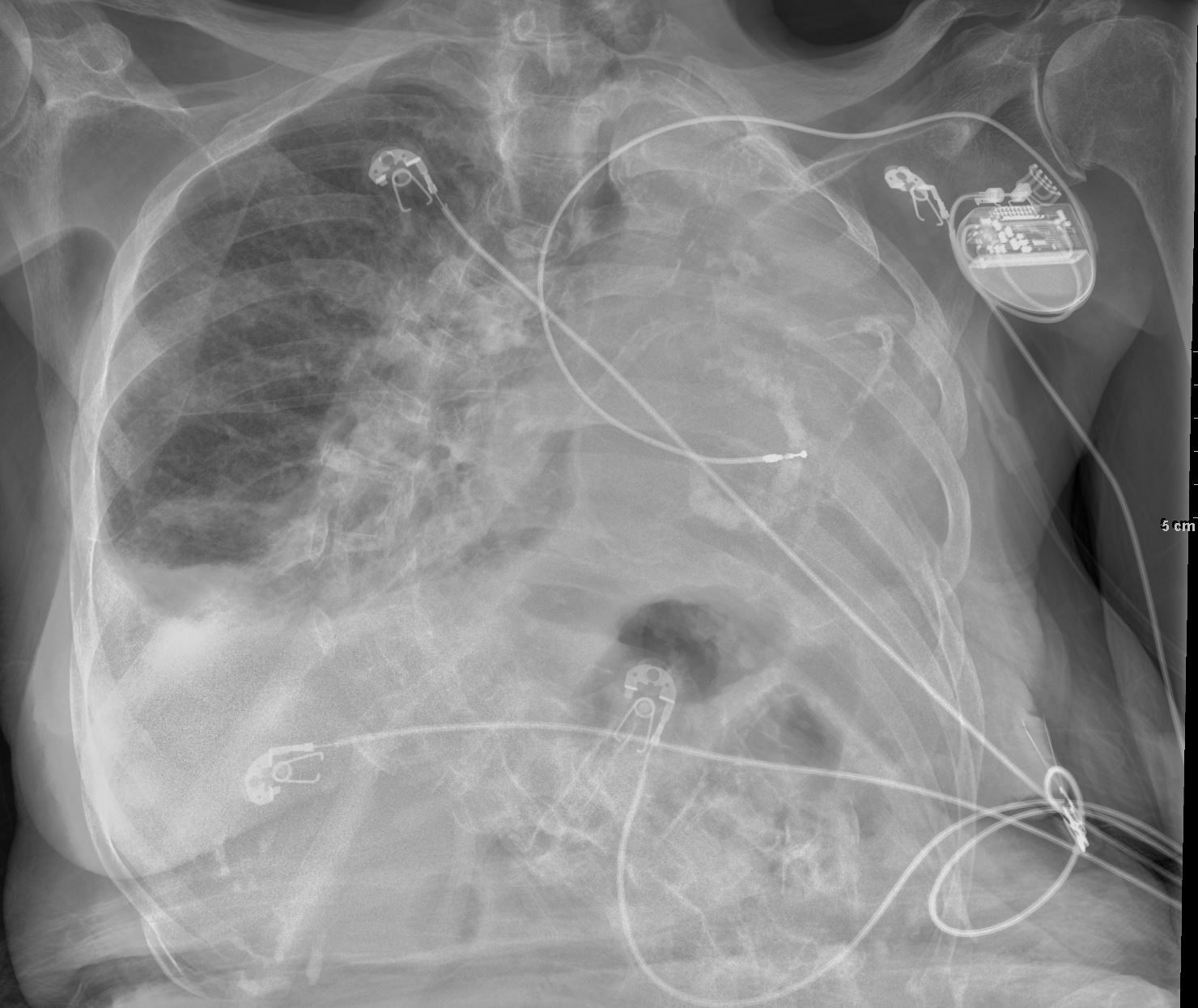
Frontal CXR of a 98-year-old woman showing a left sided white out secondary to a pneumonectomy. The soft tissue structures of the mediastinum have all shifted into the left hemithorax accounting for the white out. The calcified mitral annulus, calcified coronary arteries and right ventricle (RV pacemaker lead) confirm the diagnosis of acquired dextrocardia. There is hyperinflation of the right lung which crosses the midline associated with a small effusion . A significant dextro-thoracic scoliosis with a compensatory levoscoliosis of the lumbar spine is present
Ashley Davidoff MD TheCommonVein.net 269Lu 136234
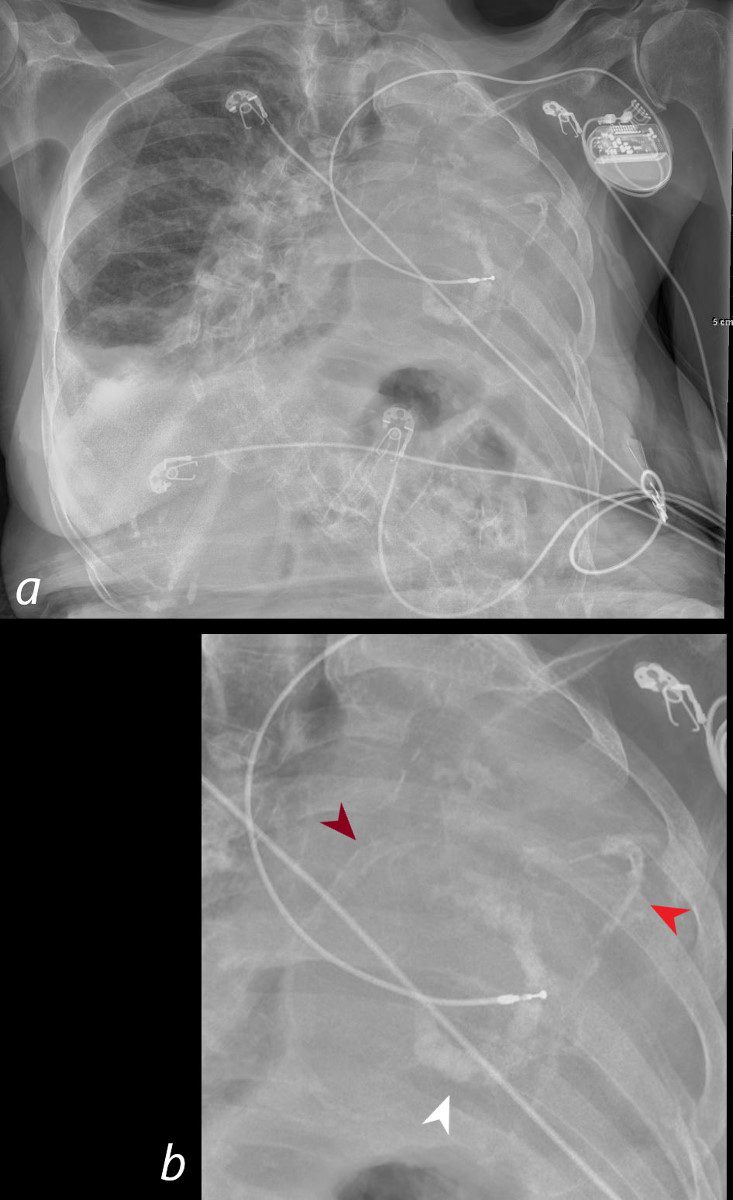
Frontal CXR of a 98-year-old woman showing a left sided white out secondary to a pneumonectomy. The soft tissue structures of the mediastinum have all shifted into the left hemithorax accounting for the white out. The calcified mitral annulus (b, white arrowhead), calcified right coronary artery – RCA (b, maroon arrowhead) and left anterior descending (LAD) – (b, bright red arrowhead) and right ventricle (RV pacemaker lead) confirm the diagnosis of acquired dextrocardia. There is hyperinflation of the right lung which crosses the midline associated with a small right effusion. A significant dextro-thoracic scoliosis with a compensatory levoscoliosis of the lumbar spine is present
Ashley Davidoff MD TheCommonVein.net 269Lu136234cL
Links and References