
Parts
Size
Shape
Position
Character
Calcification
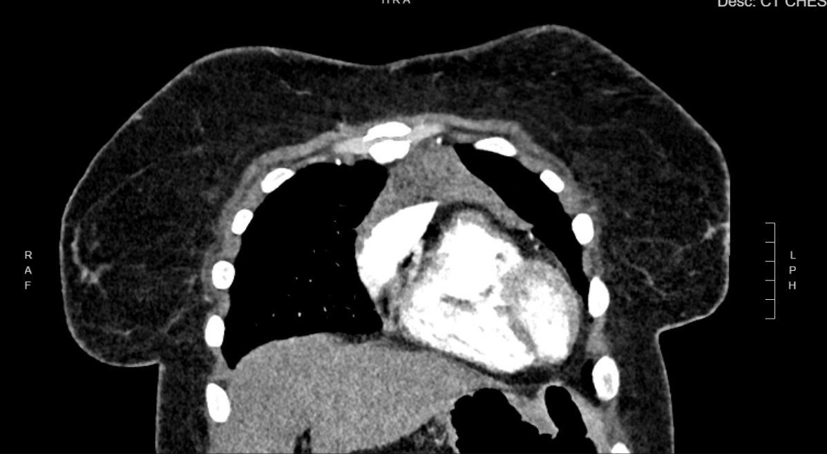
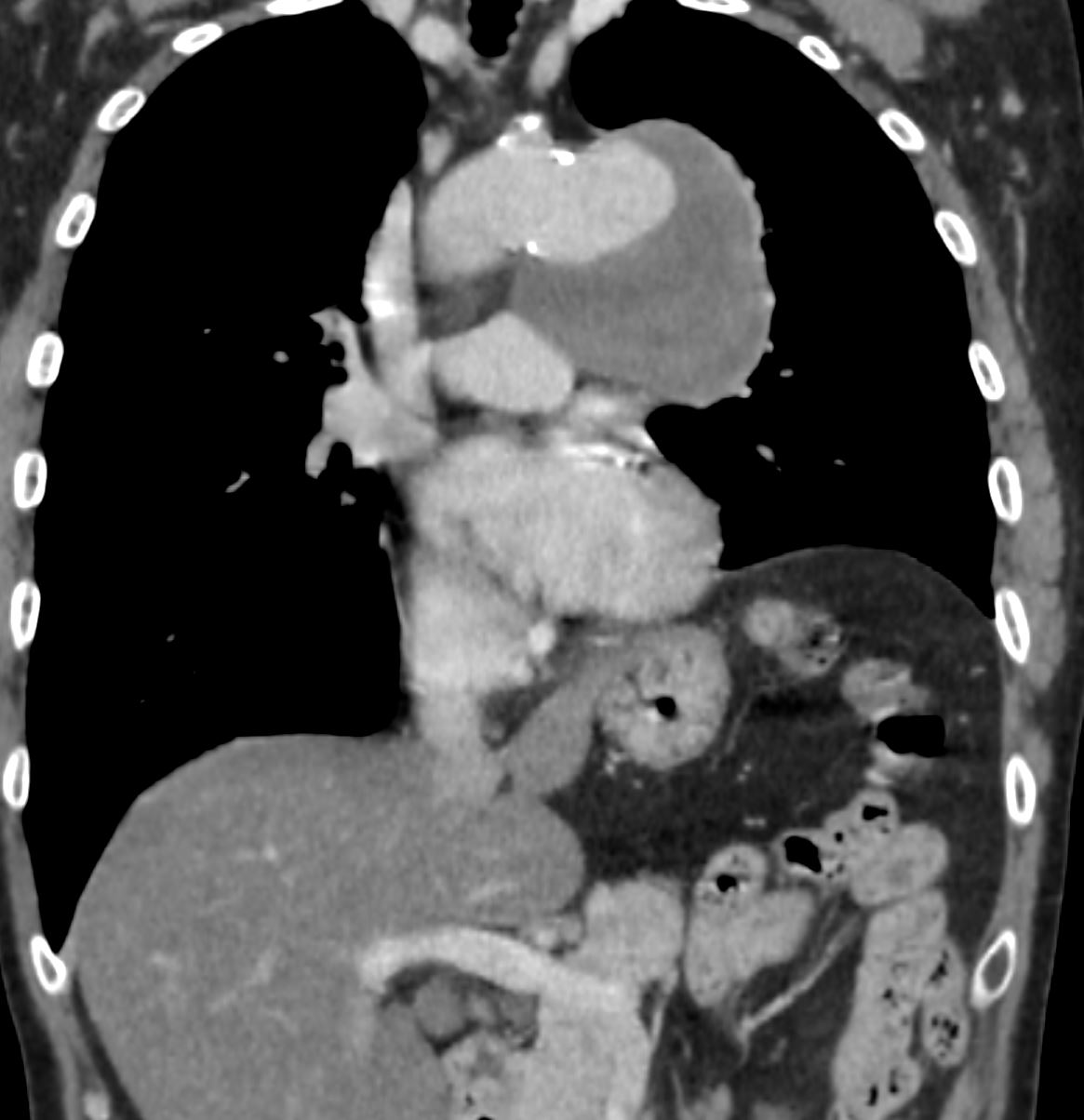
CT Right Sided Anterior Mediastinal Teratoma with Tooth Shaped Calcification
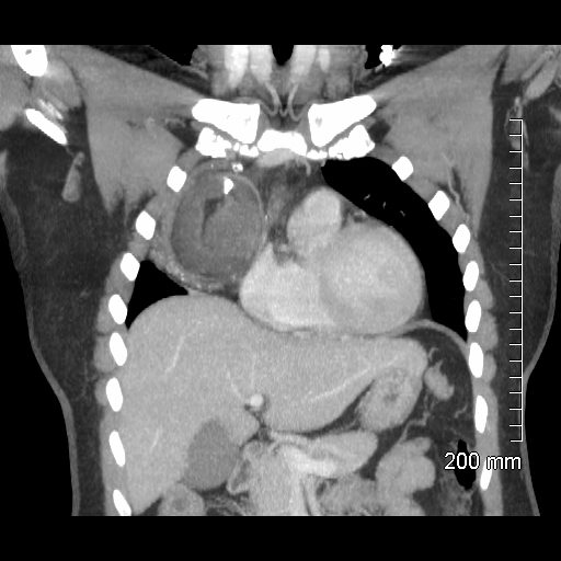
47-year-old female presents with an abnormal CXR
CT scan in the coronal projection shows a 6.3X 5.9 x 6.8cms mass in the anterior mediastinum that shows central fatty elements, surrounded by intermediate density of homogeneous soft tissue elements and a prominent calcification with a shape reminiscent of a tooth, and a more heterogeneous infero-lateral rim of soft tissue density with punctate calcifications.. These findings are consistent with a teratoma
Ashley Davidoff MD TheCommonVein.net 275Lu 136340
Fat
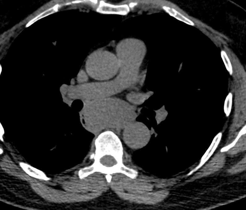
CT Anterior Mediastinal Teratoma
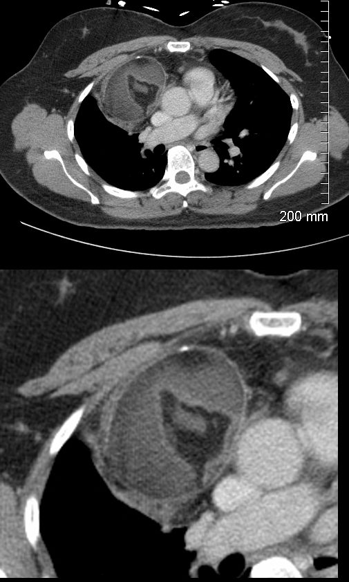
47-year-old female presents with an abnormal CXR
CT scan in the axial projection shows a 6.3X 5.9cms mass in the anterior mediastinum that shows central fatty elements, surrounded by soft tissue elements and a anterior peripheral calcification. These findings are consistent with a teratoma
Ashley Davidoff MD TheCommonVein.net 275Lu 136333c
Time
Infection
Inflammation
Malignancy
Benign Masses
Thymic Hyperplasia
Graves Disease and Thymic Enlargement
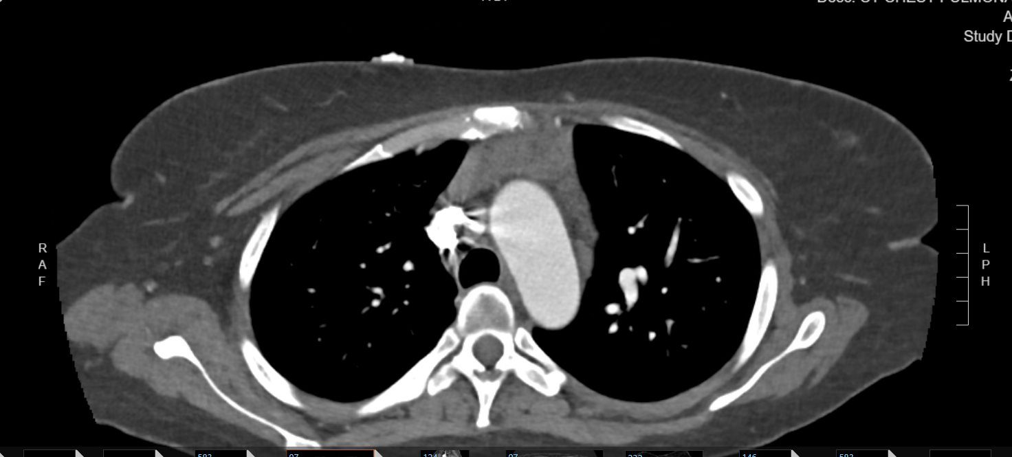
25 year old female with a history of Graves disease
CT in the axial plane shows an enlarged thymus which is a known association Graves disease
Ashley Davidoff MD TheCommonVein.net 136685
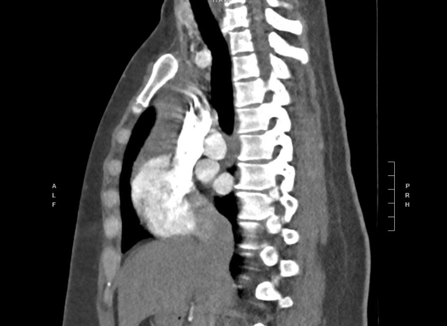
25 year old female with a history of Graves disease
CT in the sagittal plane shows an enlarged thymus which is a known association Graves disease
Ashley Davidoff MD TheCommonVein.net 136686
25 year old female with a history of Graves disease
CT in the coronal plane shows an enlarged thymus which is a known association Graves disease
Ashley Davidoff MD TheCommonVein.net 136687
Leiomyoma of the Esophagus
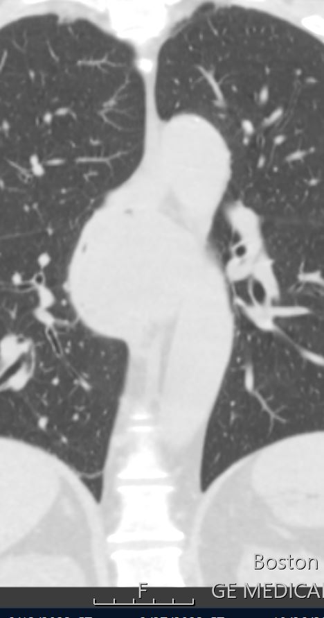
56-year-old male presents with chronic cough dyspnea and weight loss. CT scan in coronal projection shows a subcarinal esophageal mass which was diagnosed as a leiomyoma,
Ashley Davidoff MD TheCommonVein.net 267Lu 136220
56-year-old male presents with chronic cough dyspnea and weight loss. CT scan in axial projection shows an appearance reminiscent of finger in glove in the right lower lobe. Pathology of the right lower process was a squamous cell carcinoma
Ashley Davidoff MD TheCommonVein.net 267Lu 136223
Mechanical
Atelectasis
Trauma
Metabolic
Circulatory-
Scout CT shows a left sided70-year-old male presented
hoarseness and mediastinal mass
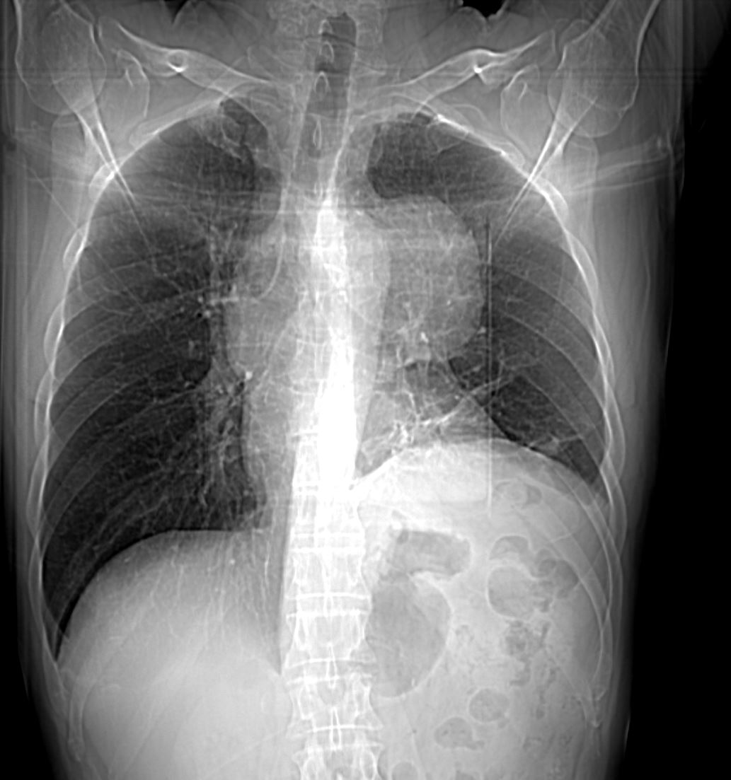
70-year-old male presented hoarseness and mediastinal mass. Scout CT shows a left sided mediastinal mass. The aortic knob left main stem bronchus and pulmonary branches can be seen – “hilum overlay” sign, indicating that the mass is not arising from the lung parenchyma. There is elevation of the left hemidiaphragm secondary to left phrenic nerve palsy.
Ashley Davidoff MD TheCommonVein.net 009A 136223
Coronal CT
Aortic Aneurysm of the Distal Aortic Arch and
Elevated Left Hemidiaphragm
Coronal CT with contrast shows a left sided aneurysm off the aortic arch with a large thrombus in the aneurysm. There is elevation of the left hemidiaphragm secondary to left phrenic nerve palsy. .
Ashley Davidoff MD TheCommonVein.net 009A 136225
Hemorrhage
Immune Infiltrative Idiopathic Iatrogenic