
- Swyer-James Syndrome
- aka
- Swyer-James-MacLeod Syndrome or
- unilateral hyperlucent lung syndrome.
- is a
- rare lung condition that usually
- occurs in childhood
- following an postinfectious bronchiolitis obliterans,
-
- The causative agents
- viruses (
- measles, respiratory syncytial virus, influenza A, Paramyxovirus morbillivirus, adenovirus
- bacteria (Bordetella pertussis, Mycobacterium tuberculosis, Mycoplasma pneumoniae)
- viruses (
- The causative agents
- inflammation and fibrosis of the
- obstructing small airways
- bronchial walls and
- bronchiolar walls
- injury of the bronchiolar epithelium
- possibly the adjacent alveoli.
- hinders the normal development of the
- alveolar ducts.
- interalveolar septa involved
- Airway changes
- repair process leads to a excess
- proliferation of granulation tissue,
- narrowing and
- luminal irregularity
- further contributes to
- airway occlusion which causes
- alveolar air trapping,
- eventually resulting in
- emphysema,
- (permanent overdistention of the airspaces distal to the terminal bronchioles.)
- emphysema,
- Alveolar changes
- emphysema as above
- also
- alveolar wall destruction leads to the
- loss of elastic recoil, causing
- airway collapse during exhalation and
- air trapping. resulting inVascular Changes
- alveolar wall destruction leads to the
- Fibrosis of the interalveolar septae leads to
- obstruction of the pulmonary capillary bed
- Also
- hyperinflation of alveoli, (emphysema),
- creates add in through the capillary beds.
- resulting
- hypoplastic pulmonary vasculature
- associated with vasoconstriction
- decreased arterial perfusion and
- results in
- reduced ventilation
- obstructing small airways
- Clinical
-
-
-
-
-
- may be asymptomatic
- chronic cough
- recurrent respiratory infections
-
-
-
-
-
-
- Imaging
-
- unilateral hyperlucency of the lung
- due to air trapping and
- increased lung volume.
- unilateral hyperlucency of the lung
-
- Lung Functions
- decreased lung function and .
- Complications
- bronchiectasis, and
- pulmonary hypertension.
- Treatment
- symptomatic
- bronchodilators,
- corticosteroids, and
- antibiotics to manage infections.
- symptomatic
Patient with Swyer James and
Anomalous Origin of LPA from Aorta
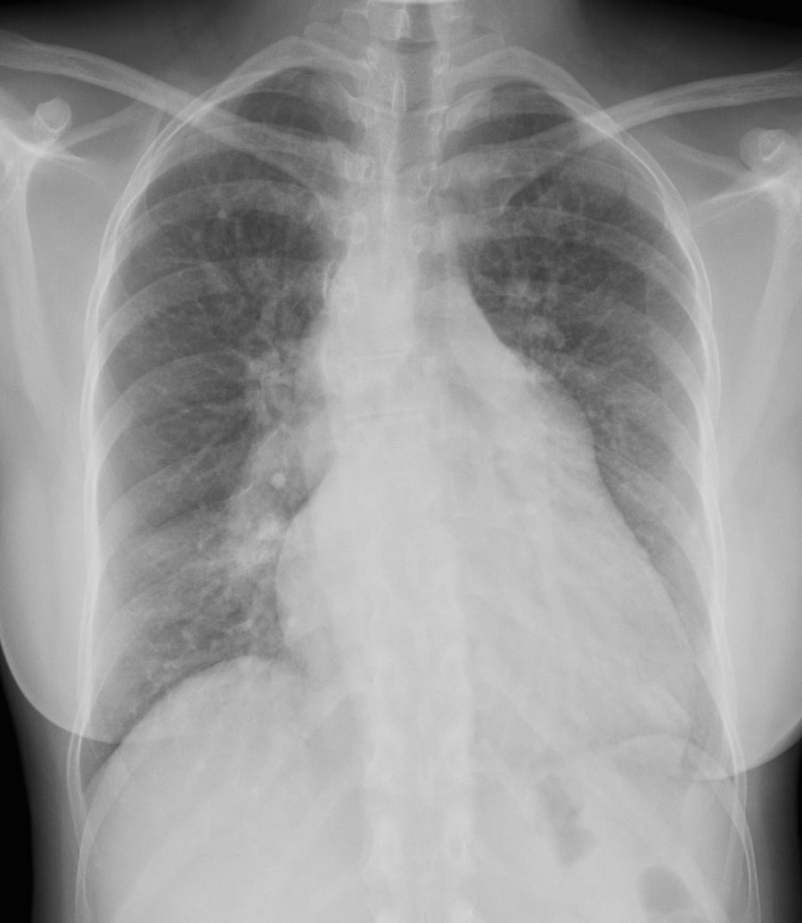
The right upper lung field is lucent consistent with the patients known Swyer James Syndrome
Ashley Davidoff MS TheCommonVein.net LPA-from-Aorta-001
Left Ventricular Hypertrophy
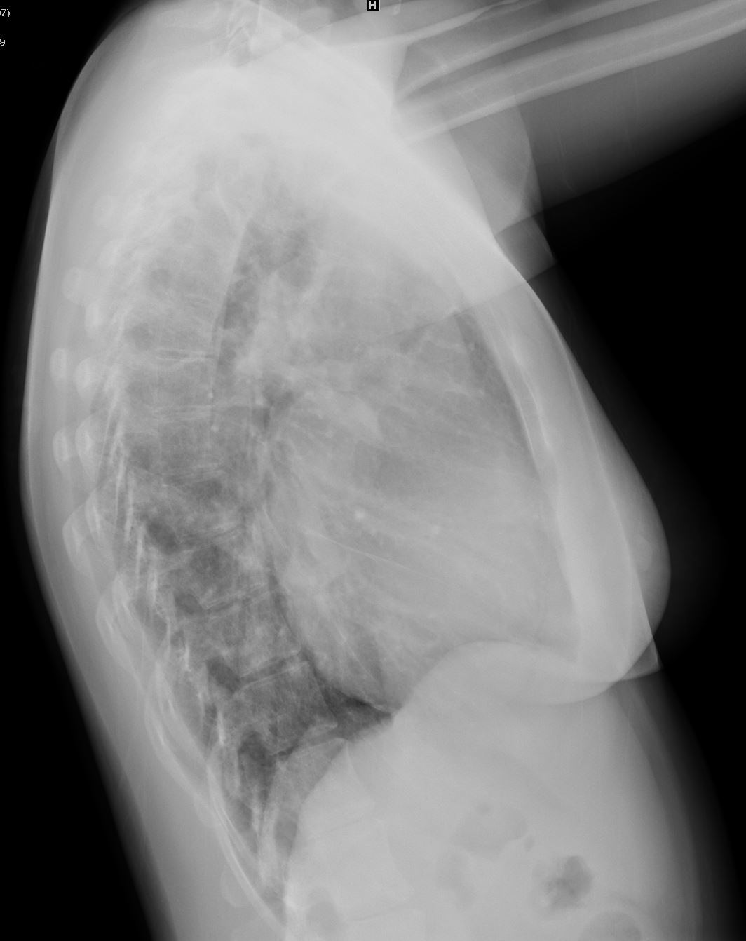
Ashley Davidoff MS TheCommonVein.net pulmonary artery off aorta 002
Swyer James
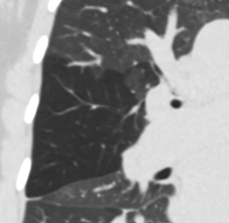
Hyperlucent Anterior Segment of the RUL
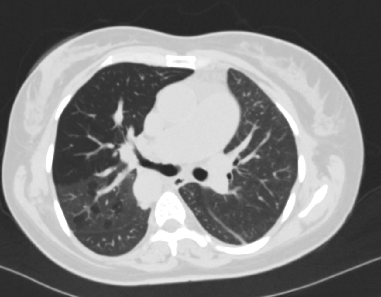
CT in axial projection suggests shows a hyperlucent anterior segment of the right upper lobe, narrowing of the segmental airway, and mosaic attenuation in the posterior segments Ashley Davidoff MD TheCommonVein.net artery-off-aorta-012
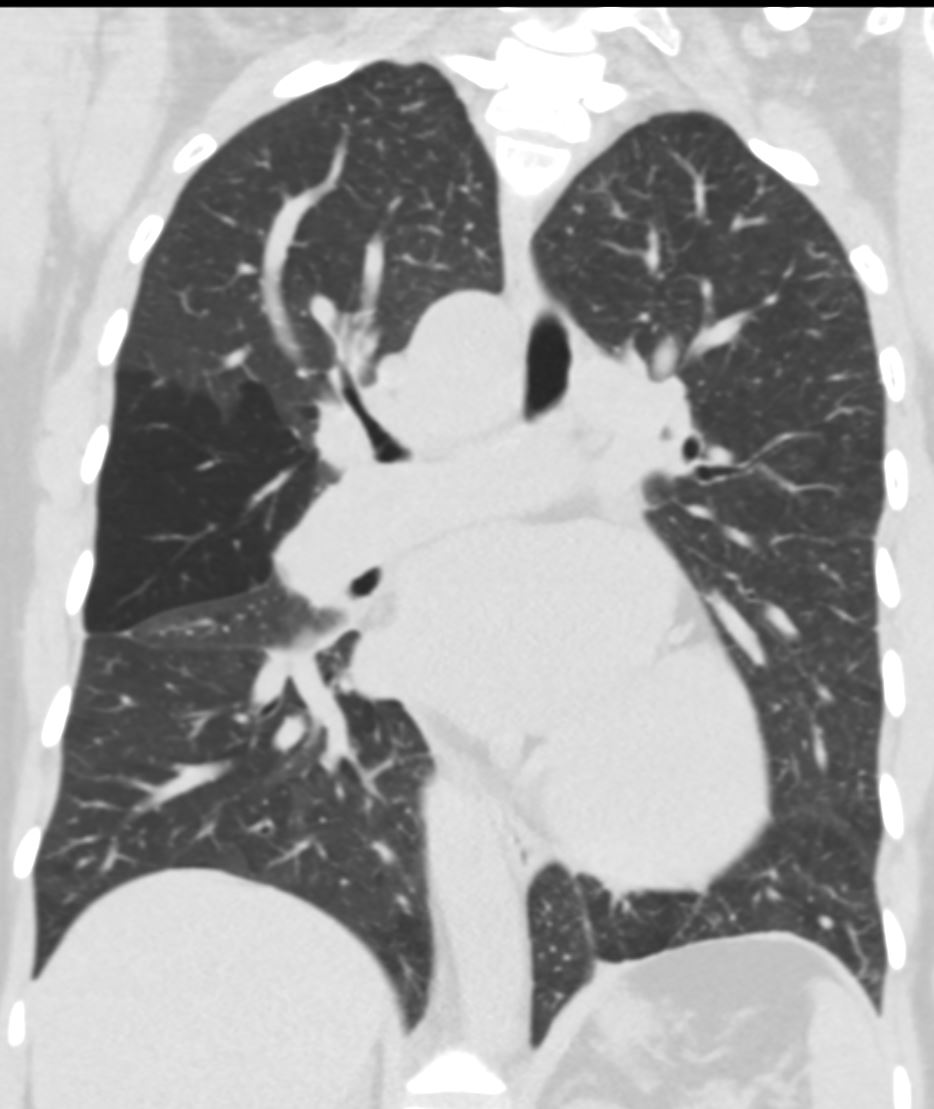
CT in coronal projection shows a hyperlucent anterior segment of the right upper lobe Ashley Davidoff MD TheCommonVein.net artery-off-aorta-013
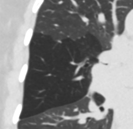
Do you see a bull made of the soft tissues looking at the lucent lung?
Keep your eye on the eye of the bull as it will get progressively smaller as the bull goes to sleep and the airway narrows
CT in coronal projection shows a hyperlucent anterior segment of the right upper lobe Note the patent segmental airway subtending the upper lobe If you look with an artistic eye you can see a bull made of soft tissues looking at the hyperlucent lung The patent segmental airway is the “eye of the bull”) Ashley Davidoff MD TheCommonVein.net artery-off-aorta-014
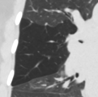
CT in coronal projection shows a hyperlucent anterior segment of the right upper lobe Note that the “eye of the bull” has become smaller as the anterior segmental airway becomes progressively narrowed Ashley Davidoff MD TheCommonVein.net artery-off-aorta-014b
CT in coronal projection shows a hyperlucent anterior segment of the right upper lobe Note that the “eye of the bull” has become smaller as the anterior segmental airway becomes progressively narrowed Ashley Davidoff MD TheCommonVein.net artery-off-aorta-015
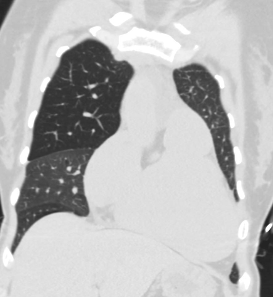
CT in coronal projection shows a hyperlucent anterior segment of the right upper lobe extending to the apex. This finding helps us understand why the ventilation in the VQ scan is deficient in the upper lung extends to the apex despite normal aeration of the superior segment Ashley Davidoff MD TheCommonVein.net artery-off-aorta-016
AP Ventilation View NM Shows Non Ventilation of the Right Upper Lung Field and Absent Ventilation of the Left Lung
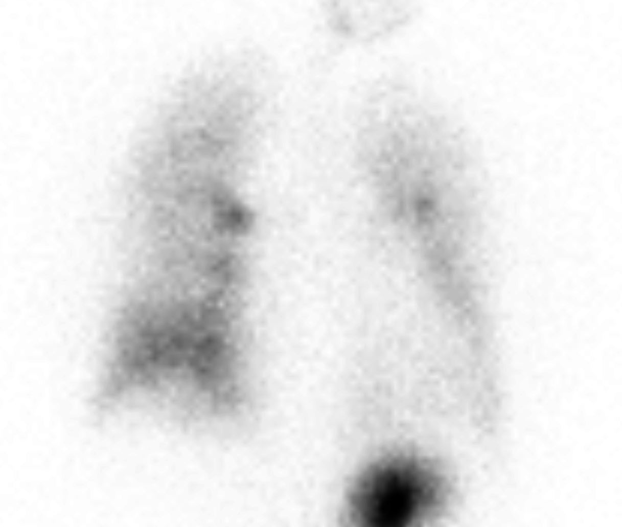
AP of the ventilation component of the VQ scan shows non ventilation of the right upper lung field and absent ventilation of the left lung. The absent ventilation of the right upper lung field is explained by the expanded hyperlucent anterior segment of the right upper lobe which reaches to the right apex (see image above). The non ventilation of the left lung is explained by the absent pulmonary blood flow and the QP QS mismatch – just like a large occlusive PE to the left lung Ashley Davidoff MD TheCommonVein.net artery-off-aorta-016
Perfusion Scan – No Perfusion of the Left Lung
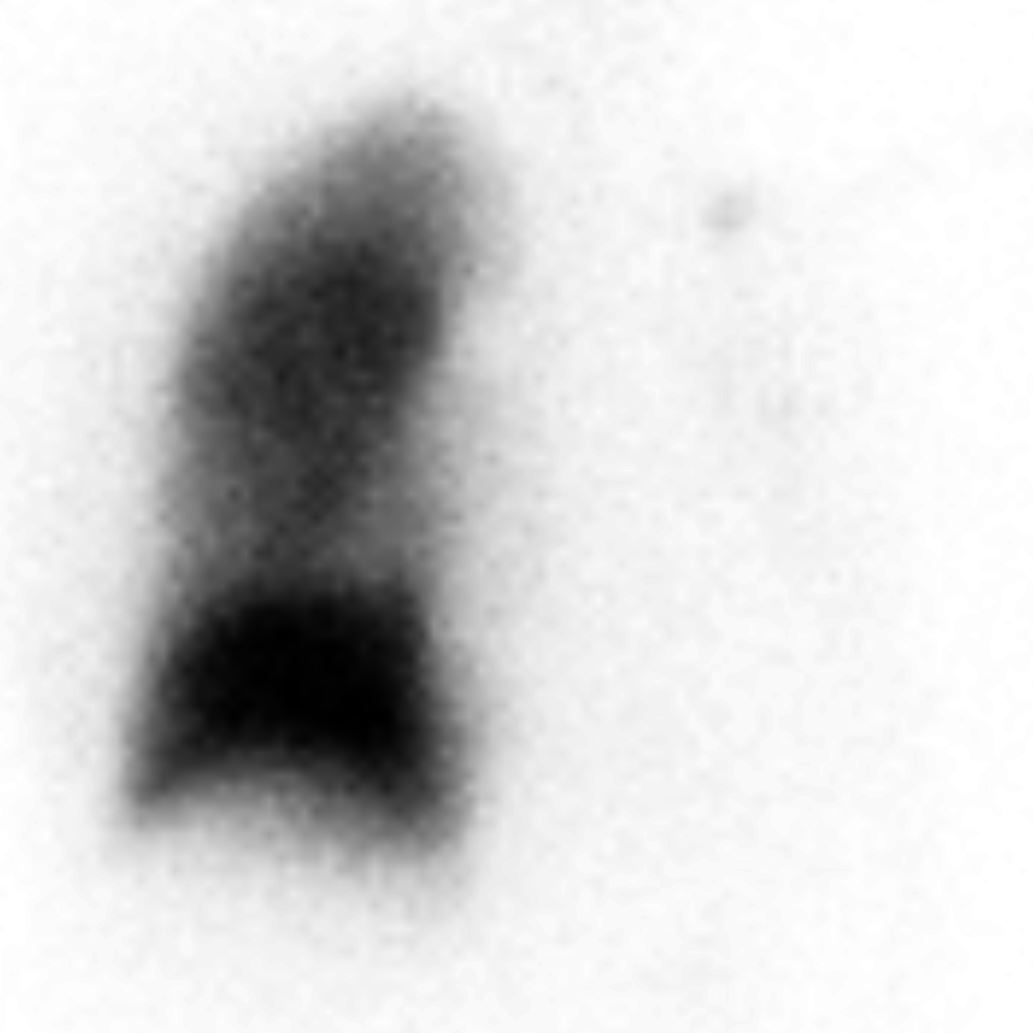
AP of the perfusion component of the VQ scan shows non perfusion of the left lung. The absent perfusion of the left lung is explained by the absent pulmonary blood flow since the blood to the left pulmonary artery is arising from the aorta – just like a large occlusive PE to the left lung Ashley Davidoff MD TheCommonVein.net artery-off-aorta-017