
80-Year-Old Male Presents with Fatigue
3cms. Mass in the RLL
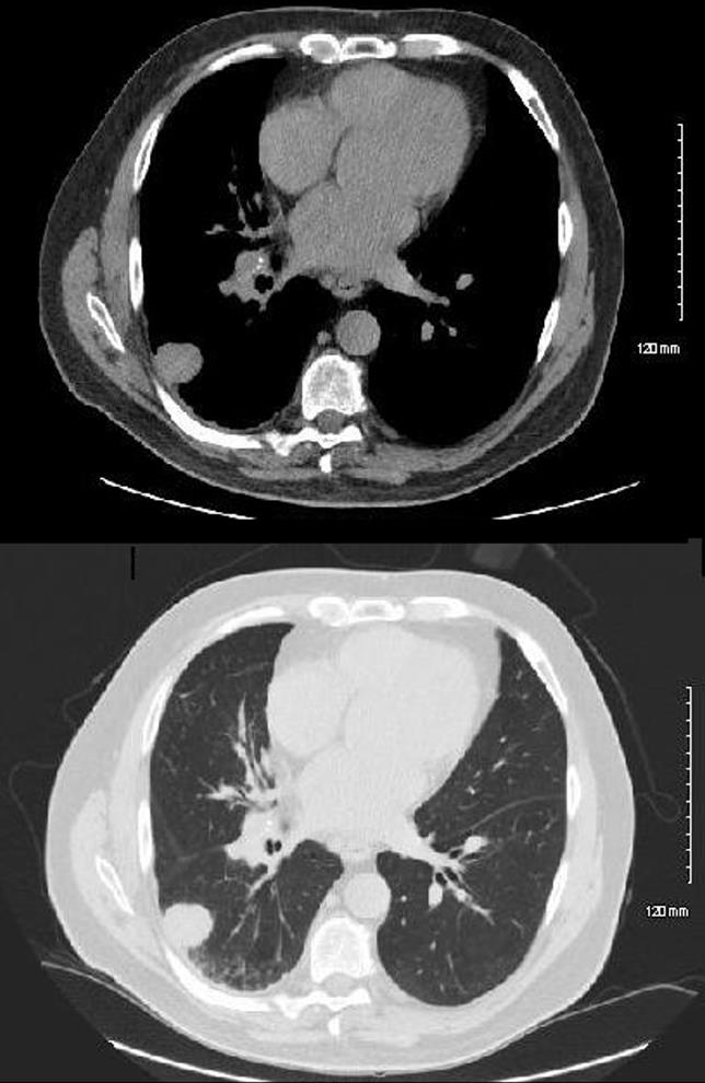
80 year old male presents with fatigue, CT in the axial projection shows a 3cms mass in the right lower lobe associated with the pleura, with mild associated pleural irregularity but without pleural effusion .
Ashley Davidoff MD TheCommonVein.net 292Lu 121889b
CT Adenocarcinoma Metastatic to Left Adrenal
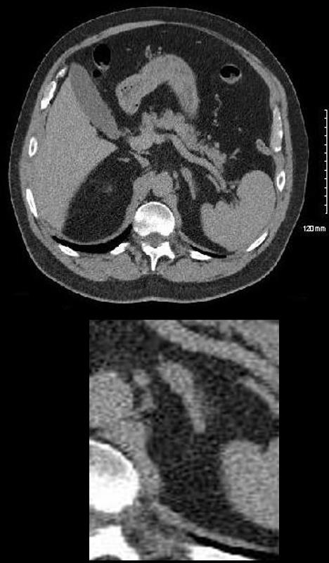
80 year old male presents with fatigue, CT in the axial projection shows a 1.5cms x .5cms nodule in the left adrenal gland
Ashley Davidoff MD TheCommonVein.net 292Lu 121889b01
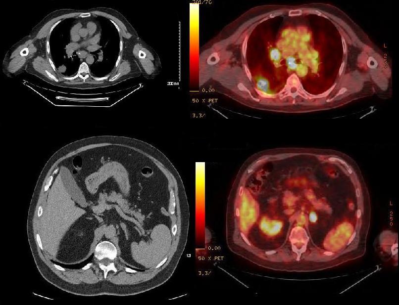
CT and PET
Adenocarcinoma Metastatic to
Left Hilum and Mediastinal Nodes
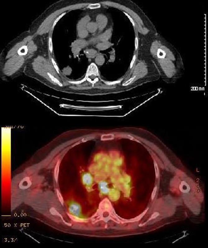
CT and PET scan of an 80 year old male with primary adenocarcinoma of the lung shows metastatic disease to the right hilum and mediastinum characterized by hypermetabolic activity in the primary mass and draining lymph nodes
Ashley Davidoff MD TheCommonVein.net 292Lu 121891c01
CT and PET
Adenocarcinoma Metastatic to the Left Adrenal Gland
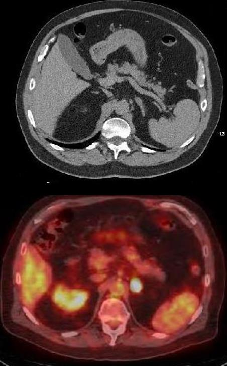
CT and PET scan of an 80 year old male with primary adenocarcinoma of the lung shows metastatic disease to the left adrenal gland characterized by hypermetabolic activity in the 1.5 x .5cms nodule
Ashley Davidoff MD TheCommonVein.net 292Lu 121891c02
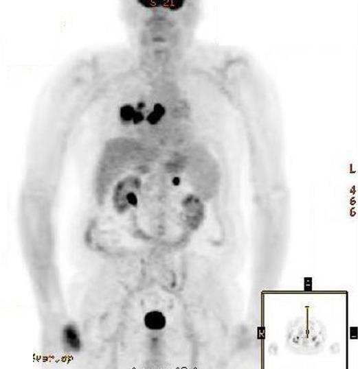
Ashley Davidoff TheCommonVein.net 292Lu 121892b
Ashley Davidoff,TheCommonVein.net 292 u121891
MRI Adenocarcinoma of the Lungs Metastatic to the Brain
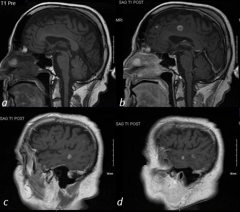
MRI of an 80 year old male with primary adenocarcinoma of the lung show metastatic disease to the brain. I mage a is a non contrast T1 weighted image prior to contrast administration. Images b, c ad are sagittal images of the brain following gadolinium administration and shows multiple enhancing lesions in the frontal parietal and temporal lobes consistent with metastatic disease
Ashley Davidoff MD TheCommonVein.net 292Lu 121893cL
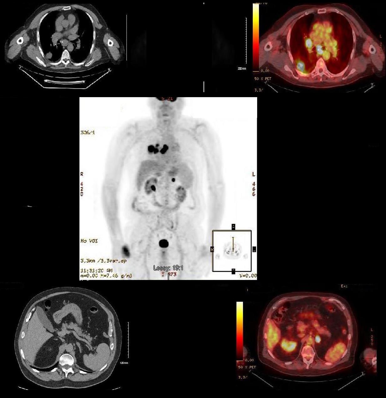
Ashley Davidoff TheCommonVein.net 292Lu 121892