
Squamous cell Carcinoma 4 Years Prior Chemo Radiation
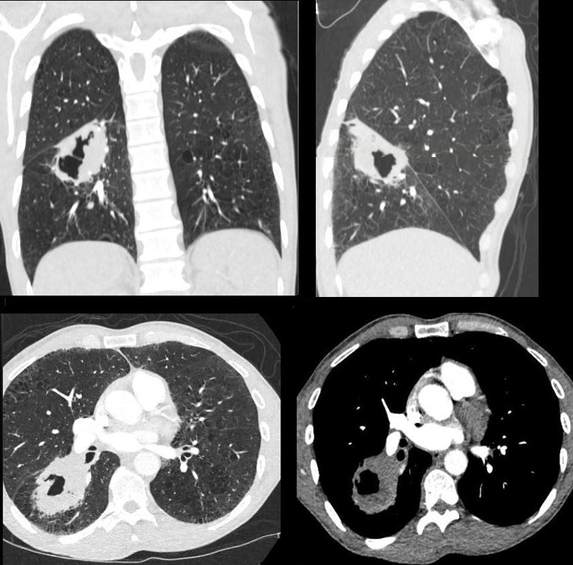
50 year old male with cough and weight loss
Coronal and sagittal CT reconstructions show a cavitating mass in the superior segment of the right lower lobe (upper images) correlated with axial images (lower panel)
Ashley Davidoff MD TheCommonVein.net 176Lu 136737
9 Months After Chemo Radiation
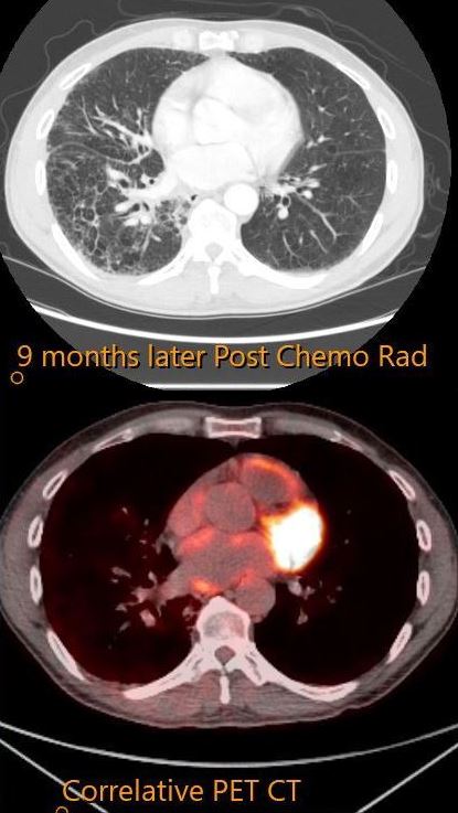
50 year old male with cough and weight loss
CT in the axial plane (upper image) shows resolution of the cavitating mass in the superior segment of the right lower lobe confirmed by the axial PET image. Post XRT fibrosis is noted in the apical segment of the RLL
Ashley Davidoff MD TheCommonVein.net 176Lu 136739
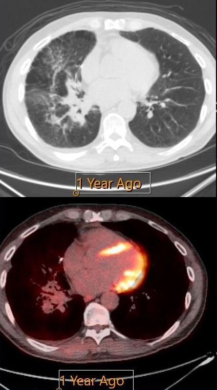
1 year after Chemo XRT
50 year old male with cough and weight loss
CT in the axial plane shows resolution of the cavitating mass in the superior segment of the right lower lobe confirmed by the axial PET image.
Post XRT fibrosis is noted in the apical segment of the RLL
Ashley Davidoff MD TheCommonVein.net 176Lu 136740
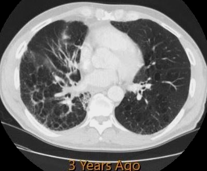
3 Years after Chemo XRT
50 year old male with cough and weight loss
CT in the axial plane shows resolution of the cavitating mass in the superior segment of the right lower lobe confirmed by the axial PET image. However there is progressive thickening of the bronchovascular bundle and close follow up would be needed. Post
XRT fibrosis is noted in the apical segment of the RLL
Ashley Davidoff MD TheCommonVein.net 176Lu 136741
4 Years after Chemo XRT
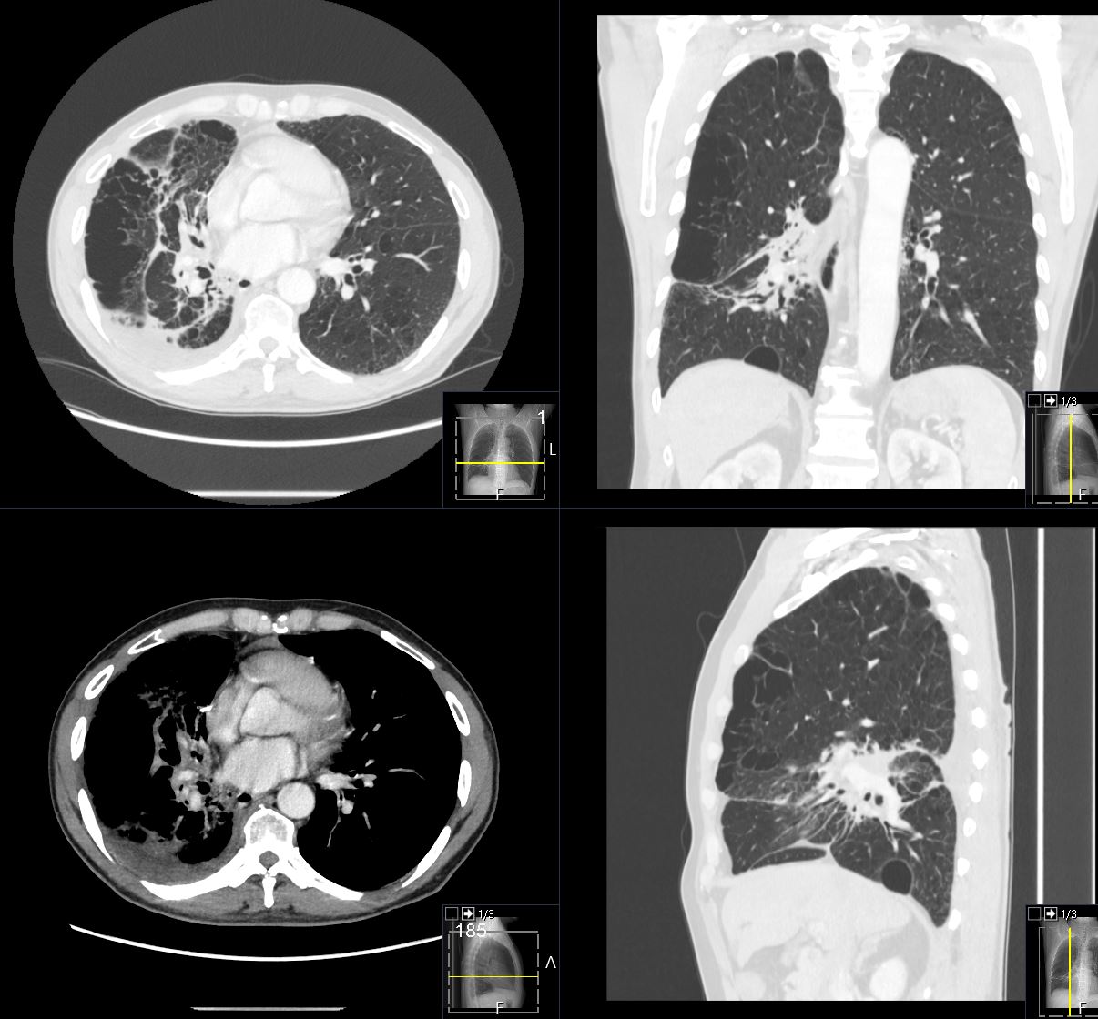
CT in the axial plane shows resolution of the cavitating mass in the superior segment of the right lower lobe confirmed by the axial PET image.
However there is progressive thickening of the bronchovascular bundle and PET scan would be indicated.
Post XRT fibrosis is progressive in the apical segment of the RLL.
There is background extensive paraseptal and centrilobular emphysema.
Ashley Davidoff MD TheCommonVein.net 176Lu 136742