
Ashley Davidoff MD TheCommonVein.net
enlarged left upper lobe superior segmental artery, culminating in a fusiform Rasmusen aneurysm. In the left apex, (image right) there are fibrotic changes which on other images had associated calcified granulomas, bronchiolectasis and and scarring reminiscent of latent TB
Ashley Davidoff MD
TheCommonVein.net
A Cavity Fibrosis A Calcification and Rasmusen Aneurysm
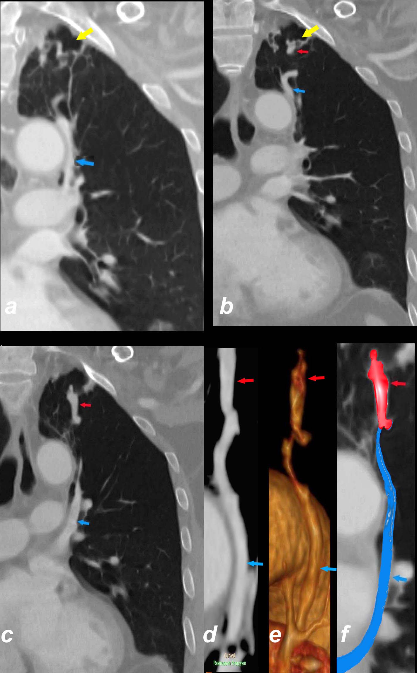
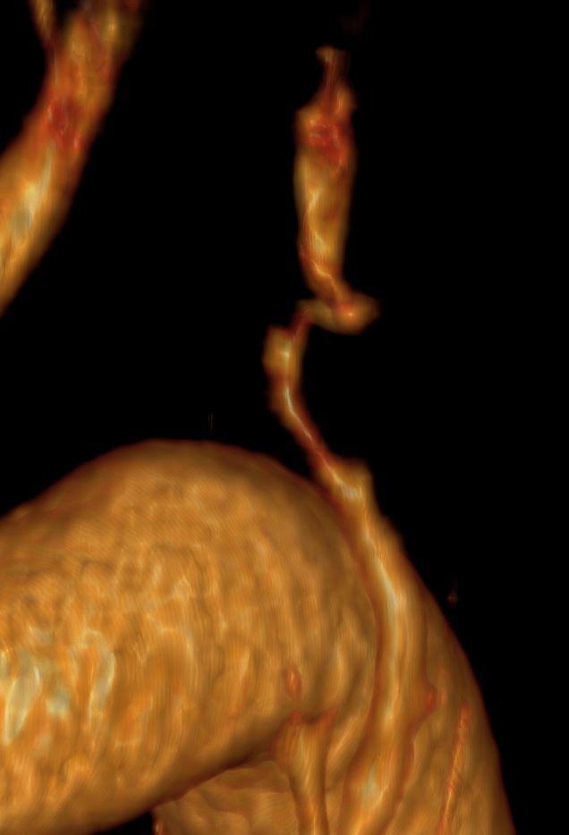
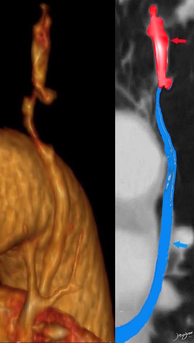
Coronally reformatted CT images through the left upper lobe shows cytic area (yellow arrow, a) likely a healed cavity in this context, in a region of fibrosis and a cluster of vessels. Image b again shows the cystic area (yellow arrow), the narrowed origin of the Rasmusen aneurysm (red arrow ) as well as a portion of the enlarged superior segmental artery (blue arrow). Image c shows the fusiform aneurysm (red arrow) and the proximal portion of the enlarged superior segmental artery (blue arrow). Image d is a multiplanar reformat of the segmental artery (blue arrow and) and the aneurysm (red arrow) reproduced in a 3D rendering in e, with color coded overlay in f.
Ashley Davidoff MD TheCommonvein.net 17a

Axial CT images through the left upper lobe show cluster of vessels in a region of fibrosis and cystic changes (yellow arrow, a). Image b shows a focal aneurysm (red arrow) and a calcified granuloma (white arrow). Image c shows the enlarged superior segmental artery (blue arrow) giving rise to the narrow neck of the Rasmusen aneurysm (red arrow, c). Image d shows the enlarged superior segmental artery (blue arrow) caught in a focal region of chronic atelectasis with a cluster of airways showing traction bronchiolectasis (orange arrows)
Ashley Davidoff MD TheCommonvein.net