
Infection Inflammation
Benign Neoplasm
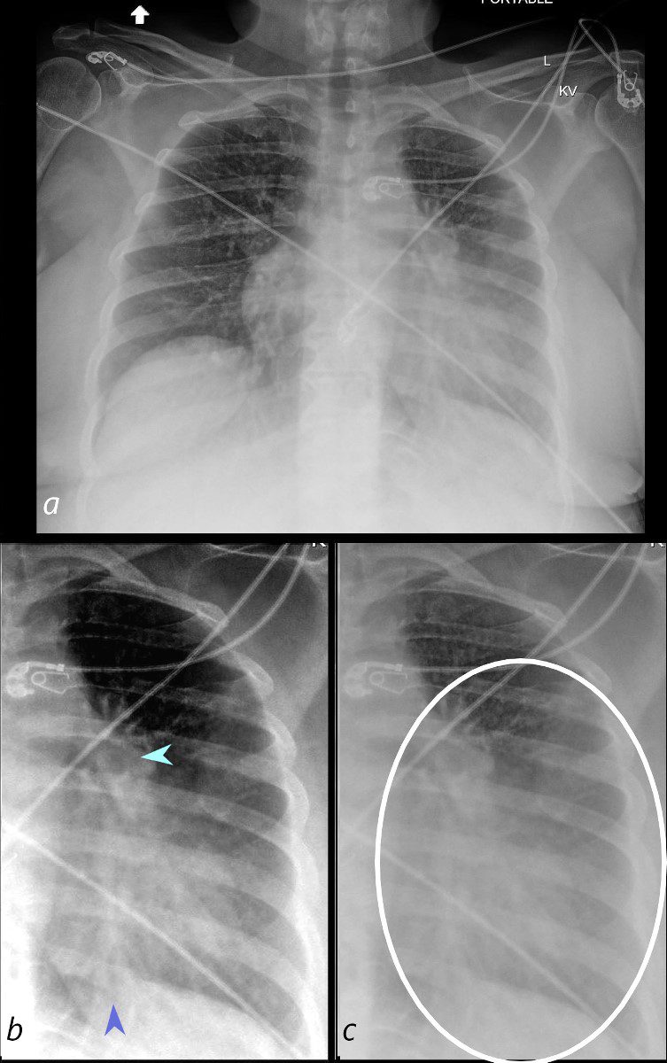
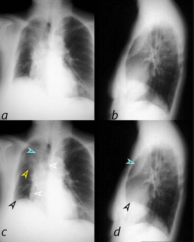
58-year-old female presents with a cough Frontal CXR shows silhouetting of the left heart border with hazy or veiling opacity extending out from the left hilum and fading out inferiorly (white circle c). The left hilum is pulled superiorly (teal arrowhead b) , resulting in an almost horizontal course of the left main bronchus and vertical orientation of the left lower lobe bronchovascular bundle (dark blue arrowhead b)
Ashley Davidoff MD TheCommonVein.net 257Lu 136109cL01
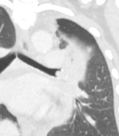
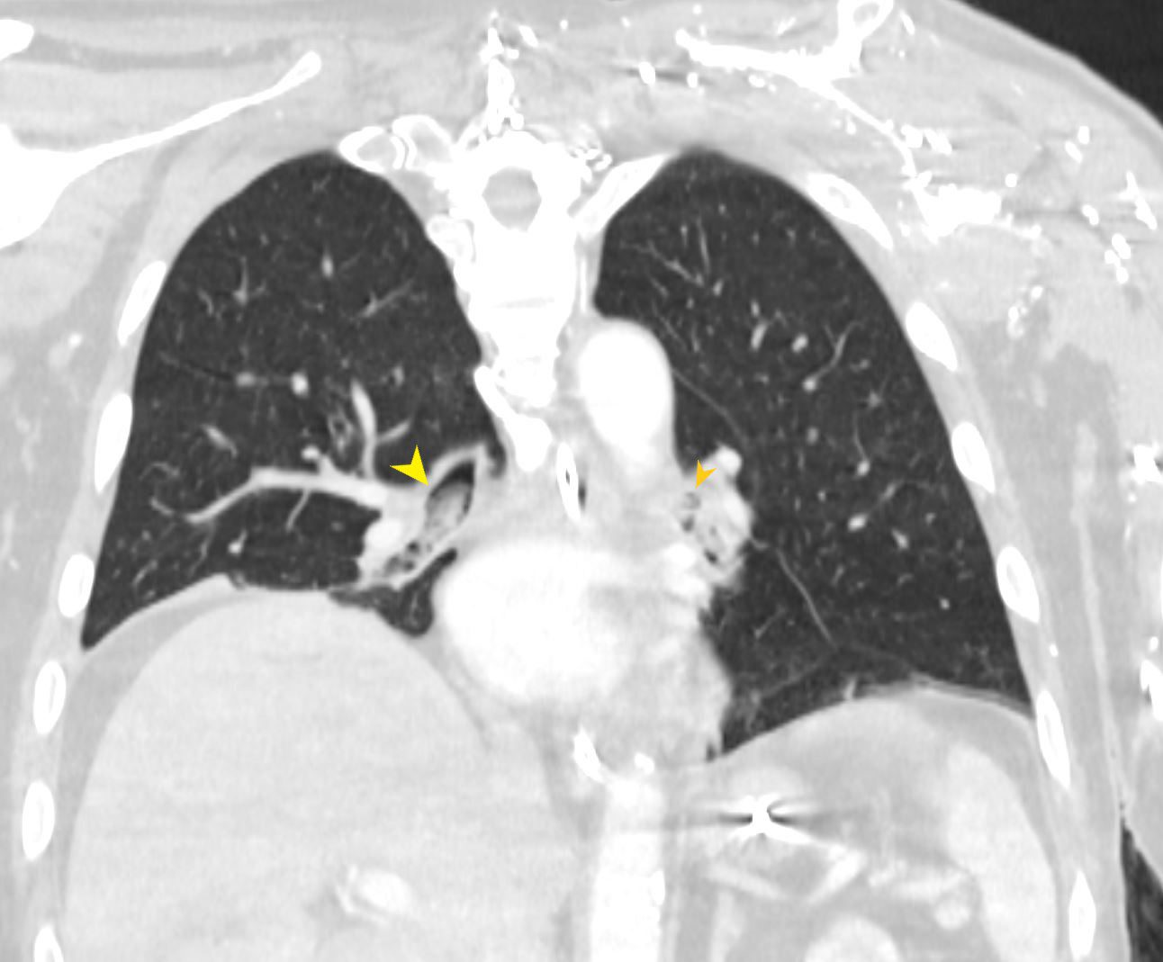
58-year-old female presents with a cough Coronal CT shows a nodule at the branching of the more horizontally oriented left mainstem bronchus with post obstructive atelectasis of the lingula and mild hyperinflation of the upper lobe segments.
Pathology revealed findings consistent with a carcinoid tumor of the left bronchus.
Ashley Davidoff MD TheCommonVein.net 257Lu 136118
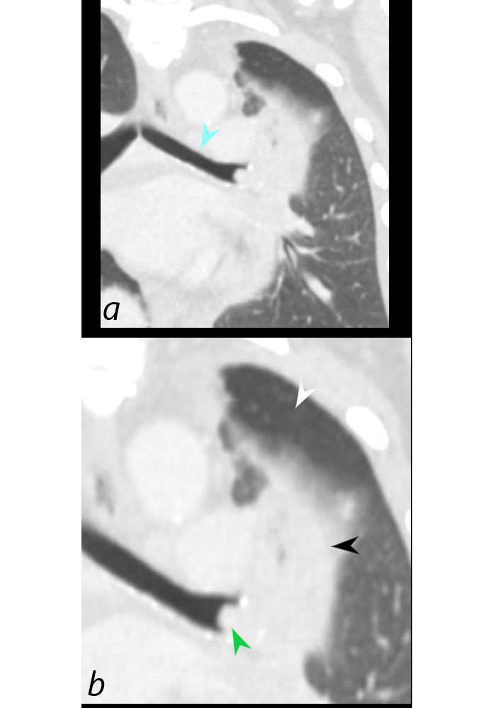
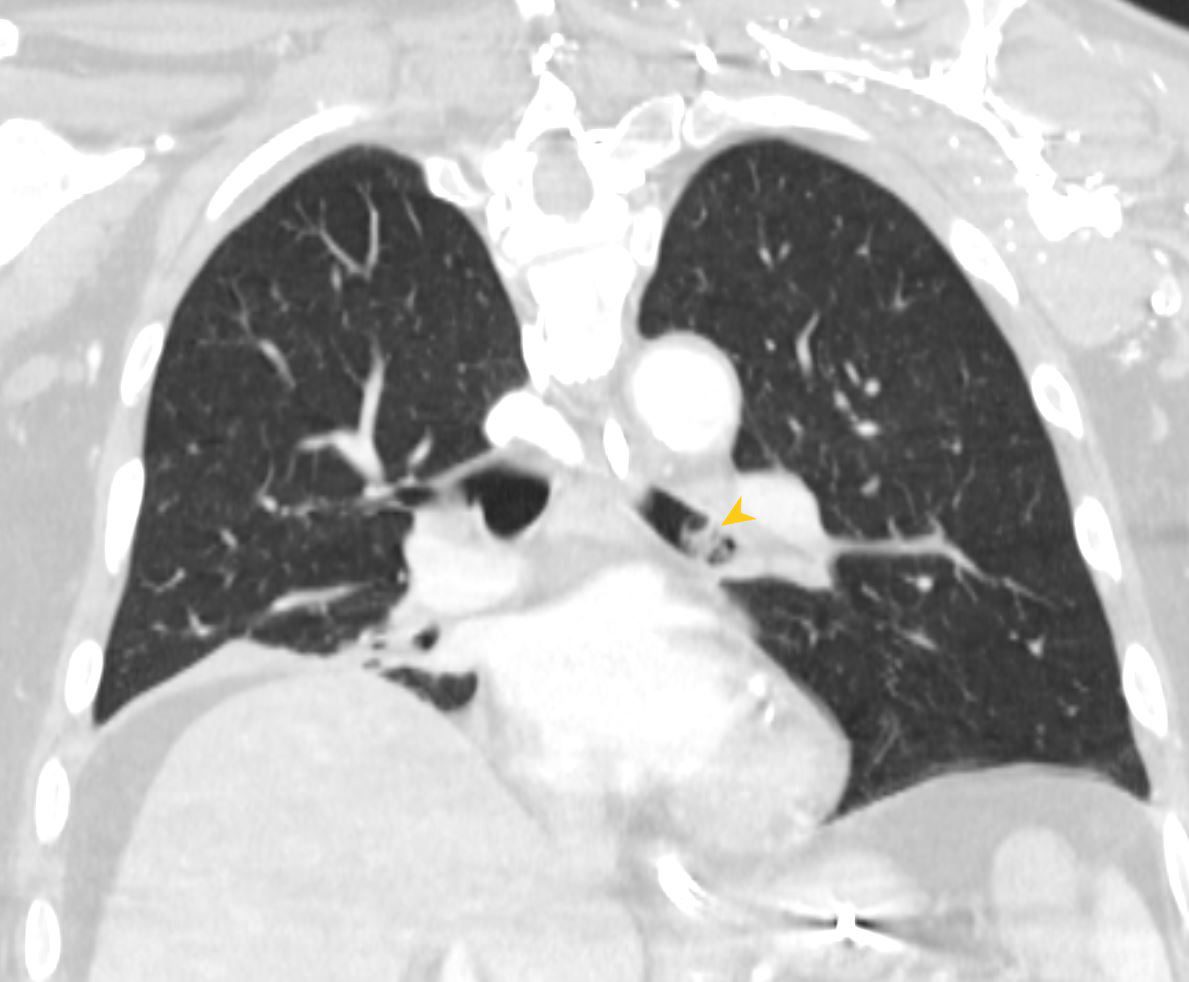
58-year-old female presents with a cough. Coronal CT shows a nodule (green arrowhead, b) at the branching of the more horizontally oriented left mainstem bronchus (teal arrow, a) with post obstructive atelectasis of the lingula (black arrowhead, b) and hyperinflation of the superior aspect of the lower lobe (white arrowhead) which occupies portion of the left apex. (Luftsichel sign)
Pathology revealed findings consistent with a carcinoid tumor
Ashley Davidoff MD TheCommonVein.net 257Lu 136118c
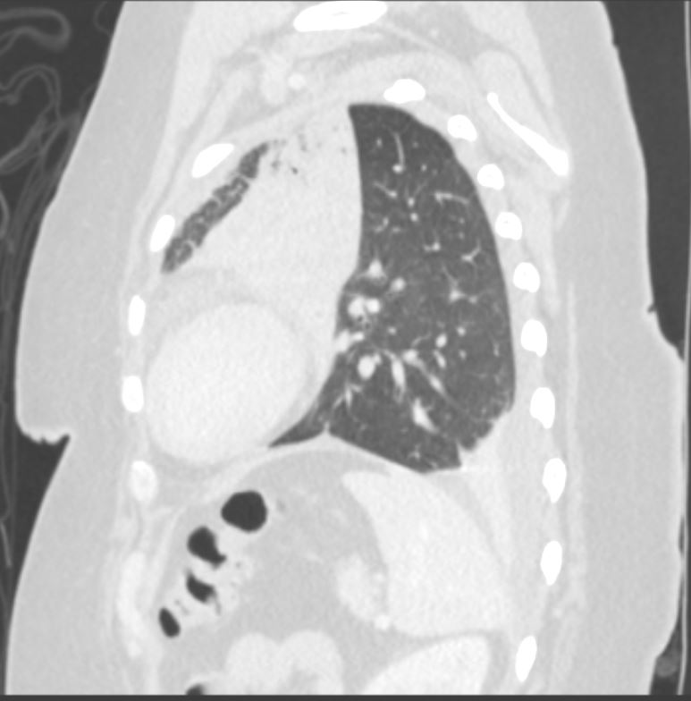
58-year-old female presents with a cough. CT in the sagittal plane shows post obstructive atelectasis of the lingula, hyperinflation of the left lower lobe, superior and anterior migration of the left major fissure, and a small portion of aerated left upper lobe anteriorly. There is a loculated effusion with subsegmental compressive atelectasis of the left lower lobe.
Pathology revealed findings consistent with a carcinoid tumor in the left mainstem bronchus
Ashley Davidoff MD TheCommonVein.net 257Lu 136121
Malignancy
Right Upper Lobe Collapse
Squamous Cell Causing
Obstruction but Airways Filled with
Tumor or Infection or Mucus

CXR shows right upper lobe (RUL) atelectasis. Final diagnosis was a central RUL proximal squamous cell carcinoma with extensive filling of the distal bronchi-ectatic segmental and subsegmental airways
Ashley Davidoff TheCommonVein.net

CXR shows right upper lobe (RUL) atelectasis. Final diagnosis was a central RUL proximal squamous cell carcinoma with extensive filling of the distal bronchi-ectatic segmental and subsegmental airways
Ashley Davidoff TheCommonVein.net

Ashley Davidoff TheCommonVein.net

Ashley Davidoff TheCommonVein.net

Ashley Davidoff TheCommonVein.net
Lobar Collapse
Right Upper Lobe Collapse
Occluded Right Main Stem Bronchus by Carcinoma with Pathology Correlation
This combination of images shows the manifestations of a malignant mass in the hilum causing compression of the right mainstem bronchus. The PA CXR shows signs of volume loss (atelectasis characterized by elevation of the right hemidiaphragm (black arrowhead), rightward tracheal and mediastinal shift (white arrowheads) and elevation of the minor fissure contributing to the reverse S sign of Golden. There is a vague infiltrate in the right upper lobe correlating with an anterior pie shaped density of the lateral (blue arrowheads), consistent with collapse of the anterior segment of the RUL
32292cL01
Ashley Davidoff MD TheCommonVein.net
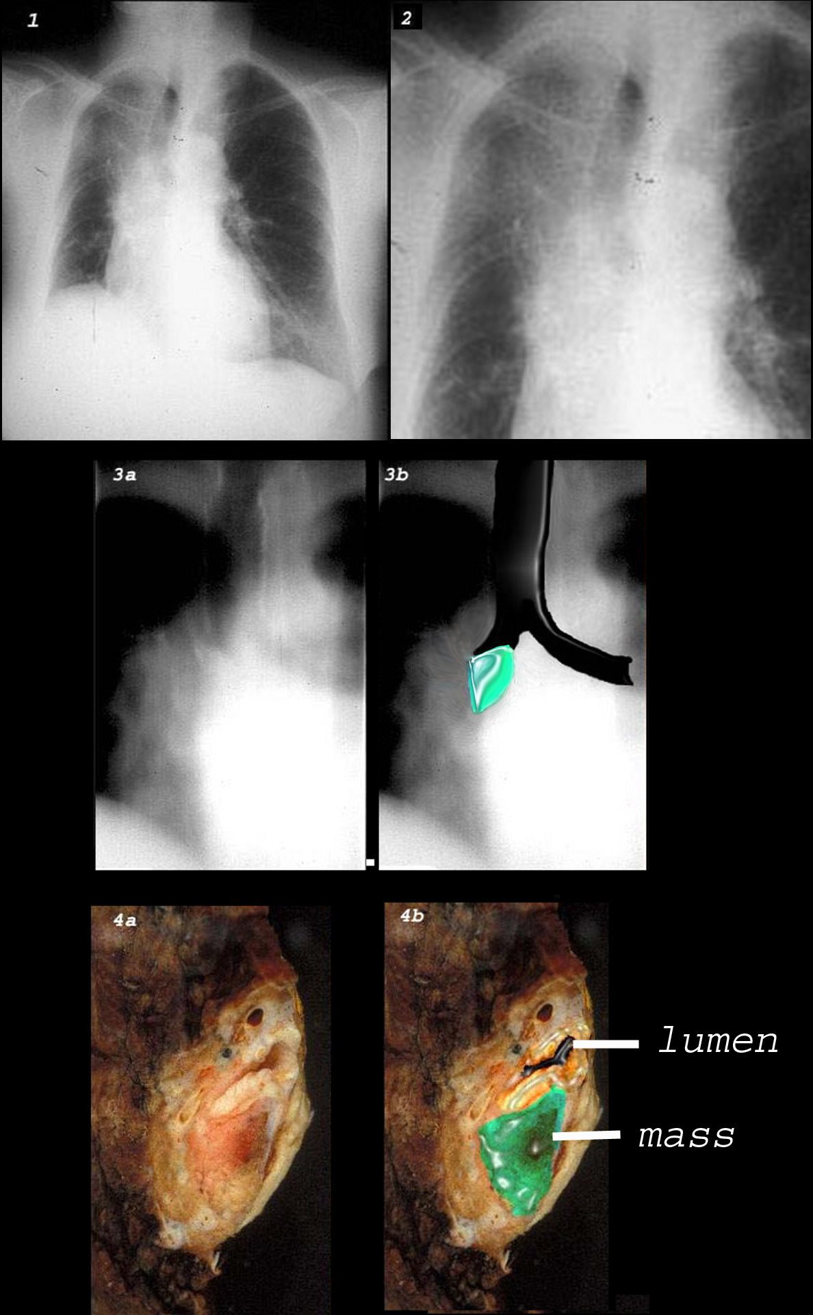
This combination of images shows the manifestations of a malignant mass in the hilum causing compression of the right mainstem bronchus. There is elevation of the right hilum on the CXR, associated with collapse of the anterior segment of the RUL seen as a vague density in the P-A . The tomogram (3a) shows an abrupt cut off of the right mainstem bronchus while the overlay in 3b shows the occlusion of the right mainstem bronchus, the implied tumor overlaid in green. Images 4a and 4b are the correlative gross pathology images showing the tumor in green pushing and occluding the right mainstem
Ashley Davidoff MD TheCommonVein.net
Right Upper Lobe Atelectasis
Reversed S Sign of Golden

The scout film performed prior to a CT scan from a 76-year-old man with chest pain and shortness of breath. The appearance suggests atelectasis of the right upper lobe with the normal position of the minor fissure (yellow) altered so that the upper portion (light green above the yellow line) is shifted upward caused by volume loss of an atelectatic right upper lobe (pink). The lower portion of the fissure (light green below the yellow line) is bulging rightward and outward caused by an implied mass (dark green). The “reversed S sign of Golden” is demonstrated in this case and infers a central mass causing obstruction and resulting in the shape described by the light green line of the minor fissure.
Courtesy: Ashley Davidoff, M.D.
The Soft Tissue Tumor is Noted in the Left Main Stem Bronchus
82-year-old female with dyspnea presents with an obstructing and infiltrating central squamous cell carcinoma of the left main stem bronchus with secondary post obstructive atelectasis of the left upper lobe of the lung. In addition, there is encasement of the left pulmonary artery and a small left effusion. A spiculated lesion at the base of the left breast in close association with the left pectoralis muscle The lesion also extends beyond the muscle to abut the rib. There is a small amount of fluid in the pericardial recess, and an small left pleural effusion.
Ashley Davidoff MD TheCommonVein.net RnD case

Female patient with central squamous cell carcinoma of the lung with left upper lobe collapse and hyperinflation of the left lower lobe resulting in a Luftsichel sign.
The left lung is relatively lucent as a result of hyperinflation . The atelectatic left upper lobe manifests as an anterior soft tissue density along the anterior mediastinum. The soft tissue tumor is noted in the left main stem bronchus
Ashley Davidoff MD TheCommonVein.net 152Lu
Endoscopic view of the Left Main Bronchus showing the
Obstructing Tumor

Ashley Davidoff MD TheCommonVein.net 152Lu
Mechanical/Atelectasis
Bilateral Lower Lobar Atelectasis with
Occlusion of the Right Main Stem Bronchus
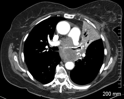
74 year old male alcoholic with bilateral basilar lobar atelectasis caused by bilateral aspiration
CT scan shows airless lower lobes with small bilateral effusions. 3D reconstruction shows total obstruction of the right mainstem bronchus, and patent proximal mainstem bronchus
Ashley Davidoff MD TheCommonVein.net
Aspiration of Solid Food Right and Left lower Lobe Bronchi
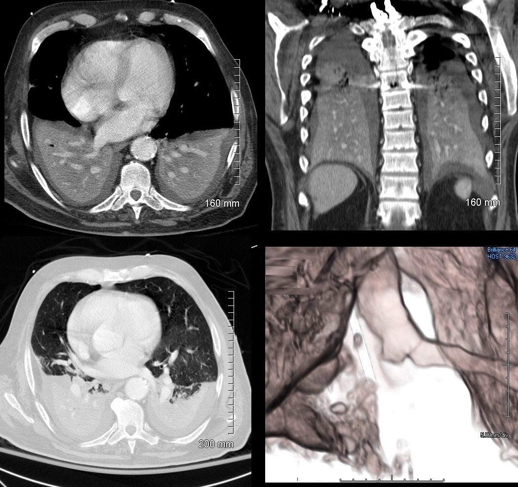
71-year-old male presents with acute respiratory difficulty. CT in the coronal plain shows solid food particles in right mainstem bronchus (yellow arrowhead), extending into the right lower lobe bronchus, as well as segmental airways to the left lower lobe (orange arrowheads). The right hemidiaphragm is elevated secondary to the atelectasis
Ashley Davidoff MD TheCommonVein.net 271Lu 136241
71-year-old male presents with acute respiratory difficulty. CT in the coronal plain shows solid food particles in left mainstem bronchus (orange arrowhead) . The right hemidiaphragm os elevated secondary to the atelectasis
Ashley Davidoff MD TheCommonVein.net 271Lu 136240
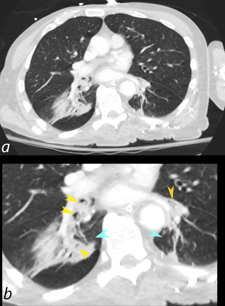
71-year-old male presents with acute respiratory difficulty. CT in the axial plain shows solid food particles in the apical segment of the right lower lobe of the lung (b, yellow arrowheads) and in the left lower lobe (b, orange arrowheads) associated with bilateral basilar subsegmental atelectasis right greater than left (b, teal arrowheads).
Ashley Davidoff MD TheCommonVein.net 271Lu 136237cL
Trauma Metabolic Circulatory- Hemorrhage Immune
Infiltrative
Amyloid
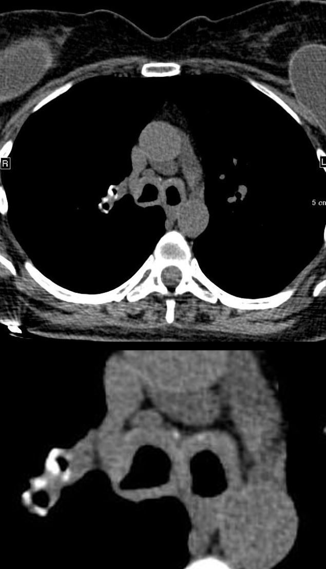
44-year-old female with immunoglobulin light chain (AL) amyloidosis of the mainstem bronchi and right upper lobe bronchi. The axial CT shows circumferential thickening of the mainstem bronchi without obstruction. The walls of the mainstem bronchi are minimally calcified anteriorly and there is circumferential calcification of the right upper lobe segmental airways.
Ashley Davidoff MD TheCommonVein.net 240Lu 135867b
Idiopathic ? Inherited
Mounier Kuhn
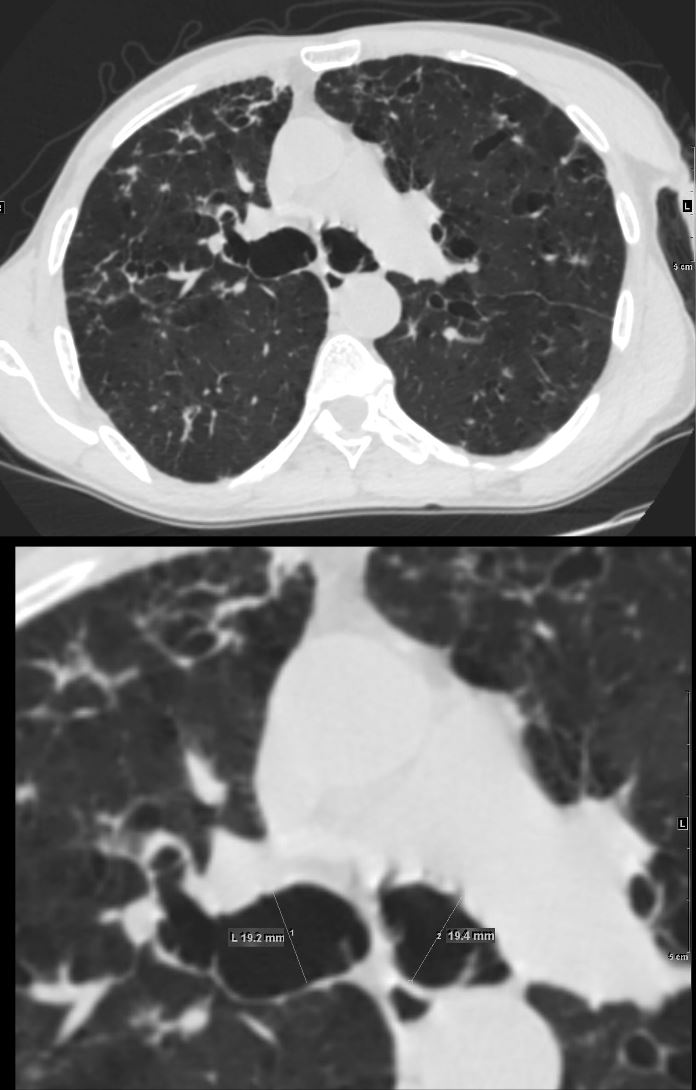
61 year old male with a history of treated mycobacterial infections and chronic cough
Axial CT at the level of the carina shows bilaterally enlarged mainstem bronchi that measure 1.9cms. each which are abnormally enlarged. There are both thin-walled cystic changes of the airways along the subsegmental arteries in the upper lobes likely reflecting bronchiectasis . Some of these cystic changes in the right upper lobe (upper panel) have thicker walls
Ashley Davidoff MD TheCommonVein.net 250Lu 135875a
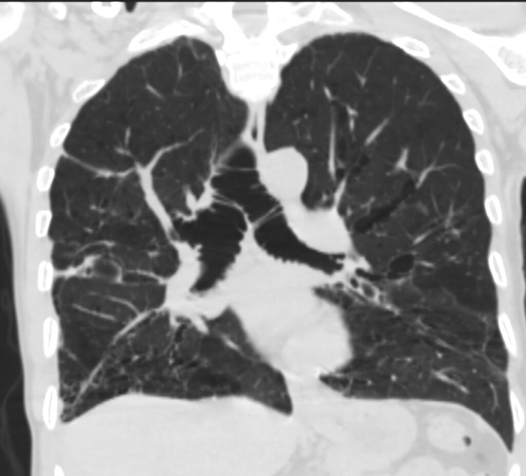
61-year-old male with a history of treated mycobacterial infections and chronic cough
Coronal CT at the level of the carina shows bilaterally enlarged mainstem bronchi that measure 1.9cms. each which are abnormally enlarged. There are both thin-walled cystic changes of the airways along the subsegmental bronchovascular bundles in the upper lobes reflecting bronchiectasis .
Ashley Davidoff MD TheCommonVein.net 250Lu 135881
Iatrogenic