
Ashley Davidoff MD TheCommonVein.net 118884
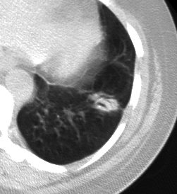
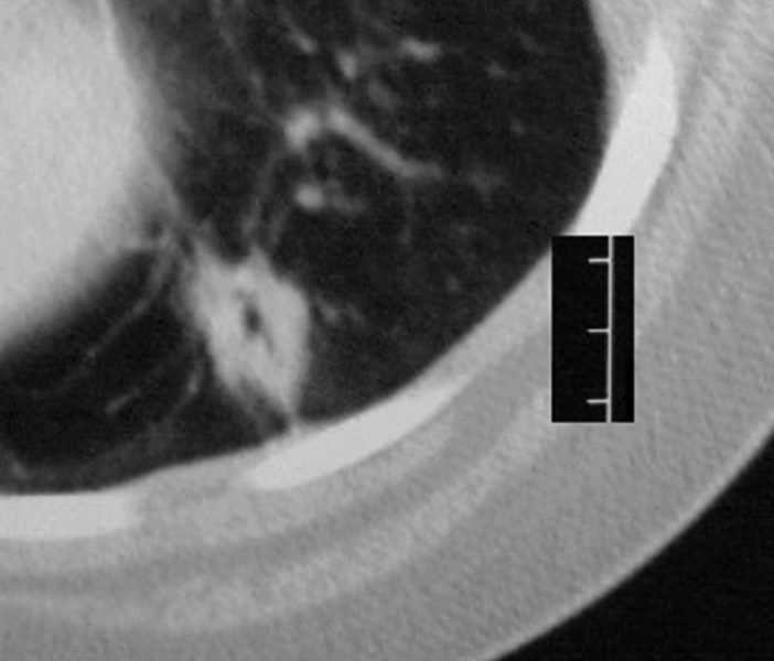
Within the posterior segment of the left upper lobe there a 2.5 cms nodule with mild reticulations likely a lymphomatous deposit
42068c03
Ashley Davidoff MD TheCommonVein.net
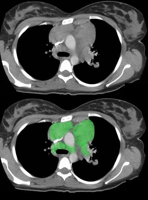
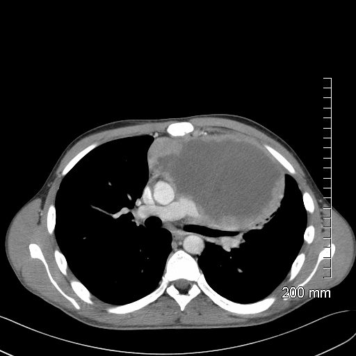
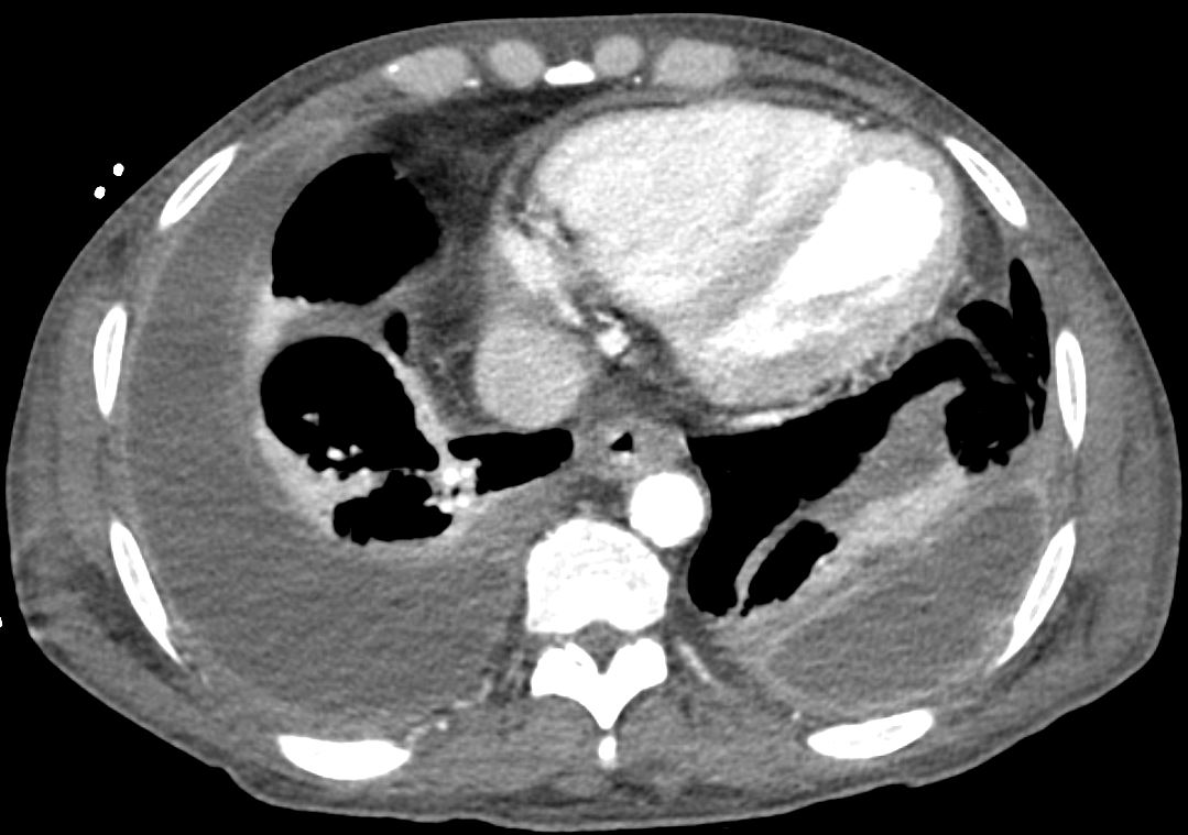
This transverse section through the top of the aortic arch shows multiple enlarged soft tissue masses in the superior mediastinum representing enlarged lymph nodes in this patient with lymphoma.
he enlarged lymph nodes are outlined in green.
Ashley Davidoff MD TheCommonVein.net 42062c01
Radiology Vol. 297, No. 3 July 2020
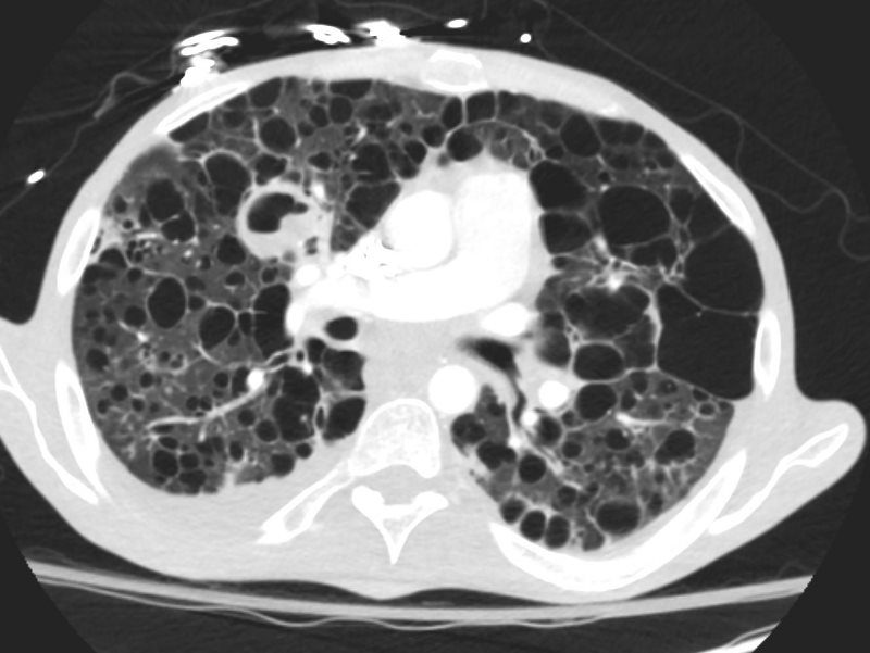
62 year old female with bronchocentric BALT lymphoma in the left lower lobe
Ashley Davidoff MD TheCommonVein.net
121259m
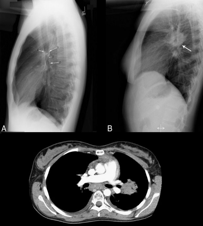
57 year old female with path proven lymphoma of the chest presenting with a broncho centric nodule
Ashley Davidoff MD TheCommonVein.net
30912m
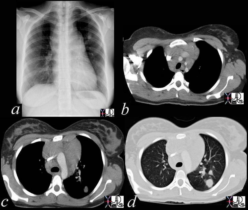
27 year old male with a history of perinatal HIV with intermittent highly active antiretroviral therapy (HAART) compliance with a CD4 count of < 50 with biopsy confirmed B cell lymphoma of the liver, s/p CHOP therapy , chronic esophageal strictures s/p dilatations, esophageal candidiasis, LIP, bronchiectasis pancreatitis, and portal vein and splenic vein thrombosis.
Initial Chest X-ray shows a diffuse reticular pattern with cystic changes dominant at the bases.
CT at that time confirmed the presence of diffuse cystic changes with the largest cysts at the lung bases. Ascites and splenomegaly are also present
He presented one month later with fever and neutropenia.
CT showed an abscess cavity in the right upper lobe in the right upper lobe, thickened distal esophagus with edematous wall, atrophic gastritis and ascites. Bronchovascular thickening along a bronchiectatic segment in the right upper lobe was present in the last CT
Ashley Davidoff MD

Source
Signs in Thoracic Imaging
Journal of Thoracic Imaging 21(1):76-90, March 2006.
Human Herpes Virus 8 and Primary Effusion Lymphoma (PEL)
Axial CT scan with contrast of 71-year-old male shows bilateral complex and loculated effusions with thickened enhancing pleura. Pleural tap of the left effusion revealed evidence of lymphoma likely related to the patients underlying herpes virus infection. An entity called primary effusion lymphoma (PEL) is a rapidly progressing non-Hodgkin’s B-cell lymphoma that develops in body cavities
Ashley Davidoff MD TheCommonVein.net 135684