
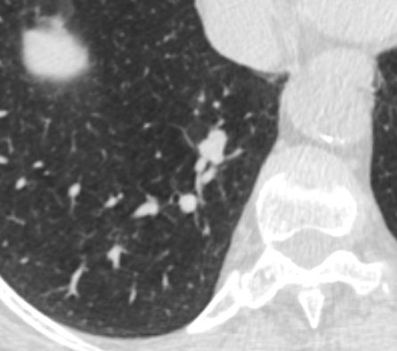
Path showed atypical adenomatoid hyperplasia
Ashley Davidoff TheCommonVein.net
atypical adenomatoid hyperplasia 001 57f
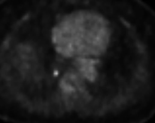
Path showed Atypical adenomatoid hyperplasia
Ashley Davidoff TheCommonVein.net
atypical adenomatoid hyperplasia 002 57f
Atypical adenomatoid hyperplasia (AAH) of the lung is a term used to describe a non-cancerous (benign) proliferation of cells in the lung tissue. It is considered a precursor lesion to adenocarcinoma, a type of lung cancer. AAH is characterized by the abnormal growth and arrangement of cells in the lung, but it lacks the features necessary to classify it as a malignant tumor.
The exact cause of atypical adenomatoid hyperplasia is not fully understood. However, it is believed to be associated with genetic mutations and exposure to risk factors such as smoking or environmental pollutants. AAH is often discovered incidentally during imaging studies or biopsies performed for other reasons.
AAH lesions are usually small and solitary, and they can occur in any part of the lung. They do not typically cause symptoms and are usually discovered during routine medical examinations or when evaluating other lung conditions. However, in some cases, AAH can mimic lung cancer on imaging studies, making it important to differentiate between the two.
Diagnosing atypical adenomatoid hyperplasia involves imaging studies, such as chest X-rays, computed tomography (CT) scans, and positron emission tomography (PET) scans. These tests help visualize the lesion and evaluate its characteristics. In some cases, a biopsy may be performed to confirm the diagnosis. During a biopsy, a small sample of the tissue is obtained and examined under a microscope.
The management of atypical adenomatoid hyperplasia depends on several factors, including the size, location, and characteristics of the lesion, as well as the individual’s overall health. In general, if the lesion is small and does not show any worrisome features, it may be monitored closely with regular imaging studies. If the lesion grows or shows concerning features, surgical removal may be recommended. The goal of treatment is to prevent the progression of AAH to adenocarcinoma.