
Lung Cancer, Tension Hydro/Hemothorax, and Atelectasis
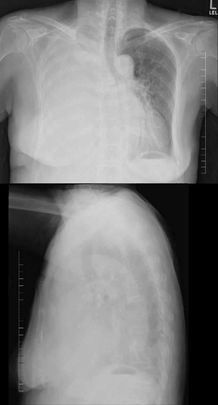
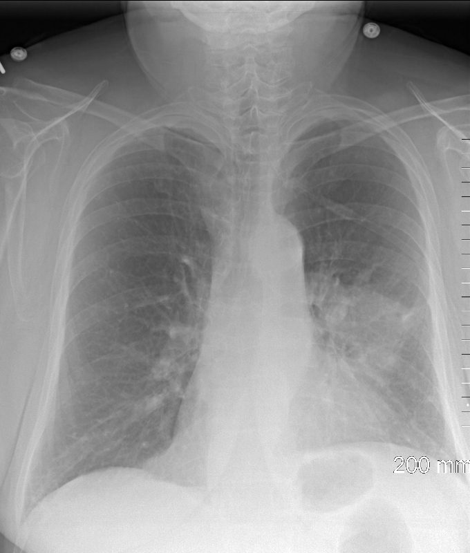
85-year-old female with a history of lung cancer, presents with a dyspnea and hypotension. CXR shows “white out” of the right hemithorax with pressure effect characterised by narrowing of the distal trachea cardio-mediastinal shift and atelectasis in the left lower lobe. The lateral examination shows silhouetting of the right hemidiaphragm.
Ashley Davidoff MD TheCommonVein.net106Lu 118469c
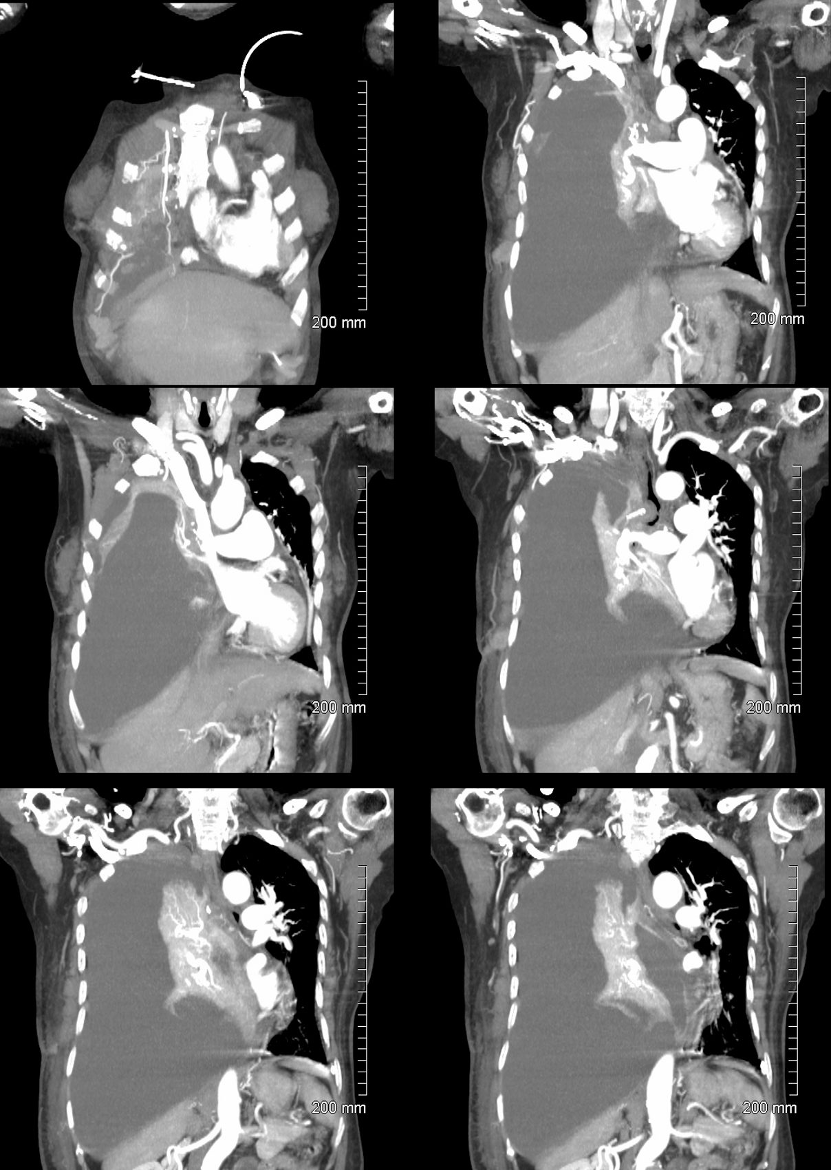
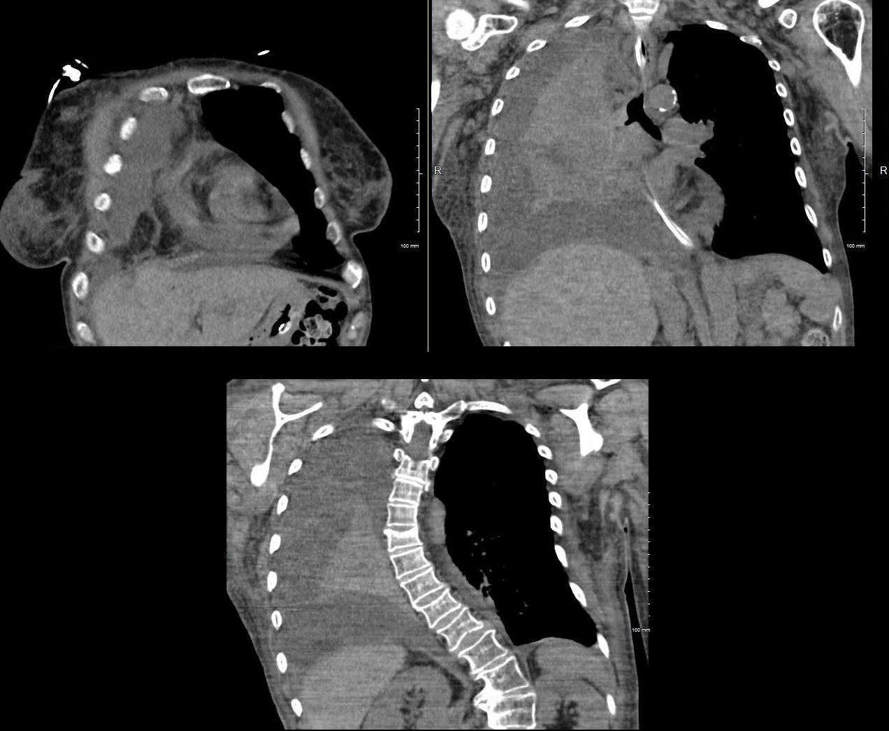
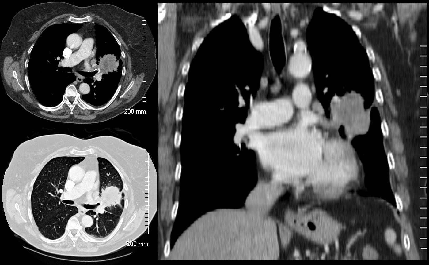
85-year-old female with a history of lung cancer, presents with a dyspnea and hypotension. CT scan shows a large right pleural effusion under pressure, with mediastinal shift to the right. In addition, there is compression of the heart with back up of venous return due the pressure effect on the heart and vascular structures. Among the structures showing venous distension are the SVC (blue arrowhead,a) right sided upper limb veins (blue arrowhead b) and the left upper pulmonary veins (red arrowhead, b. The effusion in the right pleural cavity with atelectatic lung herniates into the left hemithorax, (white arrowhead, c). There is a dense sediment in the pleural fluid (red arrowhead, d) suggesting blood in the pleural cavity. The left atrium is compressed (maroon arrowhead, d)
Ashley Davidoff MD TheCommonVein.net106Lu 118467c
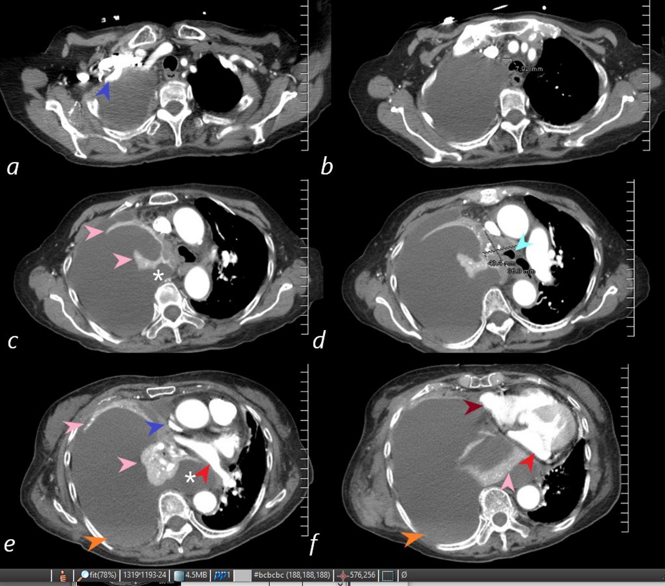
85-year-old female with a history of lung cancer, presents with a dyspnea and hypotension. CT scan shows a large right pleural effusion under pressure, suggestion of blood in the pleural cavity (orange arrowheads e, f) and mediastinal shift to the right. In addition, there is compression of the heart with back up of venous return due the pressure effect on the heart and vascular structures. Among the structures showing compressive effects are the SVC (blue arrowhead, c) with venous distension on right sided upper limb veins (blue arrowhead a). There is also pressure effect on the trachea with narrowing (light blue arrow d) The effusion in the right pleural cavity with atelectatic lung herniates into the left hemithorax, (white asterisk c, e). There is a dense sediment in the pleural fluid (orange arrowheads, e and f) suggesting blood in the pleural cavity. The left atrium is compressed (red arrowhead, e, f), and the right atrium is compressed (maroon arrow f).
Ashley Davidoff MD TheCommonVein.net 106Lu 118450cL
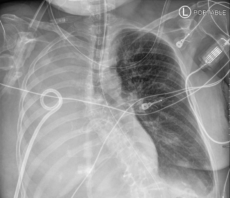
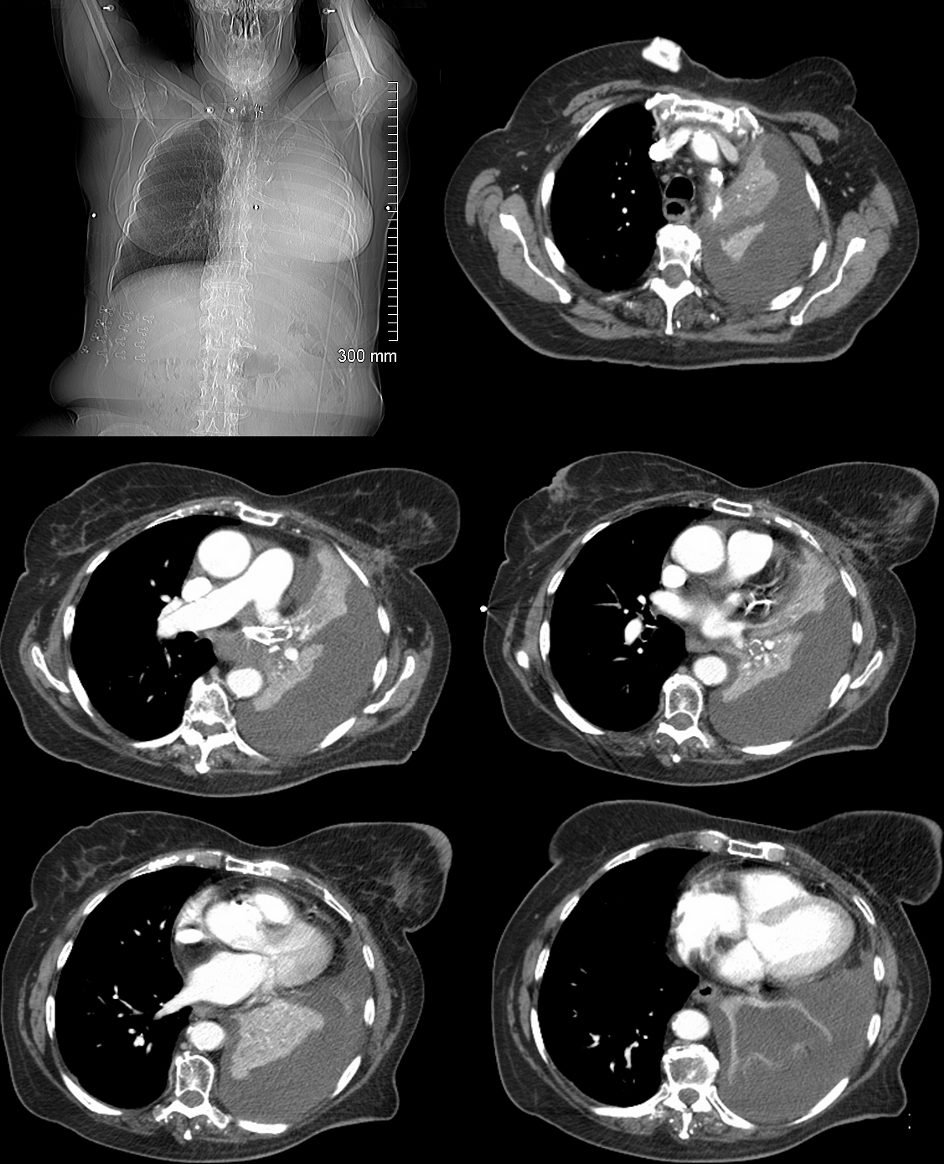
59F shows total white out caused by collapse of right lung with an
occluded right main step bronchus associated with a
large right sided effusion. The occlusion is likely due to proximal cancer. A pigtail drain has been placed to drain the effusion
Ashley Davidoff MD TheCommonVein.net 104Lu
59F shows total collapse of right lung with an
occluded right main step bronchus(top right image)associated with a
large right sided effusion. The occlusion is likely due to proximal cancer
Ashley Davidoff MD TheCommonVein.net 104Lu
82-year-old female with dyspnea presents with an obstructing lesion of the left main stem bronchus and total atelectasis of the left lung and associated effusion. White out of the left lung with mediastinal shift are noted on the scout film (top left) and the axial images confirm complete collapse of the left lung with a large effusion. A proximal malignancy was found, and the left mainstem bronchus stented, unsuccessfully.
Ashley Davidoff MD TheCommonVein.net
134720c
- Left Lung Collapse From a Mucus Plug

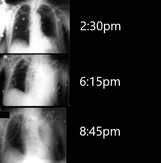
68 year old ICU patient has an Xray at 2:30pm to check tube position . 4 hours later he presents with a acute respirtatory difficulty and the re is a white out of the left lung (middle image). After suction, a repeat CXR approximately 2 hours later shows improving aeration. The rapid response to suction usually infers obstruction from a mucus plug or inhalation of a foreign body with removal .
Ashley Davidoff MD TheCommonVein.net
70231cL
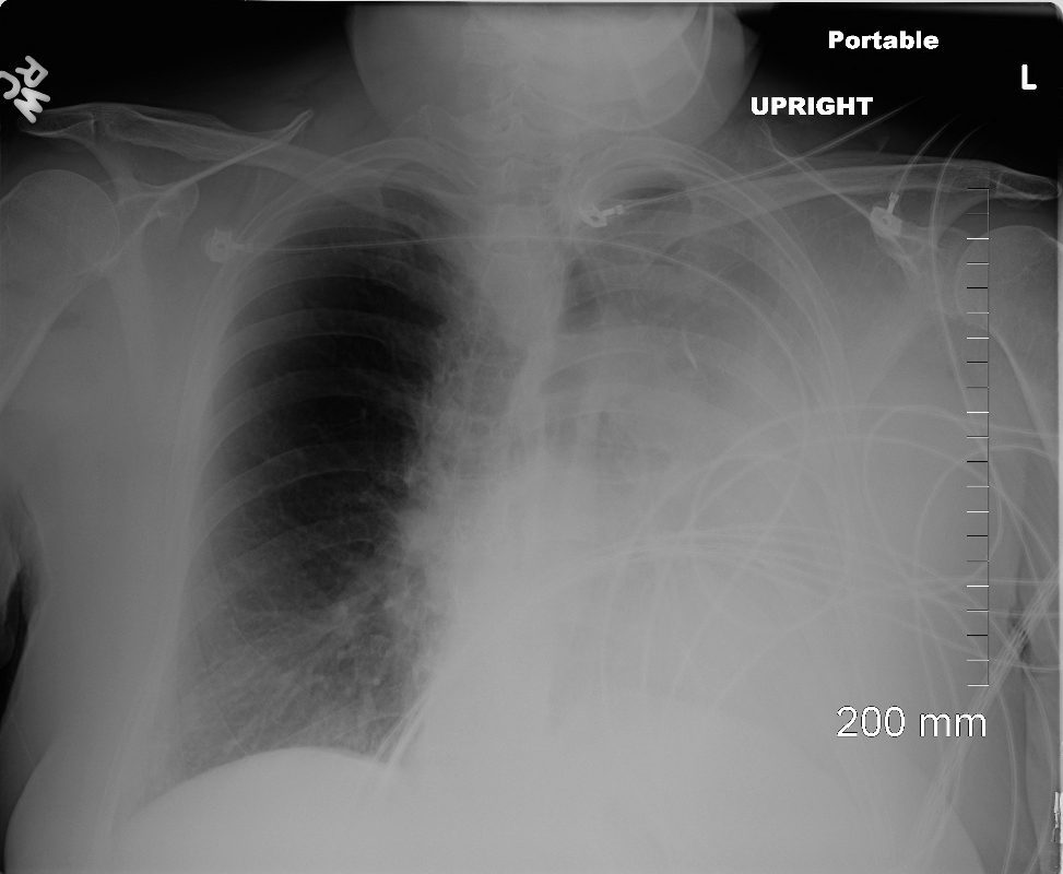
White Out After bronchoscopy for Central Cancer
Ashley Davidoff MD TheCommonVein.net
1 Day After Bronchoscopy Re- Aeration
Ashley Davidoff MD TheCommonVein.net
72 year old female presents with a cough. CT shows a suspicious 3.2cms central, spiculated left upper lobe mass.
Ashley Davidoff MD TheCommonVein.net