
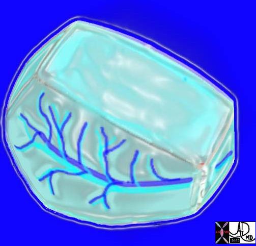
Here is a picture of the outside of the polyhedral pulmonary lobule from the side. It looked quite futuristic. Through the transparent side window we saw a couple similar to ourselves. From this vantage point the morphing did not look too different from what we had already been through – division after division – leaner and meaner. Ashley Davidoff MD. The Common Vein.net 42449b02

Ashley Davidoff MD TheCommonVein.net
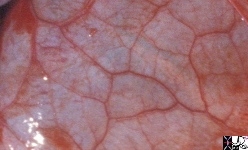
Gross anatomy specimen shows secondary lobules with interlobular septa containing pulmonary venules and the capillary components within the secondary lobules
Davidoff MD TheCommonVein.net 32557bb03.8s
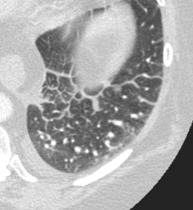
Axial CT in a patient with CHF demonstrates the heterogeneous morphology of the secondary lobule. In this patient the lobules in the periphery are larger than the centrally placed secondary lobules. In addition the peripheral lobules are more rectangular and those centrally are more of a polyhedral shape.
Ashley Davidoff MD TheCommonVein.net 135831
Small Peripheral Secondary Lobules and Varying Shapes Centrally
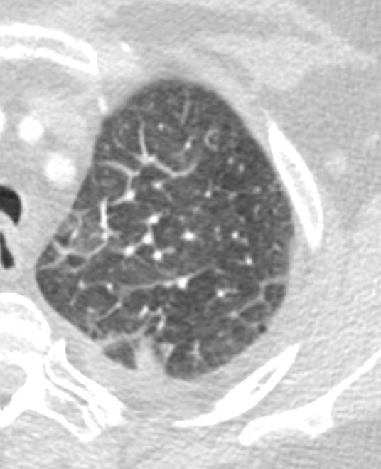
Axial CT in a patient with CHF demonstrates the heterogeneous morphology of the secondary lobule. In this patient the lobules in the periphery are smaller than the centrally placed secondary lobules. In addition the central lobules are more irregular in shape and are hexagonal or polygonal.. There is a small effusion.
Ashley Davidoff MD TheCommonVein.net 135834
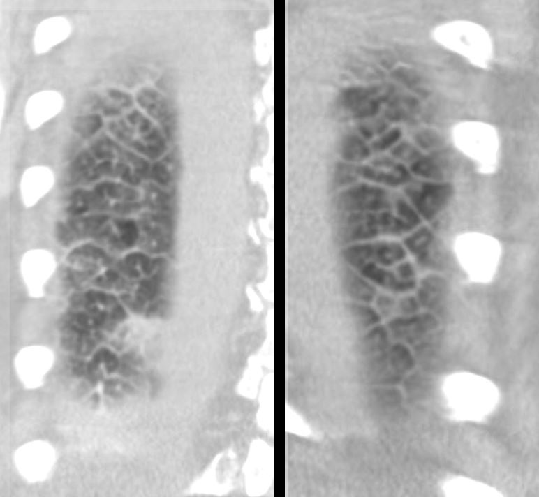
Coronal CT through the posterior chest in a patient with CHF demonstrates the heterogeneous morphology of the secondary lobule. There is a wide range of sizes and shapes, with wide variations of hexagonal and polygonal morphology. Some of the shapes are innate but consideration must be given to the orientation of the lobule and the varying angles we may catch them in a single plane of this technique.
Ashley Davidoff MD TheCommonVein.net 135832c