
Nodular amyloidosis usually presents with peripheral subpleural localisations of variable size that can be bilateral.
60 year old female with known diagnosis of AL Amyloidosis
CXR Micronodules Possibly Microcalcifications Mid and Lower Lung Zones Calcified Hilar Node
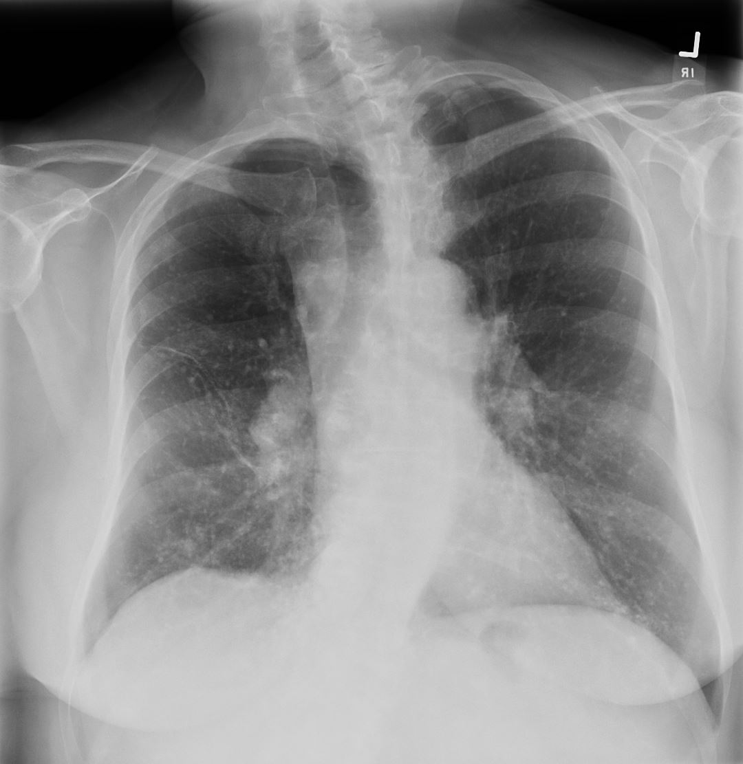
Frontal CXR of a 60 year old female with known diagnosis of AL amyloidosis shows multiple micronodules, likely calcified of varying size predominantly in the mid and lower lung zones and a suggestion of a calcified lymph node in the right
Ashley Davidoff MD TheCommonVein.net 266Lu 136178
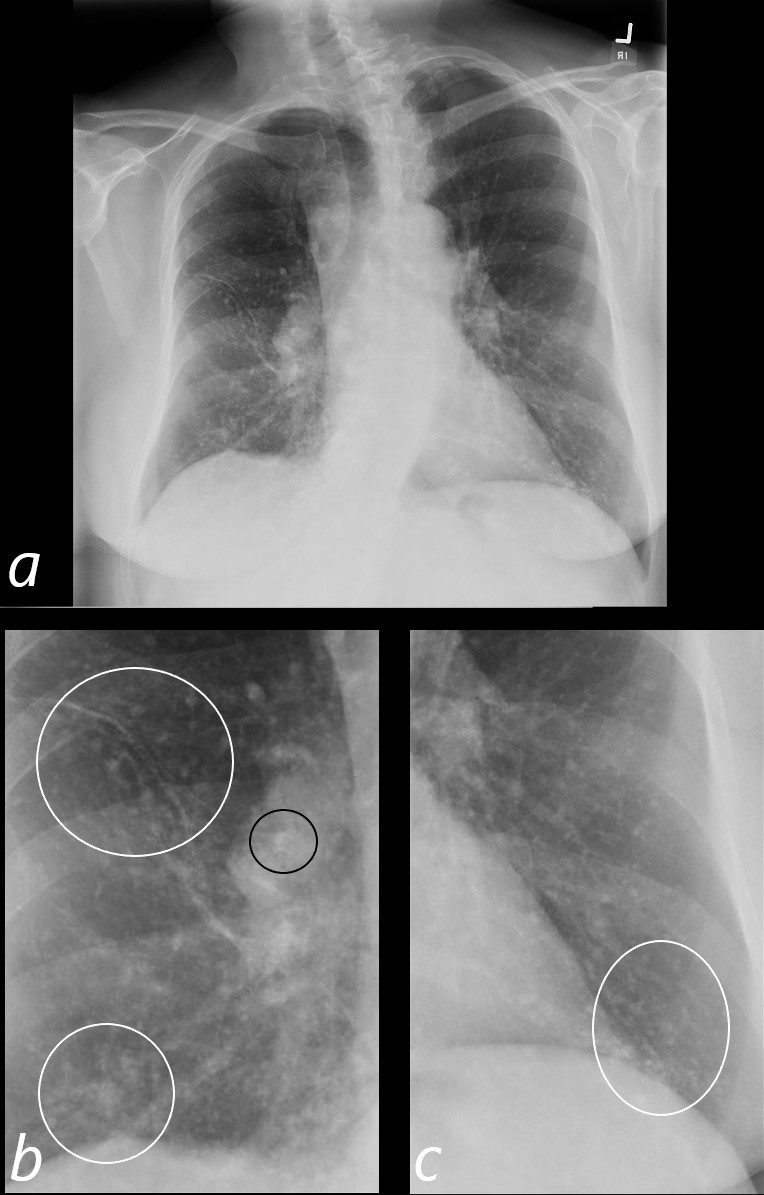
Frontal CXR of a 60 year old female with known diagnosis of AL amyloidosis shows multiple micronodules, likely calcified of varying size predominantly in the mid and lower lung zones (b, and c white rings) and a suggestion of a calcified lymph node in the right hilum (b ringed in black).
Ashley Davidoff MD TheCommonVein.net 266Lu 136178cL
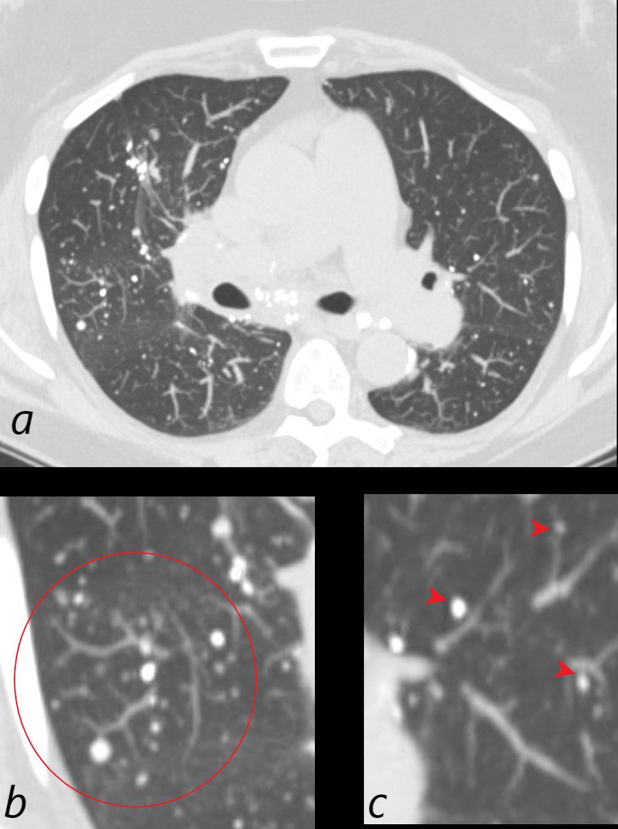
CT Microcalcifications Mid and Lower Lung Zones
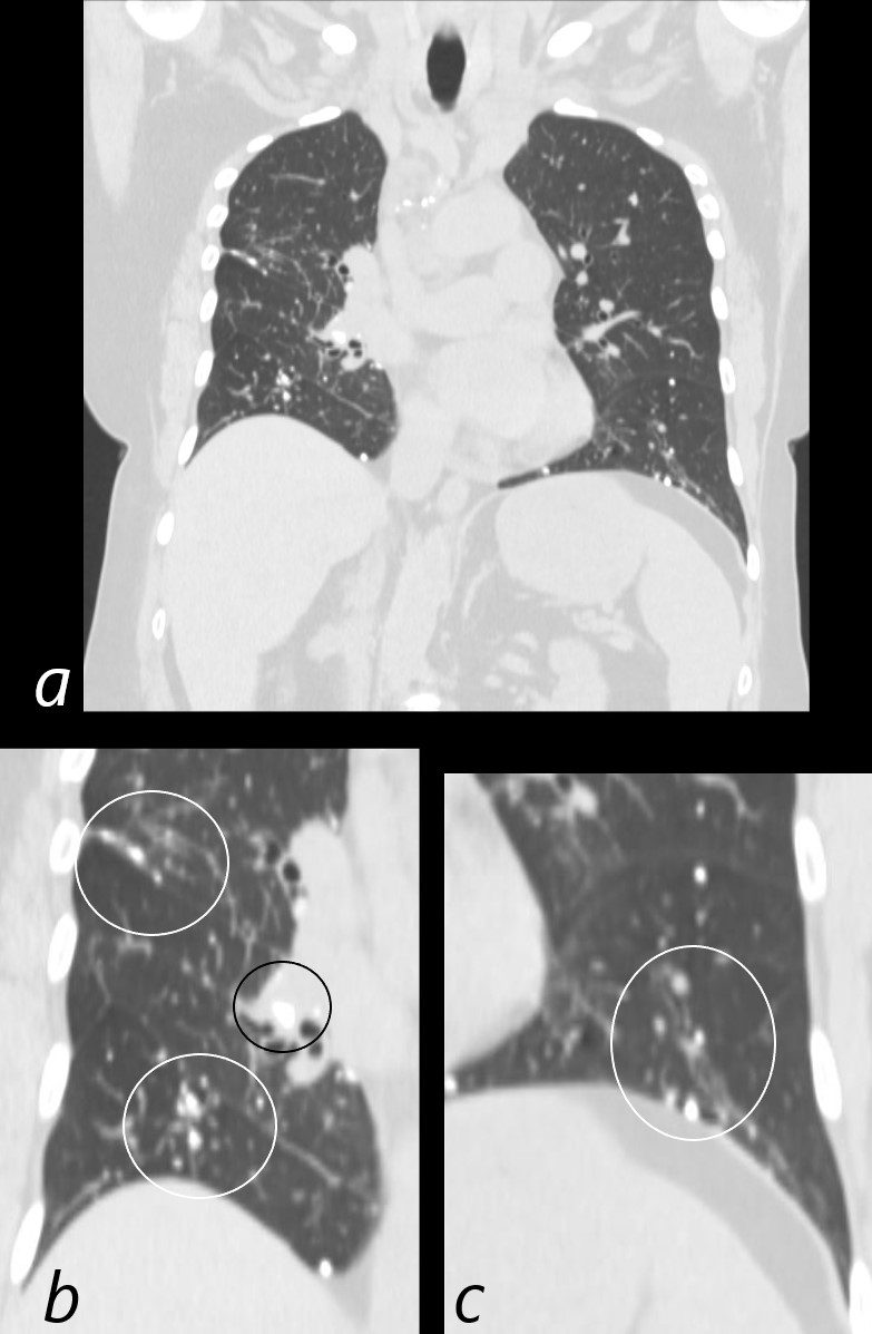
CT scan in the coronal plane of a 60 year old female with known diagnosis of AL amyloidosis shows multiple microcalcifications of varying size predominantly in the mid and lower lung zones (b, and c white rings) and a suggestion of a calcified lymph node in the right hilum (b ringed in black).
Ashley Davidoff MD TheCommonVein.net 266Lu 136179cL
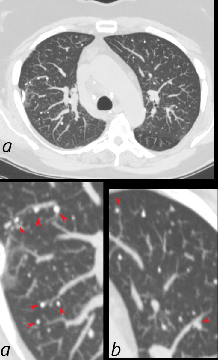
CT Microcalcifications Peripheral Mid and Lower Lung Zones Associated with Vessels and Pleura
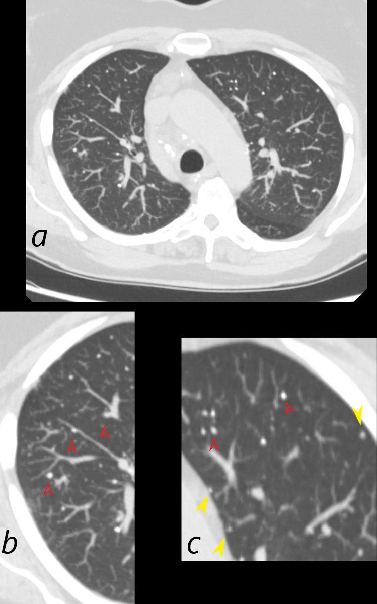
CT scan in the axial plane of a 60 year old female with known diagnosis of AL amyloidosis shows multiple microcalcifications in the periphery of the mid and lower lung zones in close association with the blood vessels (b, and c red arrowheads) and also subpleural (c, yellow arrowheads).
Ashley Davidoff MD TheCommonVein.net 136182cL
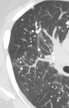
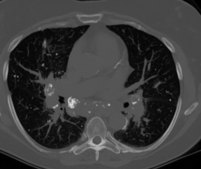
CT Microcalcifications Peripheral Mid and Lower Lung Zones Associated with Vessels and Pleura
CT scan in the axial plane of a 60-year-old female with known diagnosis of AL amyloidosis shows multiple microcalcifications in the periphery of the mid and lower lung zones in close association with the blood vessels (b, and c red arrowheads) and also subpleural (c, yellow arrowheads).
Ashley Davidoff MD TheCommonVein.net 266Lu 136183cL
CT scan in the axial plane of a 60-year-old female with known diagnosis of AL amyloidosis shows multiple microcalcifications in the periphery of the mid and lower lung zones in close association with the blood vessels (b, (red ring and c red arrowheads)
Ashley Davidoff MD TheCommonVein.net 266Lu 136184cL
CT scan in the axial plane of a 60-year-old female with known diagnosis of AL amyloidosis shows multiple microcalcifications in the periphery of the mid and lower lung zones in close association with the blood vessels
Ashley Davidoff MD TheCommonVein.net 266Lu 136185
CT Heterogeneous Calcification of Mediastinal and Hilar Nodes
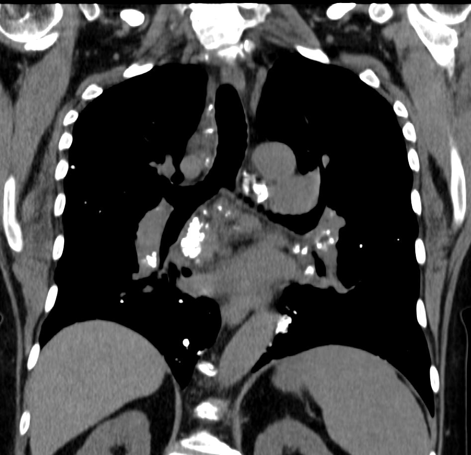
CT scan in the coronal plane of a 60-year-old female with known diagnosis of AL amyloidosis shows multiple heterogeneously calcified lymph nodes in the mediastinum and hila
Ashley Davidoff MD TheCommonVein.net 266Lu 136191
CT scan in the axial plane of a 60-year-old female with known diagnosis of AL amyloidosis shows multiple heterogeneously calcified lymph nodes in the mediastinum and hila
Ashley Davidoff MD TheCommonVein.net 266Lu 136188