
- infection
- pulmonary tuberculosis 3
- pulmonary bacterial abscess/cavitating pneumonia
- post-pneumonic pneumatocele: a thin walled pneumatocele is not really a cavity but when infected can be thick-walled
- septic pulmonary emboli
- other rare infections CCNAM
- Inflammation
- non-infective granuloma
- granulomatosis with polyangiitis
- rheumatoid nodules
- Cryptogenic organizing pneumonia
- Sarcoidosis
- non-infective granuloma
- Neoplasm
- cavitating malignancy
- primary bronchogenic carcinoma (especially squamous cell carcinoma)
- cavitating pulmonary metastases
- squamous cell carcinoma
- adenocarcinoma, e.g. gastrointestinal tract, breast
- sarcoma
- Congenital Growth Abnormalities
- Pulmonary sequestration
- cavitating malignancy
- Mechanical
- `Bullae or cysts with air fluid level
- Pneumatoceles from trauma
- Circulatory
- Vasculitis (eg, granulomatosis with polyangiitis)
- Pulmonary embolism with infarction
- Other
- Pulmonary Langerhans histiocytosis
- Foreign body aspiration
| Pulmonary embolism with infarction |
| Vasculitis (eg, granulomatosis with polyangiitis) |
| Neoplasm (eg, bronchogenic cancer, lymphoma, and metastatic head and neck, bladder, colon, pancreatic, and uterine cancer). |
| Pulmonary sequestration |
| Bullae or cysts with air fluid level |
| Bronchiectasis |
| Cryptogenic organizing pneumonia |
| Sarcoidosis |
| Rheumatoid nodules |
| Pulmonary Langerhans histiocytosis |
| Foreign body aspiration |
- Infection
- TB
-

Reactivation TB
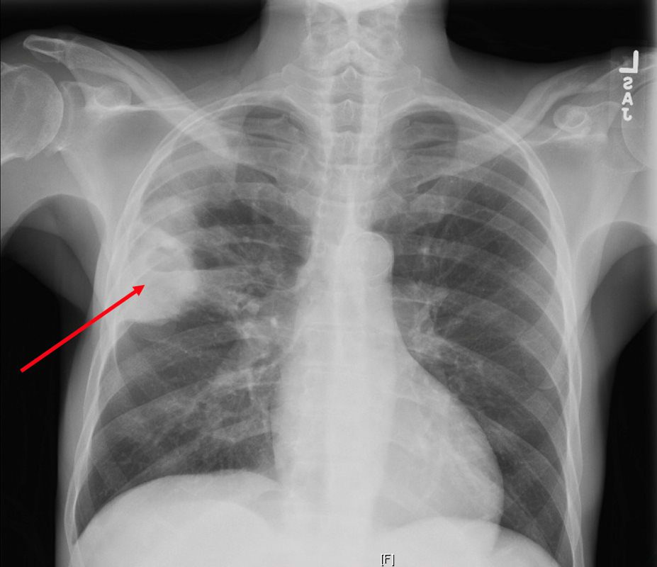
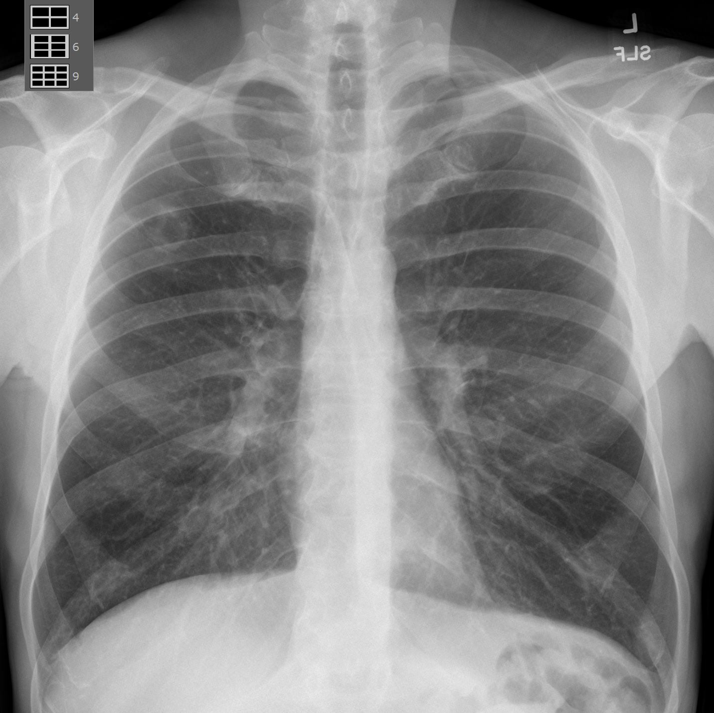
CXR reveals a dense consolidation in the right upper lobe (red arrow) with questionable air-fluid level. No pneumothorax. No pleural effusions. Differential includes right upper lobe pneumonia or tuberculosis. CT is recommended for further evaluation if there is concern for a cavity.
Courtesy Joseph Cannella,
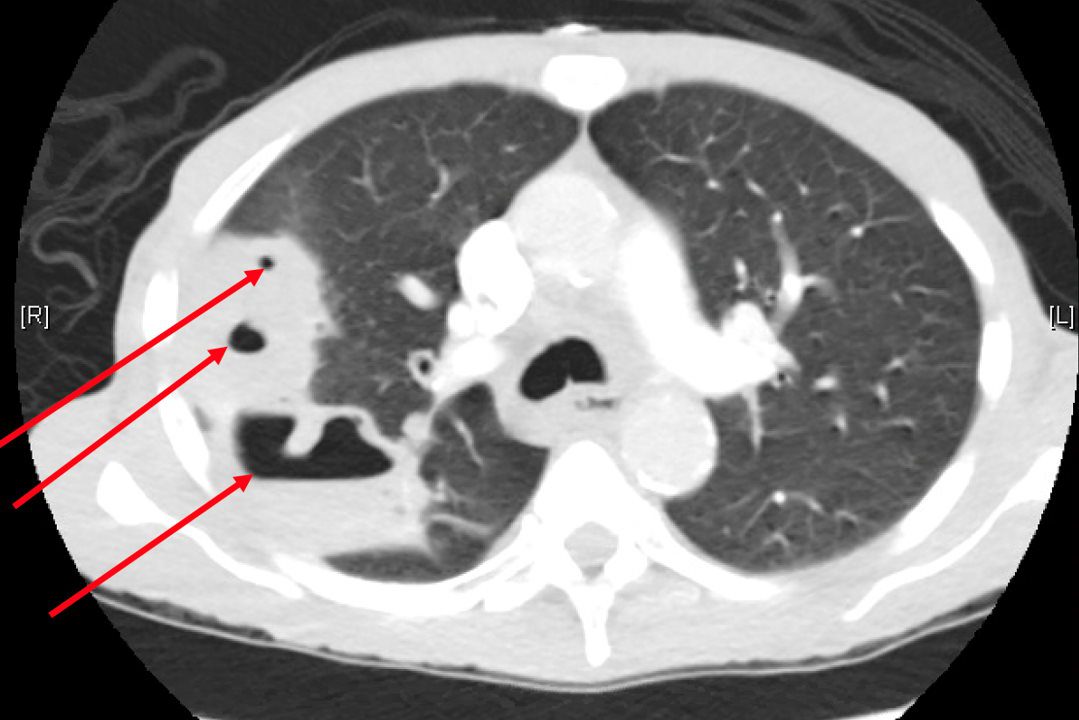
Dr. Christina LeBedis, MD, MSCTPA reveals a large consolidation in the right upper lobe and superior segment of the right lower lobe spans approximately 8.8 x 5.6 x 9.4 cm and extends to the pleura. There are multiple internal cavitations (red arrows) with air-fluid levels. These large predominately right upper lobe cavitary lesions are consistent with clinical concern for tuberculosis pneumonia, however follow-up with chest CT in 3 months post-treatment is recommended to exclude other less likely causes of cavitary lesions, such as malignancy.
Courtesy Joseph Cannella,
Dr. Christina LeBedis, MD, MS
-
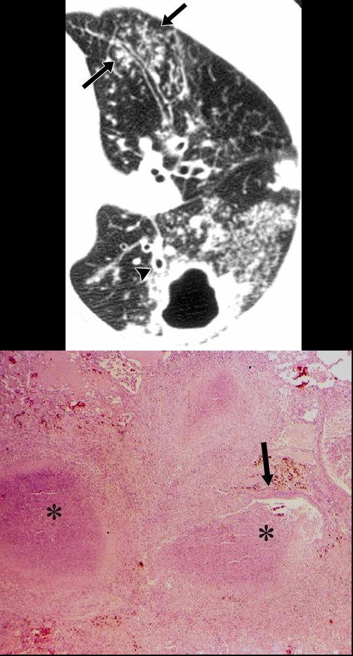
Postprimary active tuberculosis in a 34-year-old man with weight loss and a chronic cough. (a) High-resolution CT scan of the left lung shows a thick-walled cavity and multiple peripheral small nodules and branching linear structures (arrows). Note the thickening of the bronchial walls (arrowhead). (b) Photomicrograph (original magnification, ×400; hematoxylin-eosin stain) shows impacted caseous material (*) in small peripheral airways (arrow).
Rossi, SE et al Tree-in-Bud Pattern at Thin-Section CT of the Lungs: Radiologic-Pathologic Overview RadioGraphics Vol. 25, No. 3 2005 -
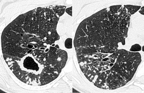
Infection with M avium-intracellulare complex in a 44-year-old woman with malaise and a chronic cough. High-resolution CT scans of the right lung show multiple peripheral small nodules connected to branching linear opacities and a thick-walled cavity in the superior segment of the lower lobe. Note the thickening of the bronchial walls, bronchial dilatation, and mucus impaction. The diagnosis was confirmed with bronchoalveolar lavage.
Rossi, SE et al Tree-in-Bud Pattern at Thin-Section CT of the Lungs: Radiologic-Pathologic Overview RadioGraphics Vol. 25, No. 3 2005 - Aspergillus
-
ASPEGILLUS Infection Cavitating Nodule
-
ASPEGILLUS Infection Cavitating Nodule
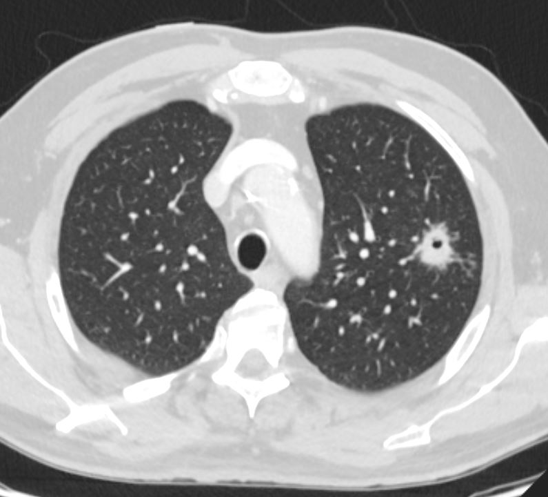
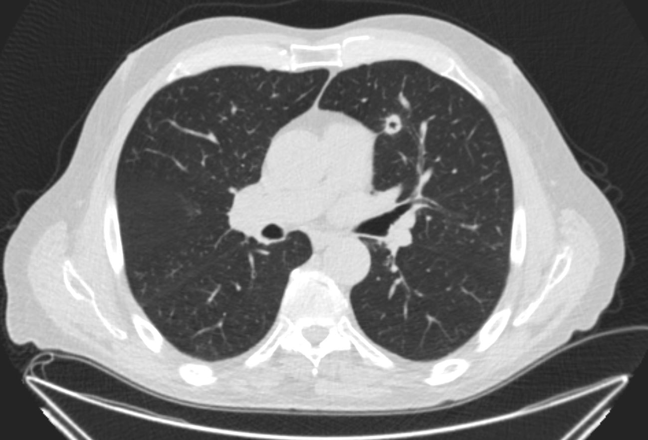
Ashley DAvidoff MD TheCommonVein.net - 62 M cavitatating nodule from necrotizing strep intermedius infection 3 months prior
62 M cavitatating nodule necrotizing strep intermedius infection 001 CT 3 months prior
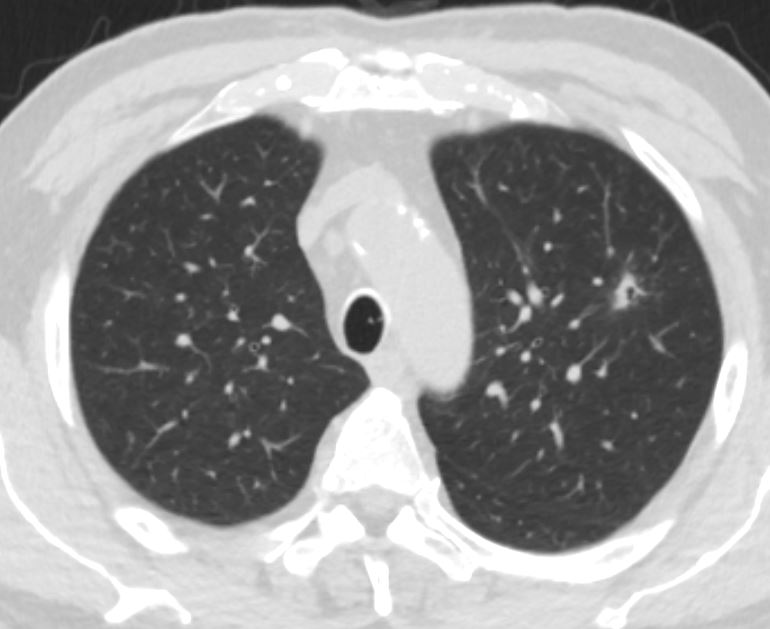
Ashley Davidoff MD thecommonvein.net3 months later following Augmentin Rx
-
62 M cavitatating nodule necrotizing strep intermedius infection
3 months later CT post Rx augmentin
Ashley Davidoff MD
thecommonvein.net -
Infection
- TB
-
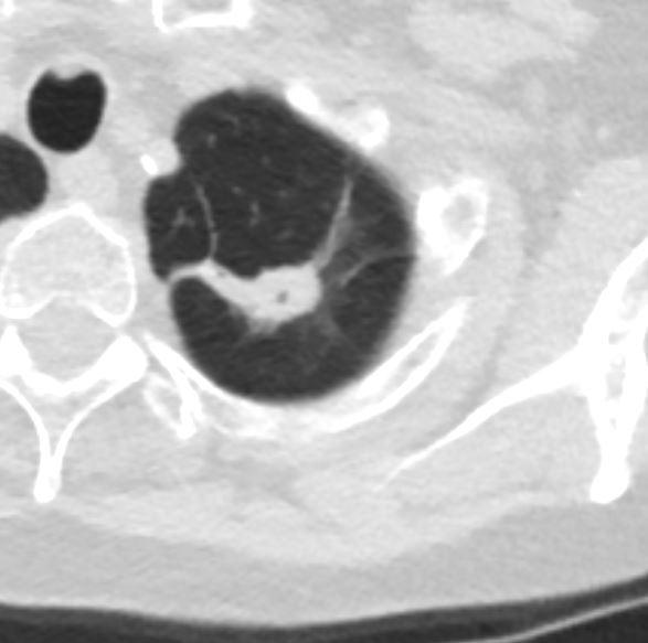
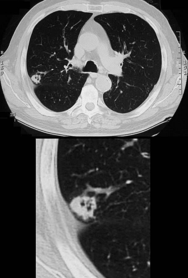
50M with Cavitating Nodule on CXR BAL yielded Mycobacterium Mycobacterium Kansasii
Ashley Davidoff MD
TheCommonVein.net50M with Cavitating Nodule on CT scan.
BAL yielded Mycobacterium Kansasii
Ashley Davidoff MD
TheCommonVein.netSeptic Emboli

Ashley Davidoff MD TheCommonvein.net 24f PE Hampton’s hump 001
Ashley Davidoff MD TheCommonvein.net 24f PE Hampton’s hump 002
Ashley Davidoff MD TheCommonvein.net 24f PE Hampton’s hump 003
Staphylococcus
Ashley DAvidoff MD TheCommonVein.net cavitating pneumonia 59M 02
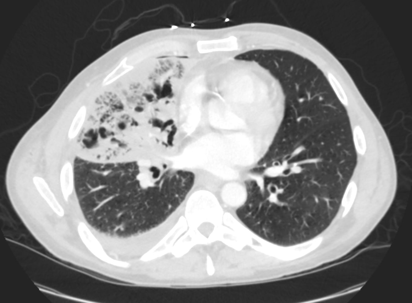
Ashley Davidoff MD TheCommonVein.net cavitating pneumonia 59M
-
Inflammation
- Wegener’s
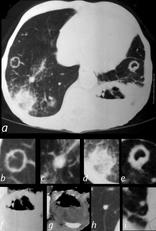
81-year-old male with weight loss, renal failure, and hemoptysis
CT axial view (a) shows cavitating masses in the upper lobes bilaterally magnified (b and e). In addition there is a large necrotic mass with an air fluid level in the left lower lobe (a), magnified ( f and g). A mass with a halo sign noted in the RLL is magnified in d, and scattered bilateral smaller nodules are magnified (c and h) in the RLL and in (i) in the LLL .
Priscilla Slanetz MPH MD
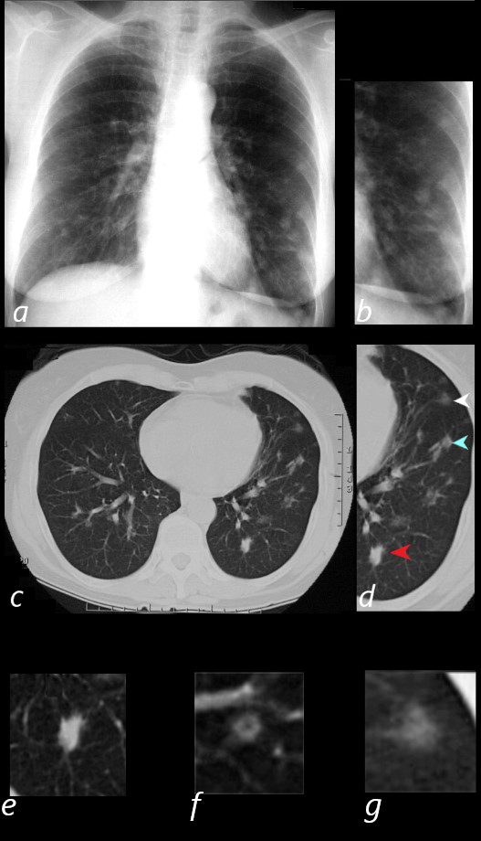
65 year old female presents with epistaxis and with nodular changes on CXR (a) magnified in b.
CT scan in axial projection (c) and magnified in d, reveals 3 types of nodules.
A spiculated solid nodule (red arrow head) is magnified in e, a bronchocentric nodule (teal arrowhead) is magnified in e. This may represent a cavitating nodule or hemorrhagic change around a bronchiole (cheerio sign) A ground glass nodule (white arrowhead) is magnified in g.
Ashley Davidoff MD
-
Cancer
- Bubble Lucencies – Pseudocavitation
-
Bubble Lucencies (Pseudocavitation)
“Nodules containing round or oval foci of air attenuation are highly suspicious for malignancy, particularly lung adenocarcinoma ( Fig. 4.9 ). These bubble lucencies (synonym: pseudocavitations) are rarely identified in benign lesions. Correlation with pathologic findings has shown that the lucencies usually represent patent airways, often ectatic, or foci of emphysema surrounded by carcinoma.”
radiologykey.comBubble Lucencies
-
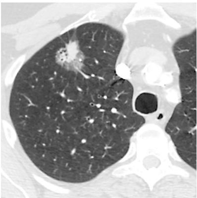
Cavitating Squamous Cell Carcinoma
65 year male with peripheral lung nodule characterized by cavitation that was not present 2 years earlier . Pathology revealed squamous cell carcinoma
Ashley Davidoff
TheCommonVein.net
Cavitating Head and Neck Squamous Carcinoma
Ashley Davidoff MD TheCommonVein.net
Cavitating Pneumonia
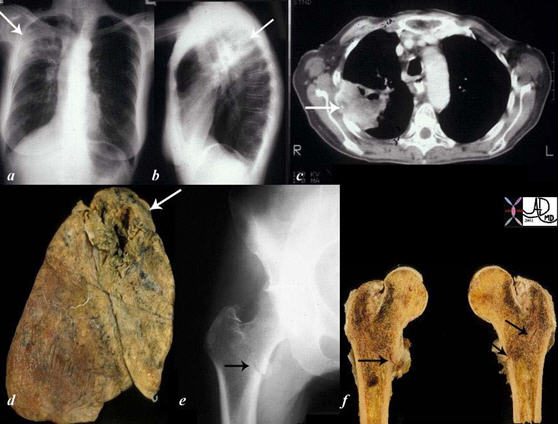
The collage of images reflects a patient with stage IV, cavitating, primary, squamous carcinoma of the right upper lobe (RUL) (a, b, c, d – white arrows) with COPD. A metastatic lesion to the right femur was complicated by a pathological fracture. (e, f black arrows).
Courtesy Ashley Davidoff, M.D. TheCommonVein.net Lung cancer P 018
References and Links
Parkar A, et al Differential Diagnosis of Cavitary Lung Lesions J Belg Soc Radiol. 2016; 100(1): 100.
-
- TCV
- TCV