
74 year old female, with a history of scleroderma.
4 months prior to presentation, a CT scan shows clear lung bases. Note the distended, fluid filled esophagus.
Ashley Davidoff MD TheCommonVein.net
74F scleroderma reflux aspiration pneumonia 004 4 months ago
4 months prior to presentation, a CTscan shows mild atelectasis.at the left base
Ashley Davidoff MD TheCommonVein.net
74F scleroderma reflux aspiration pneumonia 06 4 months ago
Presents with Acute Respiratory Distress
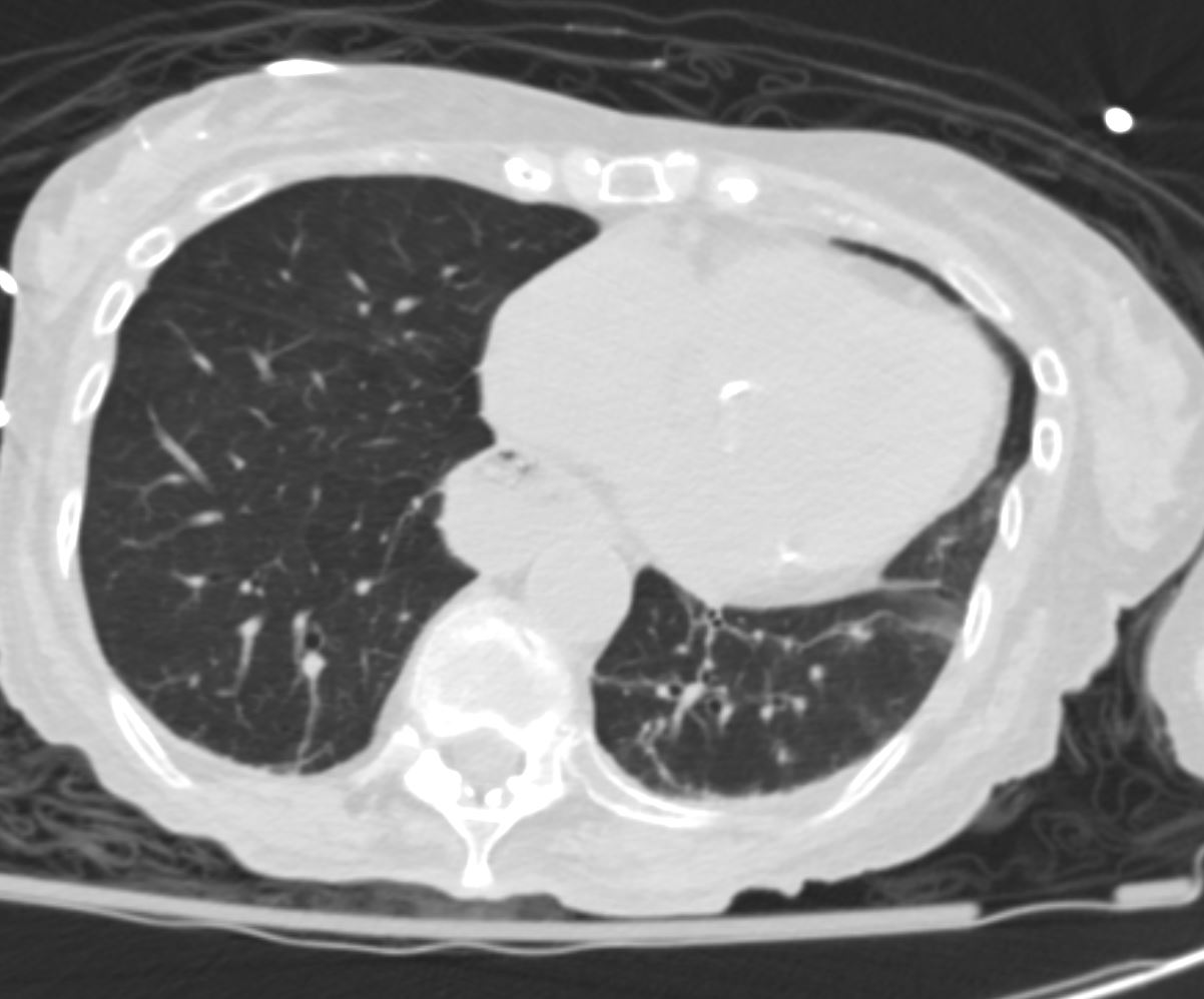
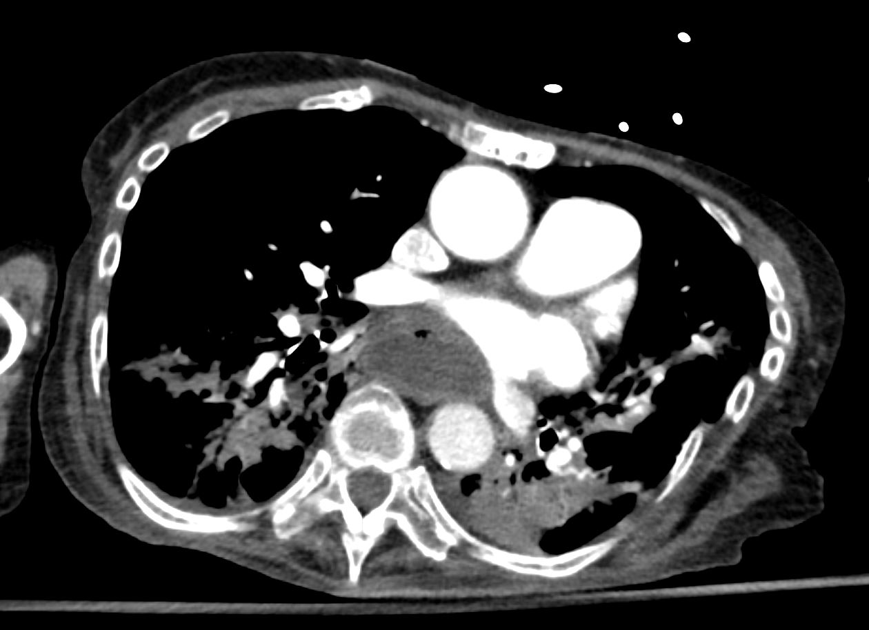
74 year old female with scleroderma presents with acute respiratory distress
CT scan shows bilateral basilar consolidation,fluid filled and significantly dilated esophagus, with fluid seen in the right lower lobe bronchus’ consistent with aspiration pneumonia
Ashley Davidoff MD TheCommonVein.net
74F scleroderma reflux aspiration pneumonia 002 esophagus
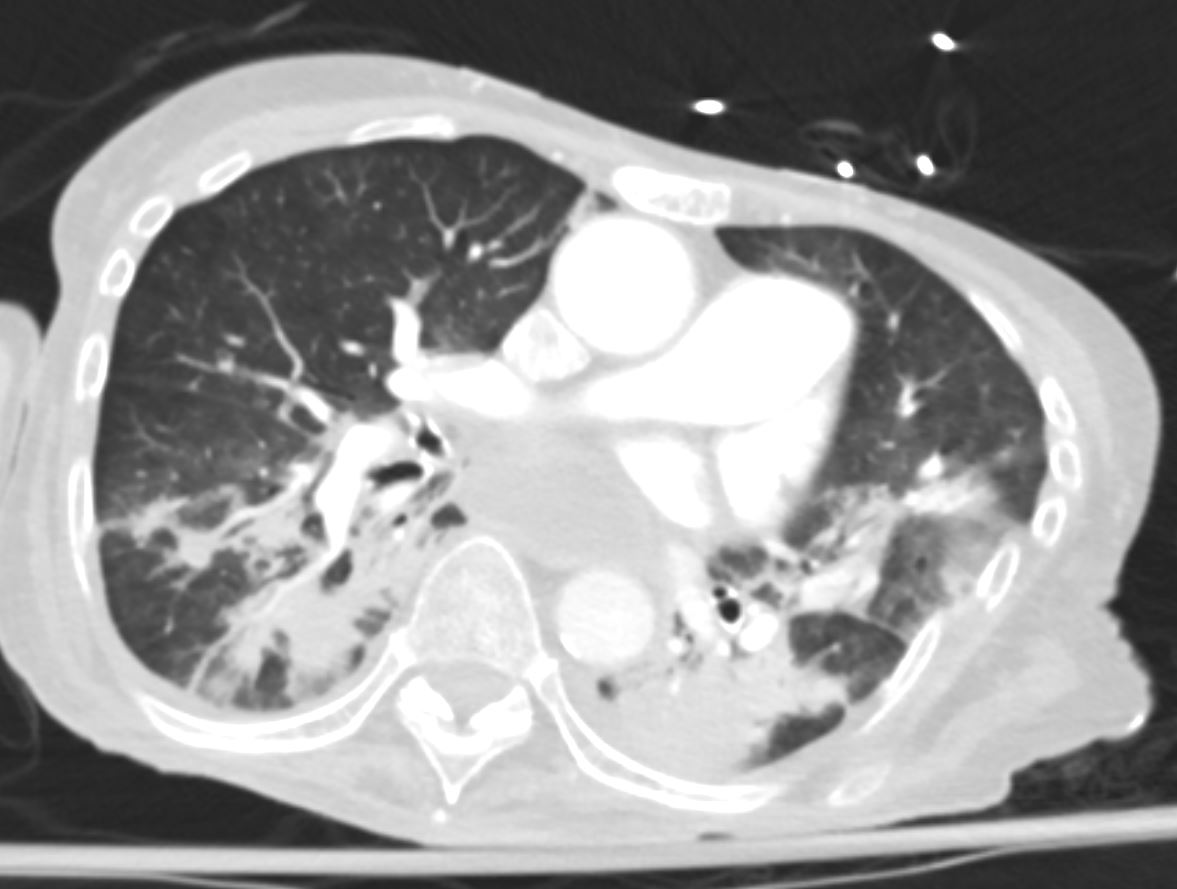
74 year old female with scleroderma presents with acute respiratory distress
CT scan shows bilateral basilar consolidation,fluid filled and significantly dilated esophagus, with fluid seen in the right lower lobe bronchus’ consistent with aspiration pneumonia
Ashley Davidoff MD TheCommonVein.net
74F scleroderma reflux aspiration pneumonia 003 esophagus
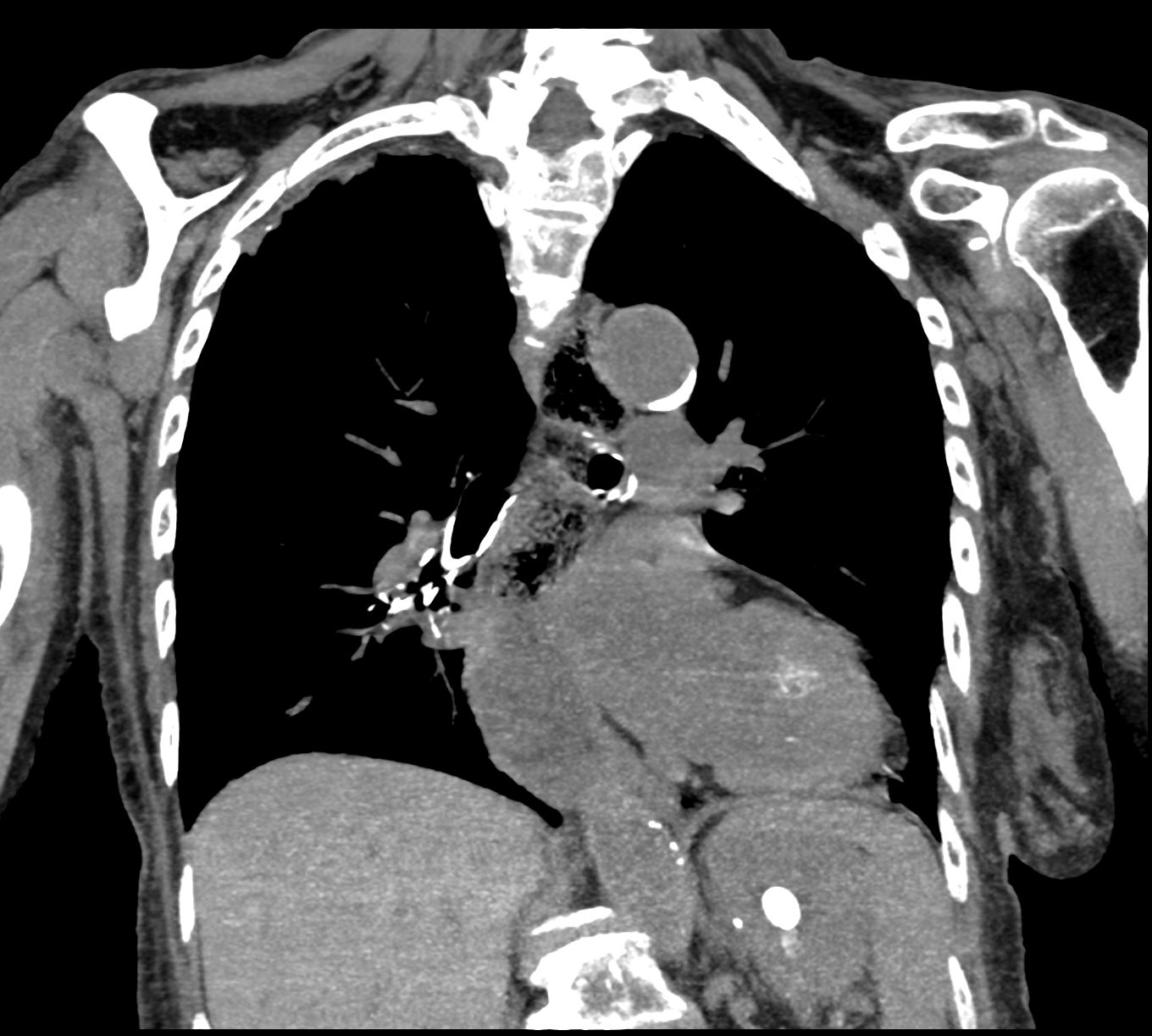
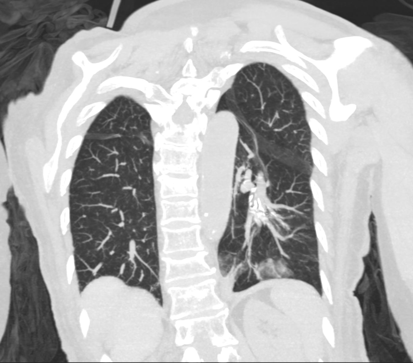
74 year old female with scleroderma presents with acute respiratory distress
CT scan reformatted in the coronal plain shows ,fluid filled and significantly dilated esophagus,
Ashley Davidoff MD TheCommonVein.net
74F scleroderma reflux aspiration pneumonia 005
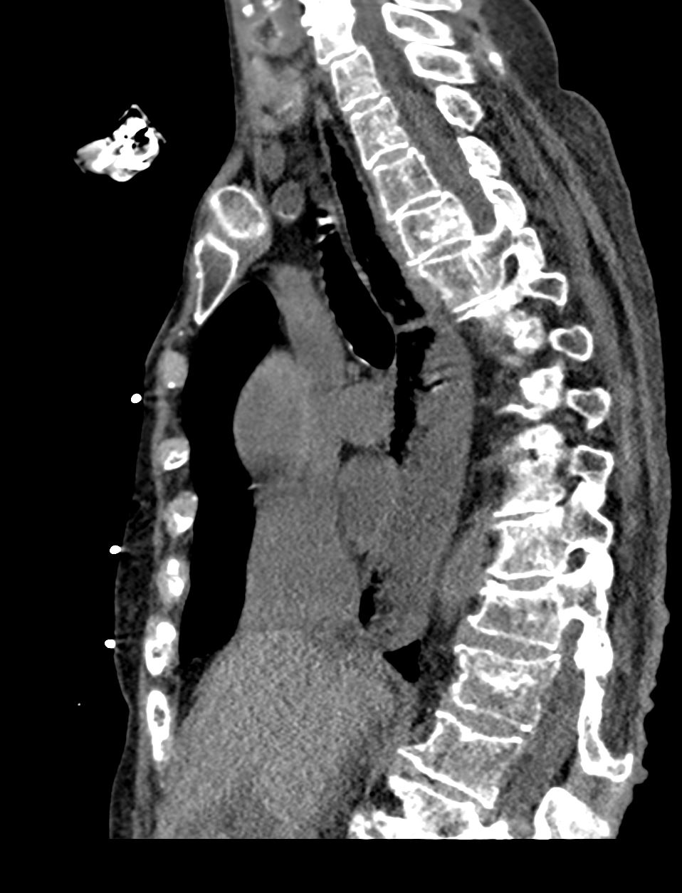
CT scan reformatted in the sagittal plain shows fluid filled and significantly dilated esophagus,
Ashley Davidoff MD TheCommonVein.net
74F scleroderma reflux aspiration pneumonia 07
1 Day Later
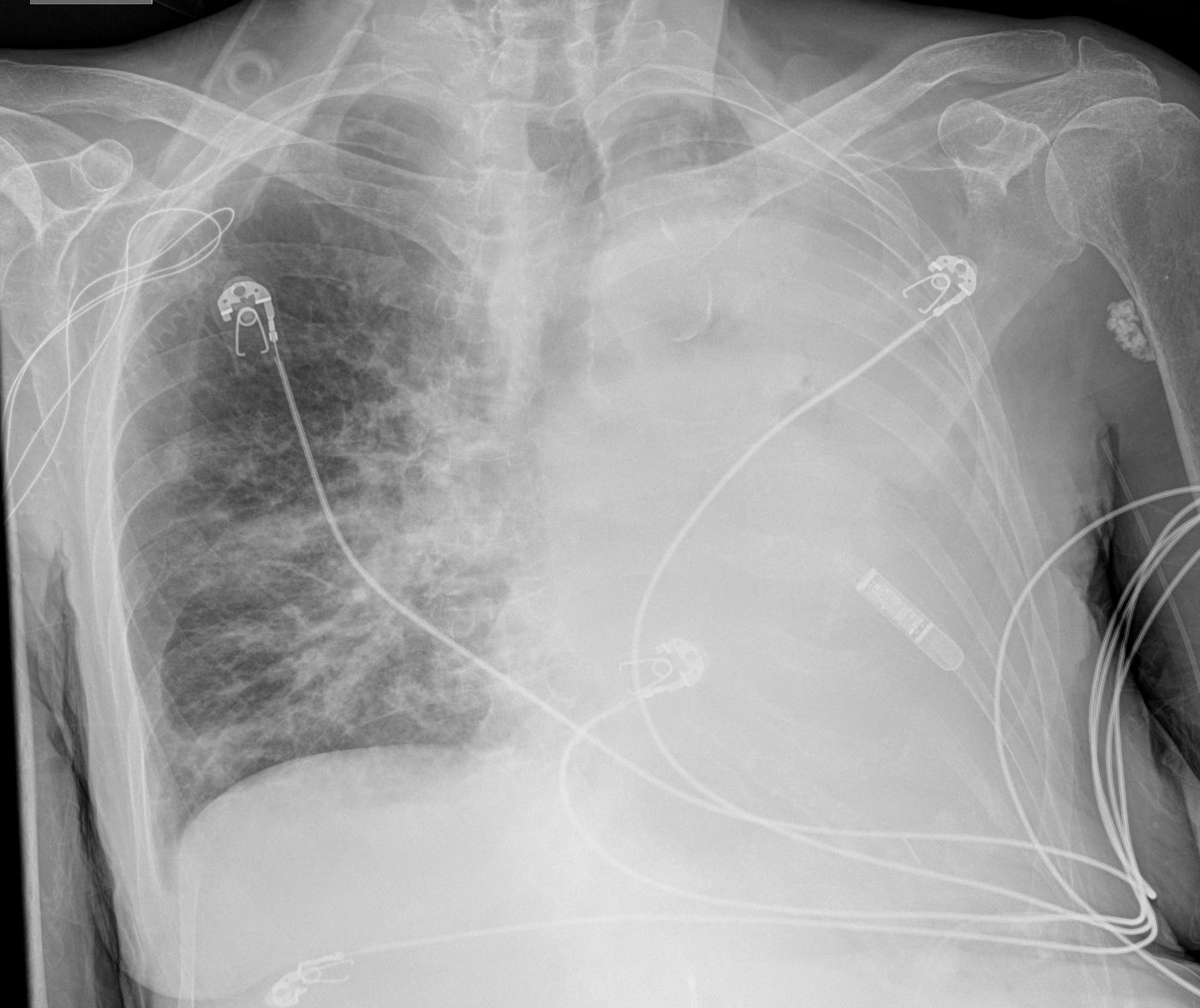
74 year old female with scleroderma presents with hyperacute respiratory distress
A CXR shows an acute white out of the left hemithorax and a right lower lobe infiltrate. There is mediastinal shift to the left suggesting left sided volume loss consistent with obstructive atelectasis and likely die t ongoing aspiration
Ashley Davidoff MD TheCommonVein.net
74F scleroderma reflux aspiration pneumonia 001 white out