
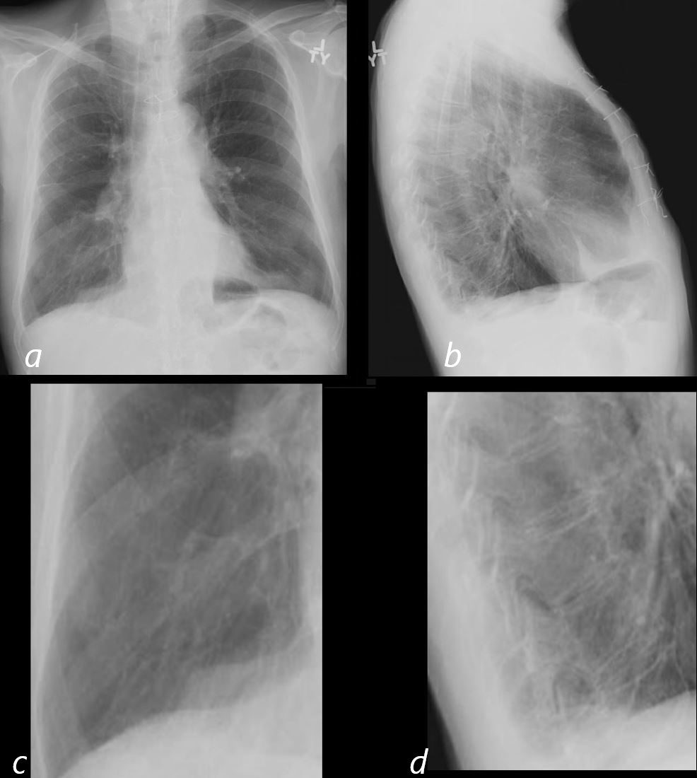
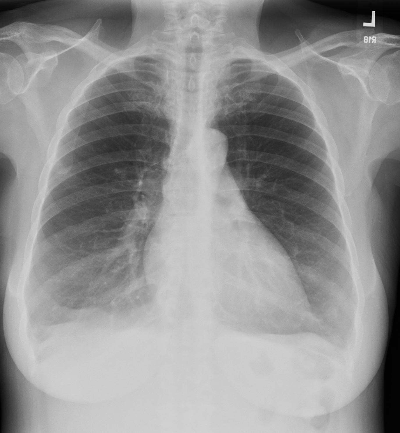
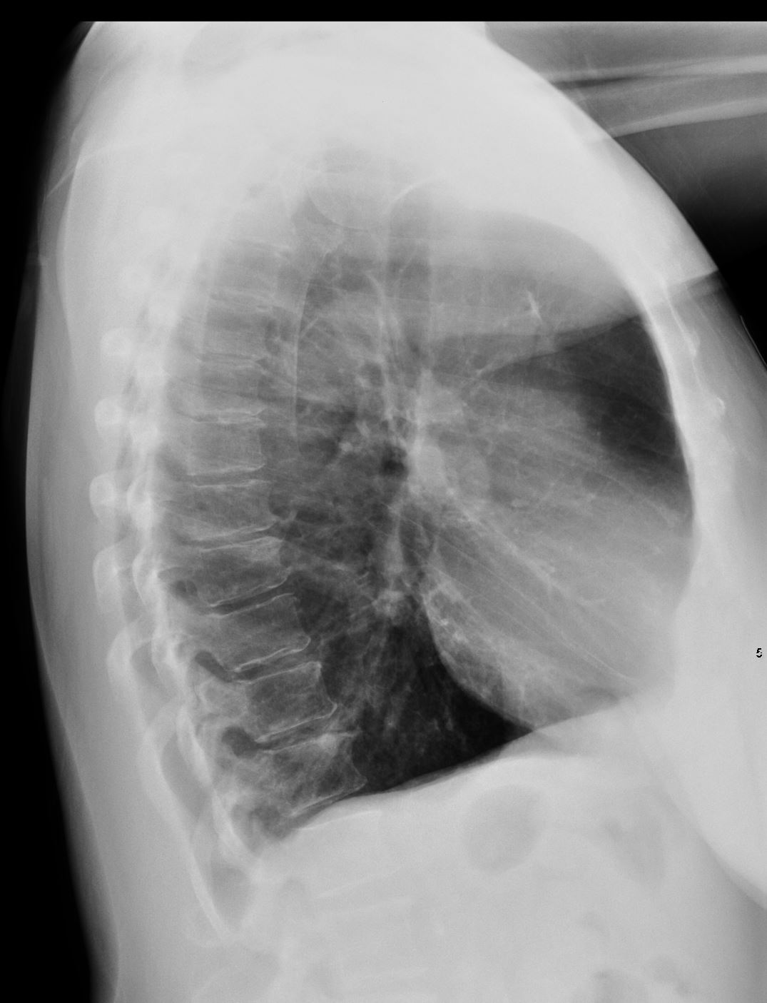
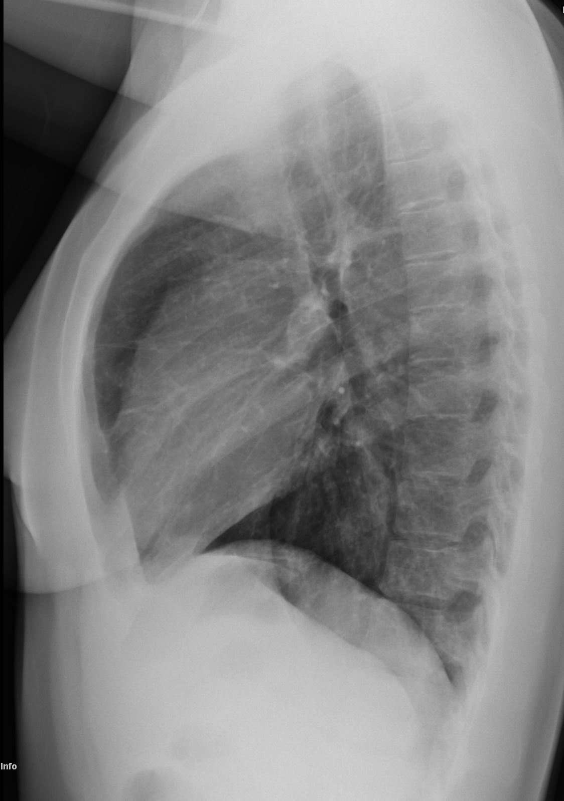
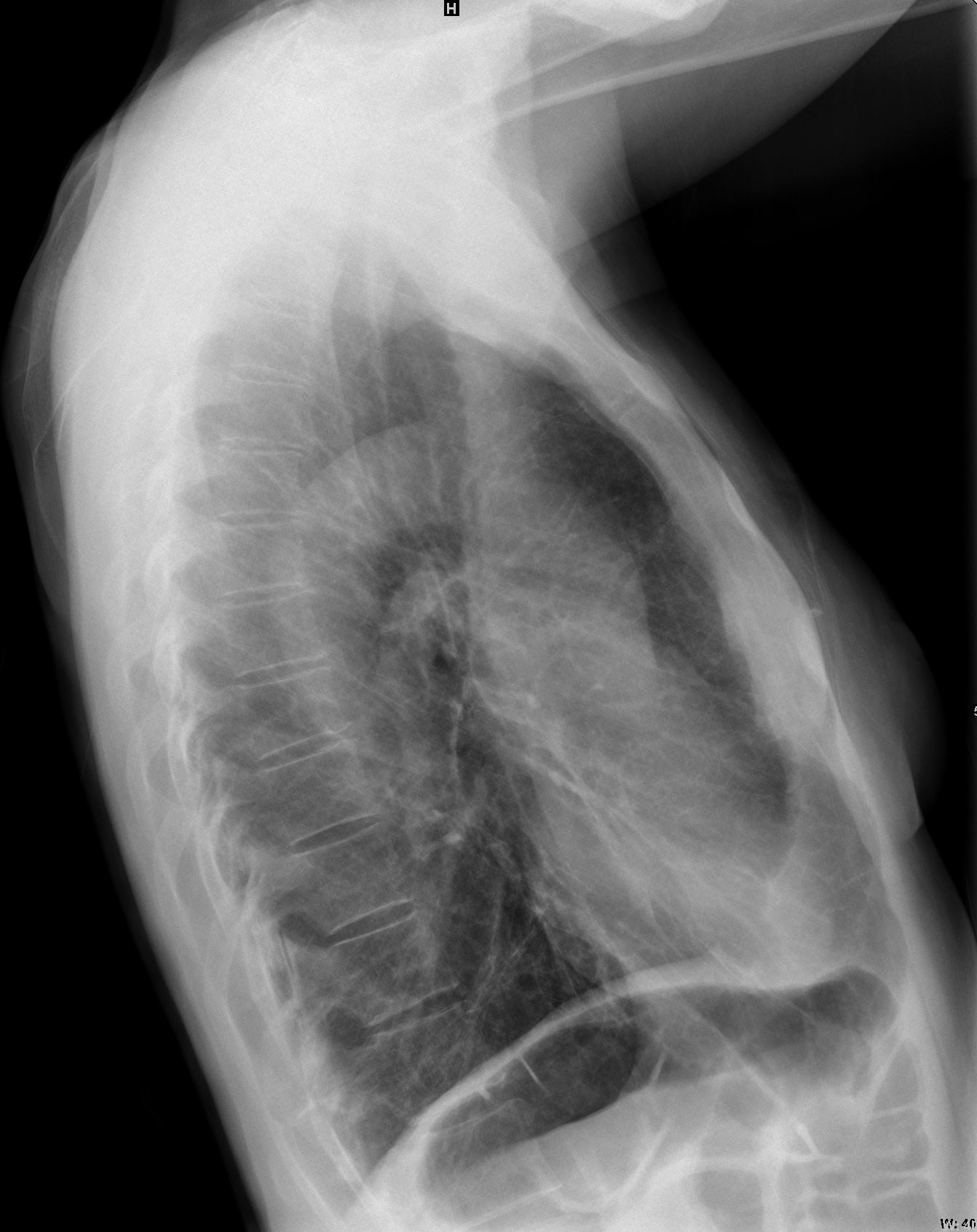
86year old patient with known emphysema and chronic bronchitis CXR 6 months prior to admission shows evidence of hyperinflation with flattened hemidiaphragms and increase in the retrosternal air space (b) On the PA view (a magnified in c) there is a suggestion of bronchovascular thickening. On the lateral view (b and magnified in d) the expected progressive lucency of the vertebral bodies is lost and there is outlining of a bronchus just anterior to the vertebral bodies confirming the bronchovascular thickening.
Ashley Davidoff MD TheCommonVein.net 30602d03cL
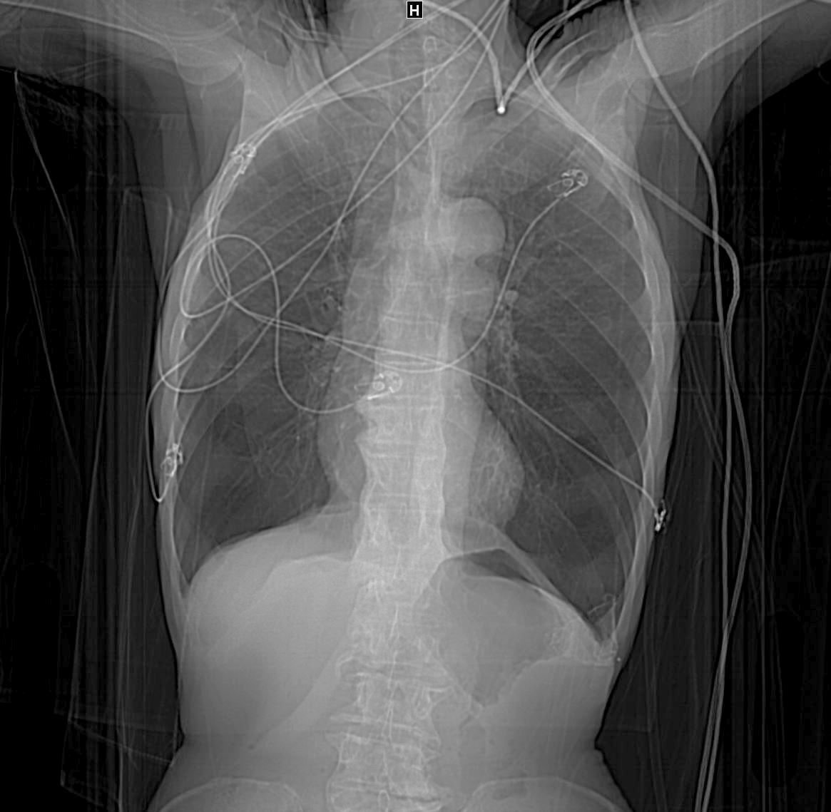
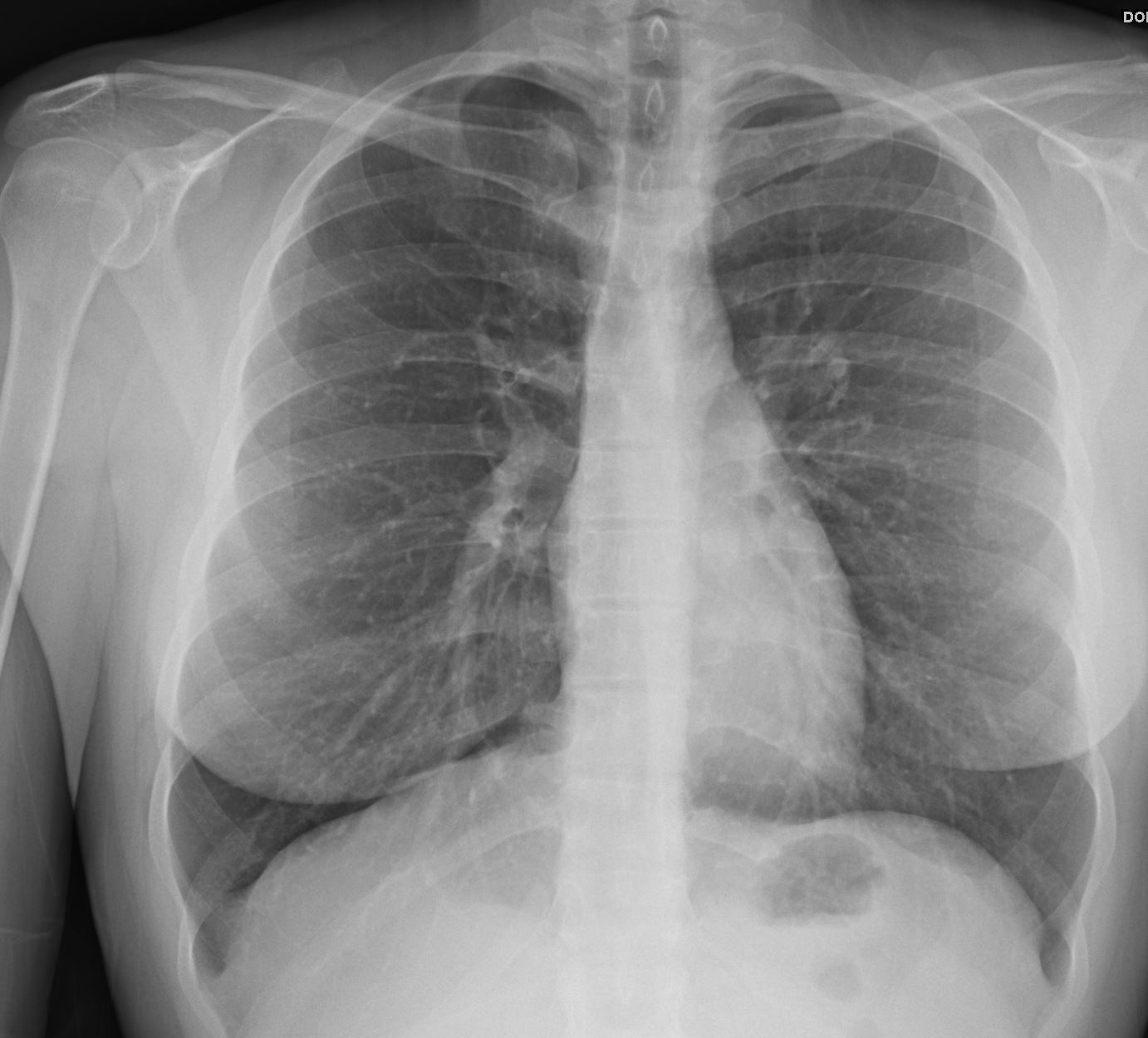
88-year-old smoker presents with shortness of breath Scout film shows hyperinflation with significantly hyperinflated lower lobes
Ashley Davidoff MD TheCommonVein.net 135538
Flattened and Partially Inverted
Ashley Davidoff MD TheCommonvein.net
Flattened and Partially Inverted
Ashley Davidoff MD TheCommonvein.net
Ashley DAvidoff
TheCommonVein.net
Asthma
Mounier Kuhn
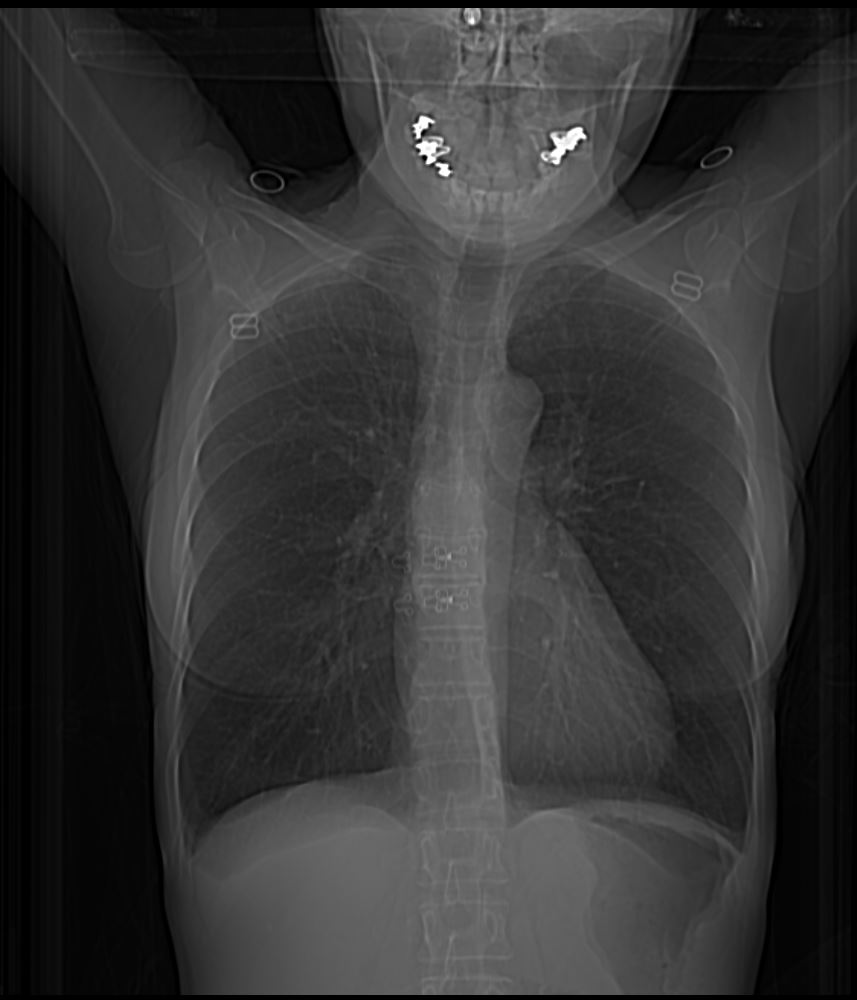
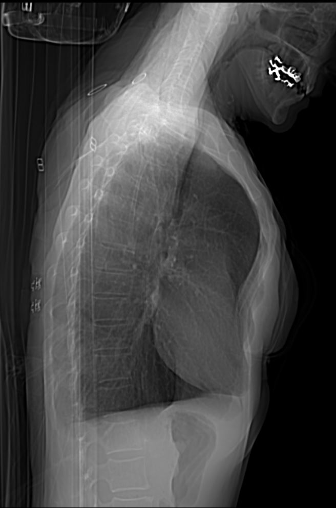
61 year old male with a history of treated mycobacterial infections and chronic cough
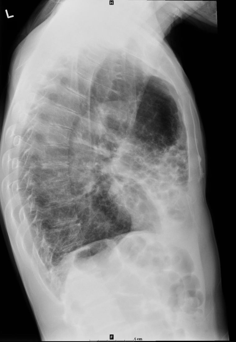
Lateral view shows an enlarged trachea and thick walled cystic changes overlying the heart consistent with known bronchiectasis. There is evidence of hyperinflation
Ashley Davidoff MD TheCommonVein.net 250Lu 135872a
Barrel Chest Lady Windermere Syndrome
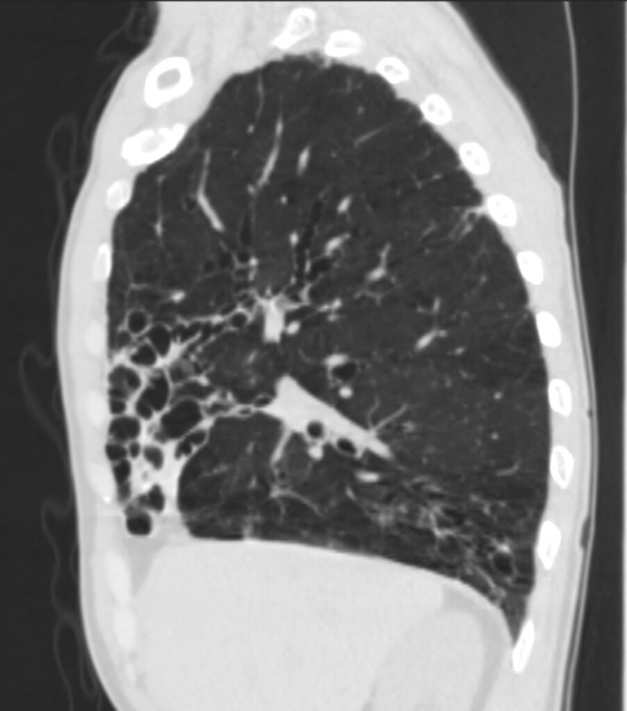
61-year-old male with a history of treated mycobacterial infections including MAC and chronic cough.
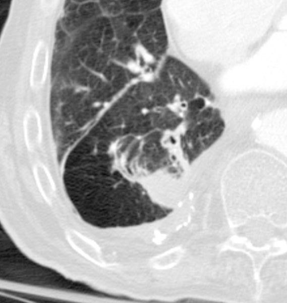
Right sagittal CT shows mildly ectatic segmental airways to the upper, middle and lower lobe airways, but significant bronchiectasis to the middle lobe subsegmental airways. There is a relative paucity of mucus in the ectatic airways. The history of MAC and the distribution of the bronchiectasis in the middle lobe and lingula are reminiscent of the diagnosis of Lady Windermere syndrome. The barrel chest reflects hyperinflation and the obstructive nature of the Mounier Kuhn Syndrome
Ashley Davidoff MD TheCommonVein.net 250Lu 135883
Alpha 1 Antitrypsin?
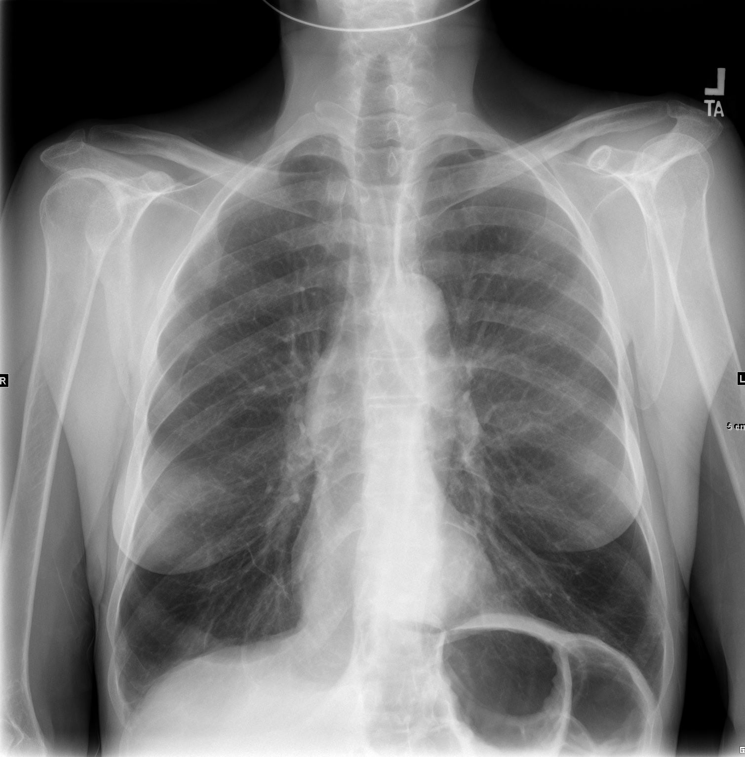
CXR shows hyperinflation with basilar prominence consistent with panlobular emphysema secondary to Alpha-1 antitrypsin deficiency
Ashley Davidoff TheCommonVein.net 216Lu
CXR shows hyperinflation with basilar prominence consistent with panlobular emphysema secondary to Alpha-1 antitrypsin deficiency
Ashley Davidoff TheCommonVein.net 216Lu
88-year-old smoker presents with shortness of breath Scout film shows hyperinflation with significantly hyperinflated lower lobes
Ashley Davidoff MD TheCommonVein.net 135538
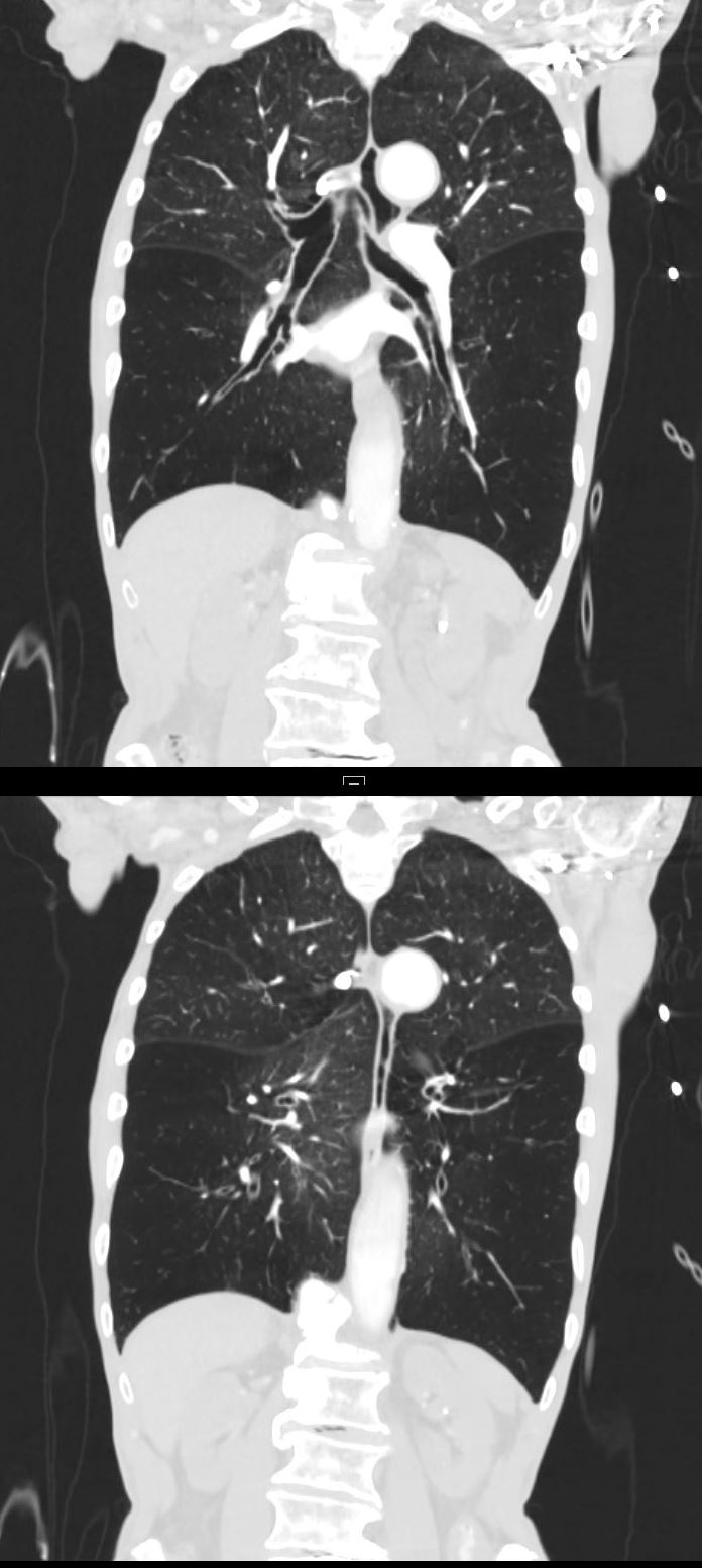
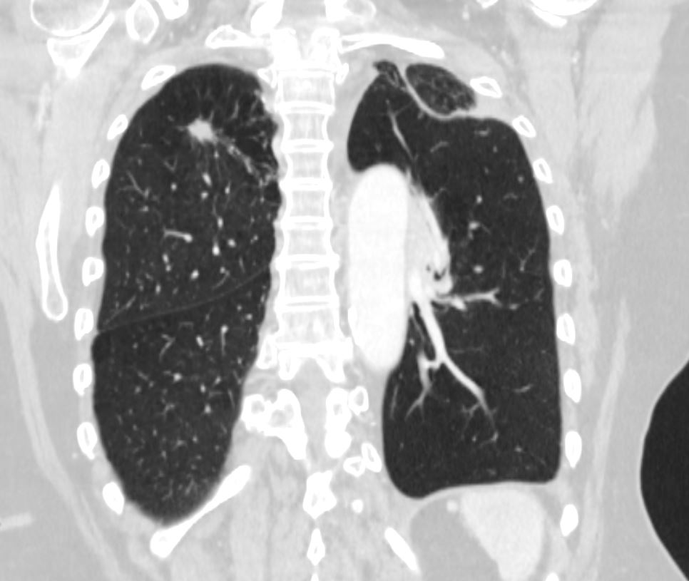
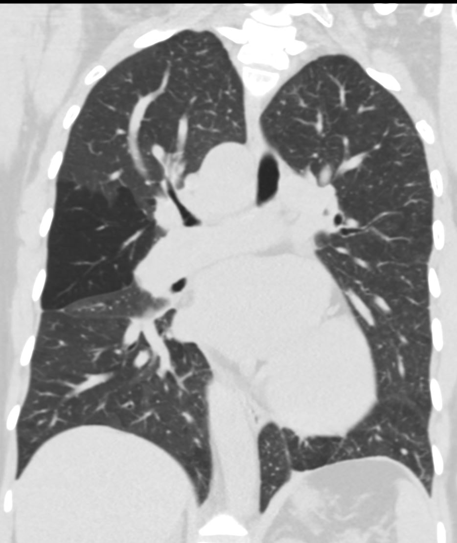
88-year-old smoker presents with shortness of breath. CT in the coronal plane shows hyperinflation with significantly hyperinflated lower lobes and suggestion of centrilobular changes in the upper lobes
Ashley Davidoff MD TheCommonVein.net 135539
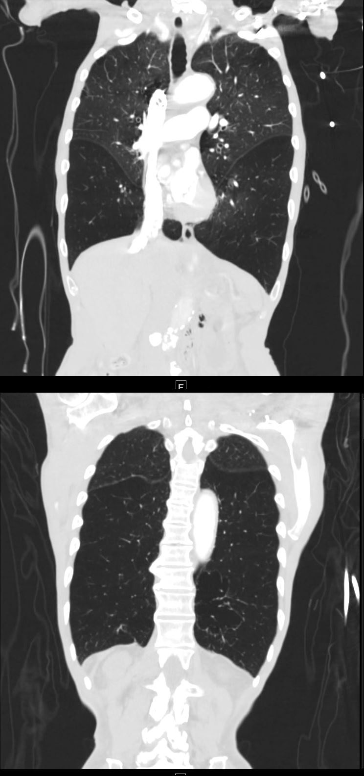
88-year-old smoker presents with shortness of breath. CT in the coronal plane shows hyperinflation with significantly hyperinflated lower lobes and suggestion of centrilobular changes in the upper lobes
Ashley Davidoff MD TheCommonVein.net 135540
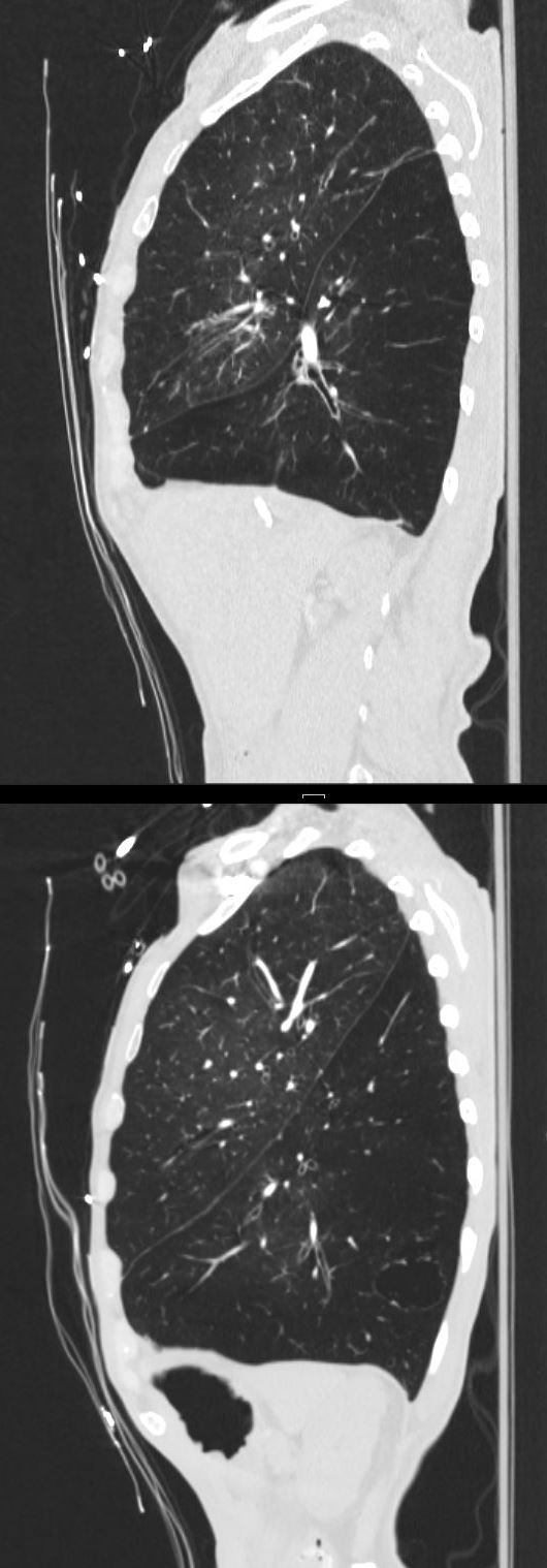
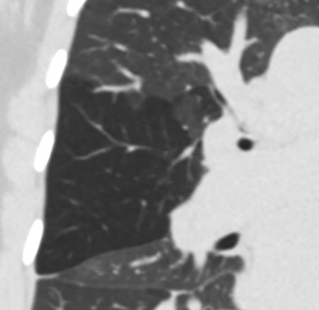
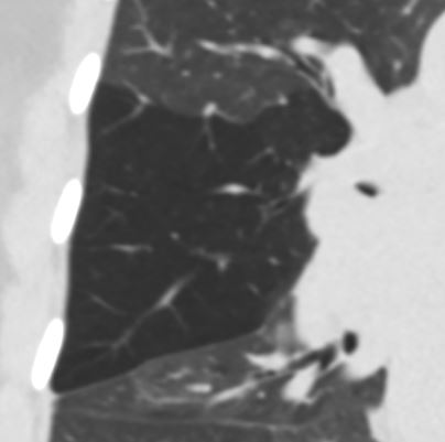
88-year-old smoker presents with shortness of breath. CT in the sagittal plane shows hyperinflation with significantly hyperinflated lower lobes and suggestion of centrilobular changes in the upper lobes
Ashley Davidoff MD TheCommonVein.net 135541
Central Obstructing LUL Squamous Cell Carcinoma
Left Upper Lobe Atelectasis
Hyperlucent Left Lower Lobe
Left Upper Lobe Atelectasis Hyperlucent Left Lower Lobe
Female patient with central squamous cell carcinoma of the lung with left upper lobe collapse and hyperinflation of the left lower lobe resulting in a Luftsichel sign
Noted spiculated lesion in the right upper lobe
Ashley Davidoff MD TheCommonVein.net 152Lu
Atelectatic Lung Collapsed Anteriorly
The Hyperinflated LLL is Seen Occupying the Left Apex
Luftsichel Sign

Female patient with central squamous cell carcinoma of the lung with left upper lobe collapse with atelectatic lung collapsed anteriorly. The hyperinflated LLL is seen occupying the left apex
Ashley Davidoff MD TheCommonVein.net 152Lu
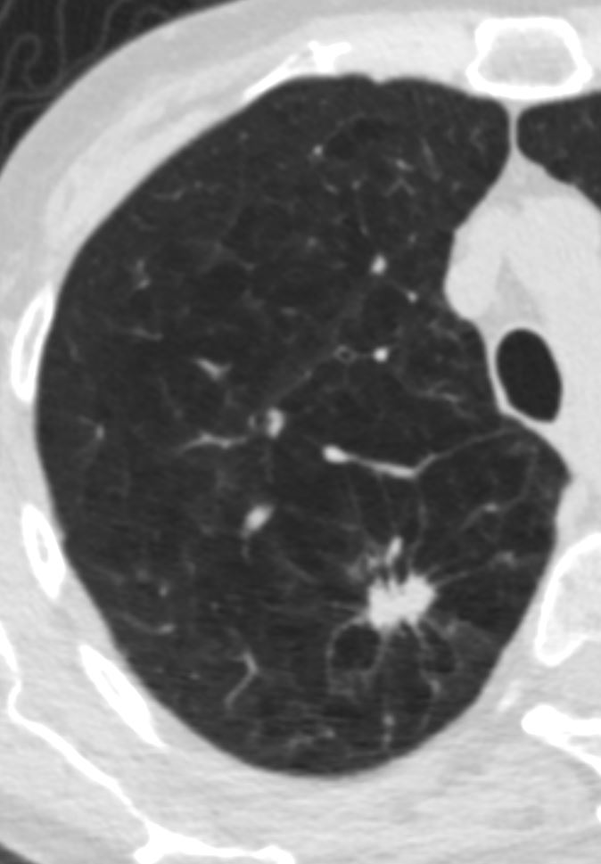
Focal Hyperinflation Secondary to Rounded Atelectasis
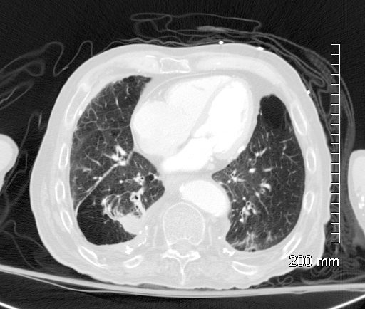
72-year-old male with a history of asbestos exposure presents with a cough. Axial CTscan shows a pleural based nodule with a comet tail and a series of lung markings folded into the nodule. There is subsegmental compensatory hyperinflation of the lateral segment of the right lower lobe Noted bilateral pleural thickening and pleural based calcification which is reminiscent of asbestos related disease. Early evolution of rounded atelectasis is also noted in the left lower lobe
Ashley Davidoff MD TheCommonVein.net RnD 240Lu
72-year-old male with a history of asbestos exposure presents with a cough. Axial CTscan shows a magnified view of a pleural based nodule with a comet tail and a series of lung markings folded into the nodule. There is subsegmental compensatory hyperinflation of the lateral segment of the right lower lobe Noted pleural thickening and pleural based calcification which is reminiscent of asbestos related disease.
Ashley Davidoff MD TheCommonVein.net RnD 240Lu
Swyer James
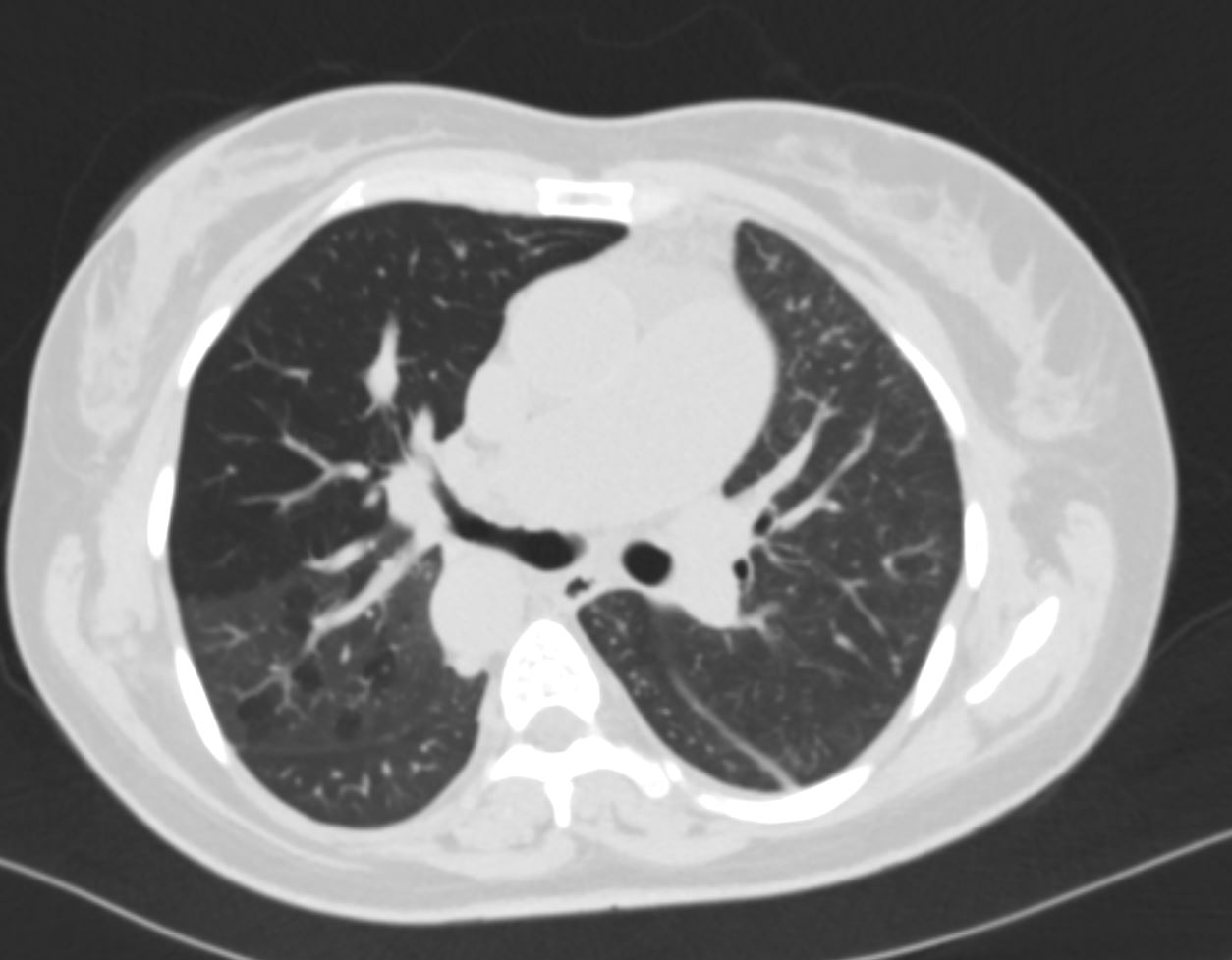
CT in axial projection suggests shows a hyperlucent anterior segment of the right upper lobe, narrowing of the segmental airway, and mosaic attenuation in the posterior segments Ashley Davidoff MD TheCommonVein.net artery-off-aorta-012
CT in coronal projection shows a hyperlucent anterior segment of the right upper lobe Ashley Davidoff MD TheCommonVein.net artery-off-aorta-013
Do you see a bull made of the soft tissues looking at the lucent lung?
Keep your eye on the eye of the bull as it will get progressively smaller as the bull goes to sleep and the airway narrows
CT in coronal projection shows a hyperlucent anterior segment of the right upper lobe Note the patent segmental airway subtending the upper lobe If you look with an artistic eye you can see a bull made of soft tissues looking at the hyperlucent lung The patent segmental airway is the “eye of the bull”) Ashley Davidoff MD TheCommonVein.net artery-off-aorta-014
CT in coronal projection shows a hyperlucent anterior segment of the right upper lobe Note that the “eye of the bull” has become smaller as the anterior segmental airway becomes progressively narrowed Ashley Davidoff MD TheCommonVein.net artery-off-aorta-014b